Introduction
Breast cancer is the second most common type of
cancer worldwide and the most common cancer type in women.
Triple-negative breast cancer (TNBC), accounting for 10–15%
(1) of all breast cancer cases, is
characterized by the absence of progesterone and estrogen receptor
as well as human epidermal growth factor receptor-2 (2). Management of TNBC is challenging due to
its aggressive behavior, lack of targeted therapies and relatively
poor prognosis (3). At present, only
few therapeutic options are available, and conventional
chemotherapy is the only effective adjuvant treatment for TNBC
patients after surgery (4), which,
however, has severe side effects. Therefore, it is important to
discover novel therapeutic agents with low toxicity for TNBC
treatment. Traditional Chinese Medicine has been used for cancer
treatment for a long time either alone or in combination with
western medicines (5). It is a
promising field providing numerous sources of anticancer agents,
which deserve further investigation for being developed into novel
clinical treatments.
Xihuang pill (XHP) is a well-known Chinese medicine
formula first mentioned in in a document from 1740 in the Qing
dynasty period of China (6). XHP
contains Niuhuang (Bos Taurus domesticus Gmelin), Shexiang
(Moschusmoschiferus Linnaeus), Ruxiang (Boswellia
carterii Birdw) and Moyao (Commiphora myrrha Engl.). Its
main uses are detoxification as well as relief of swelling and
pain. XHP is mainly used for the treatment of furunculosis,
scrofula and neoplasms. In the clinical practice, the anticancer
activities of XHP have been documented for malignancies including
breast and liver cancer as well as leukemia (7). While evidence for the antitumor effects
of XHP against TNBC is rare, a recent study by Pan et al
(8) was the first to assess the
effects of aqueous extract of XHP (AEXHP) on a TNBC cell line,
MDA-MB-231, revealing a cytotoxic effect, the possible mechanism of
which was indicated to be associated with induction of apoptosis;
however, the molecular mechanisms were not further investigated.
The aim of the present study was to investigate the effects of
AEXHP on another TNBC cell line, namely Hs578T, with the aim of
assessing its anti-proliferative activity and the underlying
molecular mechanisms. To the best of our knowledge, the present
study was the first to demonstrate that the cytotoxic effects of
AEXHP in TNBCs may be associated with induction of cell cycle
arrest.
Materials and methods
Chemicals and antibodies
XHP was purchased from Tong Ren Tang Technologies
Co., Ltd. (Beijing, China). Dimethyl sulfoxide (DMSO) and
3-(4,5-dimethyl-2-thiazolyl)-2, 5-diphenyl-2-H-tetrazolium bromide
(MTT) were purchased from Sigma-Aldrich (St. Louis, MO, USA).
Caspase-3 monoclonal antibody (cat. no. ab32351; 1:3,000 dilution)
was purchased from Abcam (Cambridge, UK). Cyclin dependent kinase 2
(CDK2) (cat. no. 2546; 1:5,000 dilution), cyclin A (cat. no. 4656;
1:5,000 dilution), cyclin E (cat. no. 4129; 1:5,000 dilution),
p21Cip1 (cat. no. 2947; 1:5,000 dilution), caspase-8
(cat. no. 9746; 1:4,000 dilution), B-cell lymphoma 2 (Bcl-2; cat.
no. 2870; 1:5,000 dilution) and Bcl-2-associated X protein (Bax;
ca. no. 5023; 1:5,000 dilution) monoclonal antibodies were
purchased from Cell Signaling Technology (Beverly, MA, USA).
β-actin rabbit monoclonal antibody (cat. no. TDY051; 1:5,000
dilution) was purchased from TDYbio (Beijing, China).
Peroxidase-conjugated goat anti-rabbit immunoglobulin (Ig) G (cat.
no. ZB-2306) and goat anti-mouse IgG (cat. no. ZF-0312; both
1:5,000 dilution) were purchased from the Zhongshang
Goldenbridge-BIO (Beijing, China).
Extract preparation
XHP (3 g) was immersed in 15 ml cold distilled water
and stirred for 2 h at 4°C. Subsequently, XHP was extracted for 2 h
at 37°C in an ultrasound oscillator and centrifuged at 1,500 × g
for 5 min at 4°C. The supernatant was centrifuged again at 4,800 ×
g for 15 min at 4°C, and the final supernatant was filtered through
a sterile microporous membrane (0.45 µm) and stored at −20°C. For
treatment of the cells, AEXHP was diluted with Dulbecco's modified
Eagle's medium (DMEM) to the desired concentrations.
Cell culture
The Hs578T human TNBC cell line was obtained from
the cell bank of Chinese Academy of Medical Sciences (Shanghai,
China). The cells were cultured in DMEM (Gibco, Thermo Fisher
Scientific, Inc., Waltham, MA, USA) with 10% fetal bovine serum
(Hyclone, Logan, UT, USA) and antibiotics (100 U/ml penicillin and
100 µg/ml streptomycin).
The MCF-10A human breast epithelial cell line also
obtained from the cell bank of Chinese Academy of Medical Sciences.
The cells were grown, maintained and treated in medium containing
DMEM/F-12 (Gibco) 1:1 mix, supplemented with human insulin (10
µg/ml), epidermal growth factor (20 ng/ml), cholera toxin (100
ng/ml), hydrocortisone (0.5 µg/ml), 5% horse serum (Hyclone) and
antibiotics (100 U/ml penicillin and 100 µg/ml streptomycin).
The cell lines were incubated in a humidified
atmosphere containing 5% CO2 at 37°C.
MTT assay
Cells (7.5×10³ or 1×10⁴/well)
were seeded in 96-well plates (Corning Inc., Corning, NY, USA) and
treated on the following day with various concentrations of AEXHP
(0, 4, 8, 12 or 16 mg/ml) for 12, 24 or 48 h [the units ‘weight per
volume’ refer to the quantity of solid XHP and the volume of water
used for extraction, which was performed according to a previously
published procedure (8)].
Subsequently, the cells were subjected to an MTT assay (9) and the optical density (OD) was determined
at 570 nm using a 96-well microplate reader (Applied Biosystems;
Thermo Fisher Scientific, Inc.). Since reduction of MTT only
occurred in metabolically active cells, the OD was a measure of the
viability of the cells. The viability rate was calculated according
to the following formula:
ODtreatment/ODcontrol × 100%.
Apoptosis assay
Hs578T Cells (2.4×10⁵) were seeded in a
30-mm culture dish (Corning Inc.). On the following day, cells were
treated with AEXHP (0, 4, 8 or 12 mg/ml). After 24 h of incubation,
cells were trypsinized, washed with cold phosphate-buffered saline
(PBS) and stained using an annexin V/FITC kit (BestBio, Shanghai,
China). The cells were then analyzed on a FACSCalibur flow
cytometer (Becton Dickinson, Franklin Lakes, NJ, USA).
Mitochondrial membrane potential
(Δψm) assay
Δψm loss was detected using a
mitochondrial membrane potential assay kit (Beyotime Institute of
Biotechnology, Haimen, China). In brief, Hs578T cells
(2.4×10⁵) were seeded into a 30-mm culture dish. On the
following day, the cells were treated with AEXHP (0, 4, 8 or 12
mg/ml). After 24 h of incubation, the cells were trypsinized and
incubated with JC-1 at 37°C for 15 min. Subsequent to washing with
JC-1 staining buffer twice, the cells were immediately analyzed by
flow cytometry.
Cell cycle distribution assay
Hs578T cells (6×10⁵) were seeded in a
50-mm culture dish. On the following day, the cells were treated
with AEXHP (0, 4, 8 or 12 mg/ml) for 48 h. Subsequent to
trypsinization and washing with PBS, cells were fixed in 1 ml
ice-cold 70% ethanol overnight at 4°C. The cells were centrifuged
(1,000 × g, 5 min, 4°C), washed with cold PBS and treated with
propidium iodide (PI)/RNase Staining Buffer (BD Pharmingen, San
Diego, CA, USA) for 15 min at room temperature in the dark. The
cell cycle distribution was then determined by flow cytometry. The
percentage of cells in G1, S and G2/M phase
was calculated using ModFit software v 3.2 (Becton Dickinson, San
Diego, CA, USA).
Western blot analysis
Hs578T cells (2.25×10⁶) were seeded in a
100-mm culture dish. On the following day, the cells were treated
with 0, 4, 8 or 12 mg/ml AEXHP. Following incubation for 24 or 48
h, the cells were harvested, washed with cold PBS and homogenized
with radioimmunoprecipitation assay lysis buffer (TDYbio). Total
protein was extracted and the protein concentration was determined
using a BCA protein assay kit (Thermo Fisher Scientific, Inc.).
Subsequently, the proteins were mixed with 5X loading buffer
(CWBIO, Beijing, China) and boiled for 5 min. Samples of 35 µg
protein were separated by 10% sodium dodecyl sulfate-polyacrylamide
gel electrophoresis, and transferred onto polyvinylidene difluoride
membranes (Millipore, Billerica, MA, USA). Membranes were blocked
for 2 h at room temperature with 5% fat-free milk (YiLi Inc.,
Huhehaote, China) or 5% bovine serum albumin (Amresco, Inc.,
Framingham, MA, USA) in 10 mM Tris-HCl, 0.1 M NaCl and 0.1%
Tween-20, pH7.4 (TBST), and incubated with specific primary
antibodies at 4°C overnight with gentle agitation. The membranes
were washed with cold TBST and incubated with peroxidase-conjugated
secondary antibody for 1 h. Visualization of the protein bands was
accomplished using an Immobilon Western Chemiluminescent HRP
Substrate (Millipore). The intensity of protein bands was measured
using ImageJ software 1.48 (National Institutes of Health,
Bethesda, MD, USA), normalized to that of β-actin and presented as
a percentage of the control.
Statistical analysis
Values are expressed as the mean ± standard
deviation of at least three independent experiments. Statistical
analyses were performed using the two-tailed Student's t-test with
SPSS 13.0 (SPSS, Inc., Chicago, IL, USA). P<0.05 was considered
to indicate a statistically significant difference.
Results
AEXHP reduces the viability of Hs578T
cells
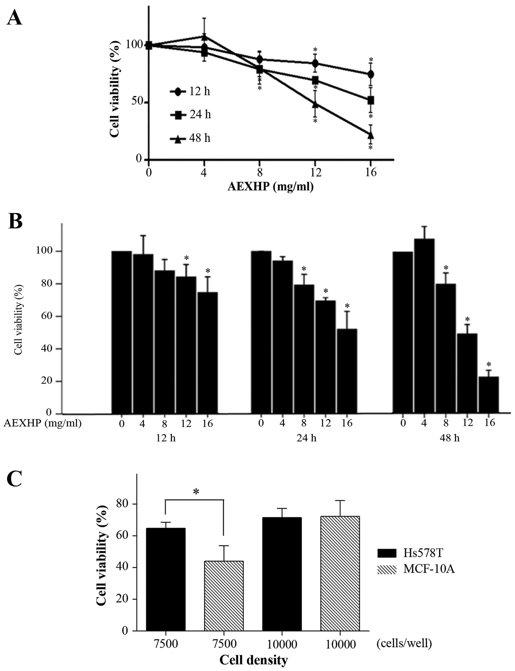
To evaluate the cytotoxic effects of AEXHP on Hs578T
cells, the viability of cells (7.5×10³/well) treated
with various concentrations of AEXHP (0–16 mg/ml) was determined
using an MTT assay. The results showed that AEXHP decreased the
number of viable cells in a dose- and time-dependent manner
(Fig. 1A and B). The inhibition of
Hs578T cell viability by AEXHP started relatively early at 12 h. At
the highest concentration used (16 mg/ml), incubation with AEXHP
for 12, 24 or 48 h significantly decreased the cell viability rate
to 74.67±9.63, 52.07±10.89 and 22.21±8.30%, respectively, while
there was no statistical difference between the control and the
treatment group at the lowest concentration (4 mg/ml) at each
time-point.
In order to investigate the difference in
cytotoxicity/anti-proliferative effects of AEXHP on MCF-10A normal
breast epithelial cells and Hs578T cells, each cell line was
treated with 12 mg/ml AEXHP for 24 h. At a seeding density of 7,500
cells/well, the cell viability rate of Hs578T and MCF-10A decreased
to 64.75±3.82 and 43.93±9.96%, respectively (Fig. 1C). There was a significant difference
in the viability rate between these two cell lines (P<0.05).
However, when the cell density was increased to 10,000 cells/well,
the decreases in cell viability were similar between Hs578T and
MCF-10A cells (71.67±10.60 and 72.41±9.80%, respectively, vs.
control) (Fig. 1C).
AEXHP induces apoptosis in Hs578T
cells
In order to assess whether the anti-proliferative
effects of AEXHP on Hs578T cells were due to induction of
apoptosis, annexin V/PI double staining and flow cytometric
analysis were used. After treatment with AEXHP (4, 8 or 12 mg/ml)
for 24 h, the percentage of early apoptotic cells increased from
1.6% in the control group to 3.9, 5.4 and 8.6%, respectively, and
the percentage of cells in late apoptosis increased from 8.0% in
the control group to 12.2, 13.8 and 15.7%, respectively (Fig. 2A). At 8 and 12 mg/ml, AEXHP
significantly increased the overall apoptotic rate. In conclusion,
AEXHP induced apoptosis in Hs578T cells in a dose-dependent manner
(Fig. 2B).
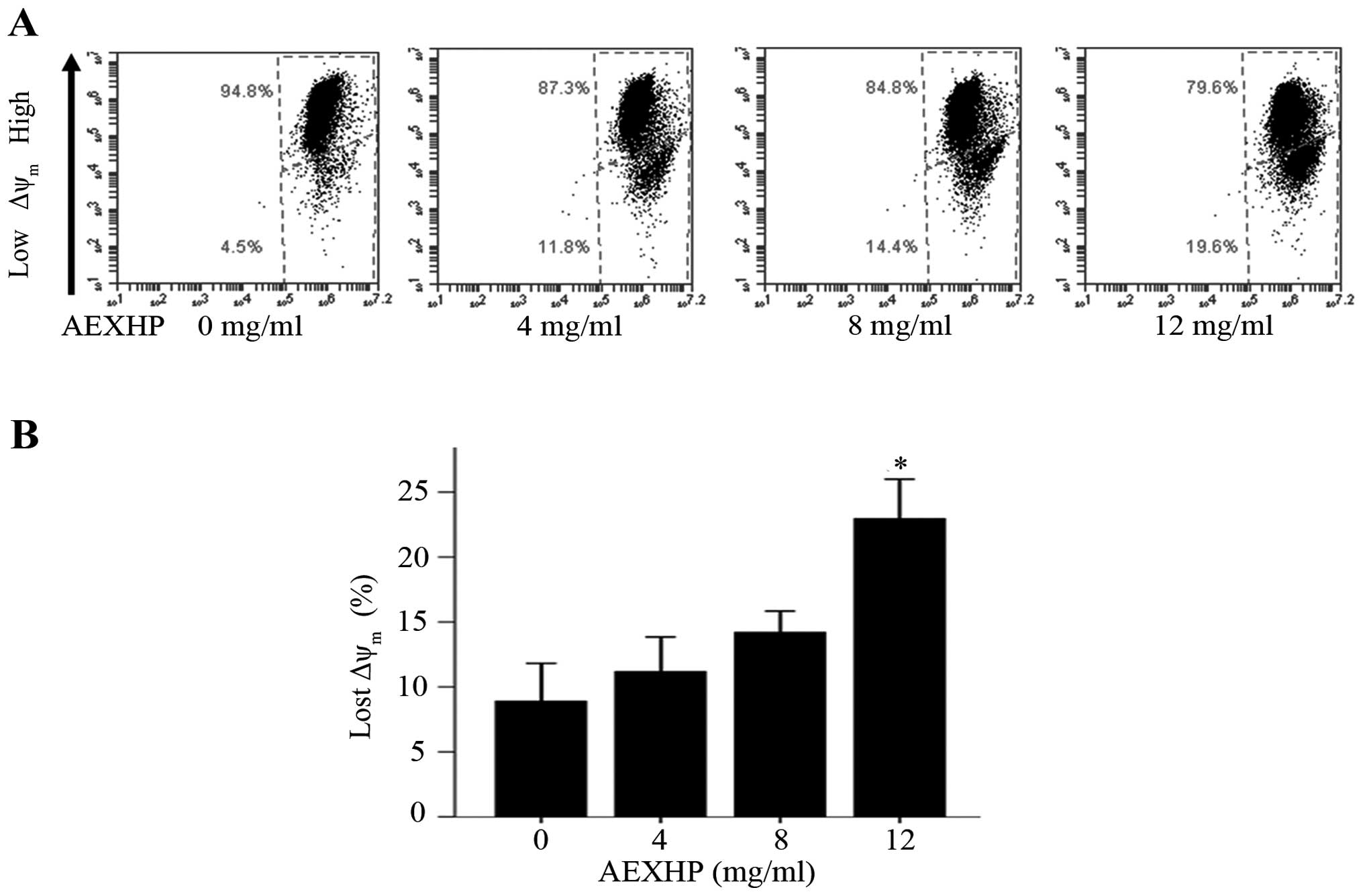
AEXHP treatment leads to depletion of
the Δψm in Hs578T cells
To assess whether the apoptosis of Hs578T cells was
associated with the depletion of the Δψm, Hs578T treated
with various concentrations of AEXHP were assessed by JC-1 staining
and flow cytometric analysis. After treatment with AEXHP (4, 8 and
12 mg/ml) for 24 h, the percentage of cells with Δψm
loss increased from 4.5% in the control group to 11.8, 14.4 and
19.6%, respectively, which was significant at the highest
concentration (P<0.05) (Fig. 3A and
B). Thus, AEXHP induced the depletion of the Δψm in
Hs578T cells in a dose-dependent manner.
AEXHP induces apoptosis via the
intrinsic pathway
Apoptosis may proceed via two major pathways, namely
the intrinsic and the extrinsic pathway (10), each of them leading to caspase-3
activation. The caspase family is the most prominent protease
family in apoptosis (11), and is
divided into two functional groups, namely the apoptosis initiators
(caspase-8, −9 and −10) and the apoptosis executors (caspase-3, −6
and −7) (12).
Bax is a well-known pro-apoptotic protein, while
Bcl-2 is an anti-apoptotic protein, and the Bcl-2/Bax ratio has a
decisive role regarding the induction of apoptosis (13,14). To
determine whether caspase-3, caspase-8, Bax and Bcl-2 proteins were
involved in apoptosis of Hs578T cells induced by AEXHP, the levels
of these proteins were assessed by western blot analysis. The
results showed that after treatment with AEXHP (4, 8 and 12 mg/ml)
for 24 h, levels of cleaved caspase-3 were increased to 1.70-,
1.81- and 1.84-fold of that of the control (P<0.05), while no
significant impact on the levels of procaspase-3, cleaved
caspase-8, procaspase-8, Bcl-2, Bax, and the Bcl-2/Bax ratio was
observed (Fig. 4A and B). In
conclusion, the results indicated that AEXHP induces apoptosis via
the intrinsic pathway, but not the extrinsic pathway, as only an
involvement of caspase-3 cleavage but no association with
caspase-8, and Bcl-2/Bax ratio was found.
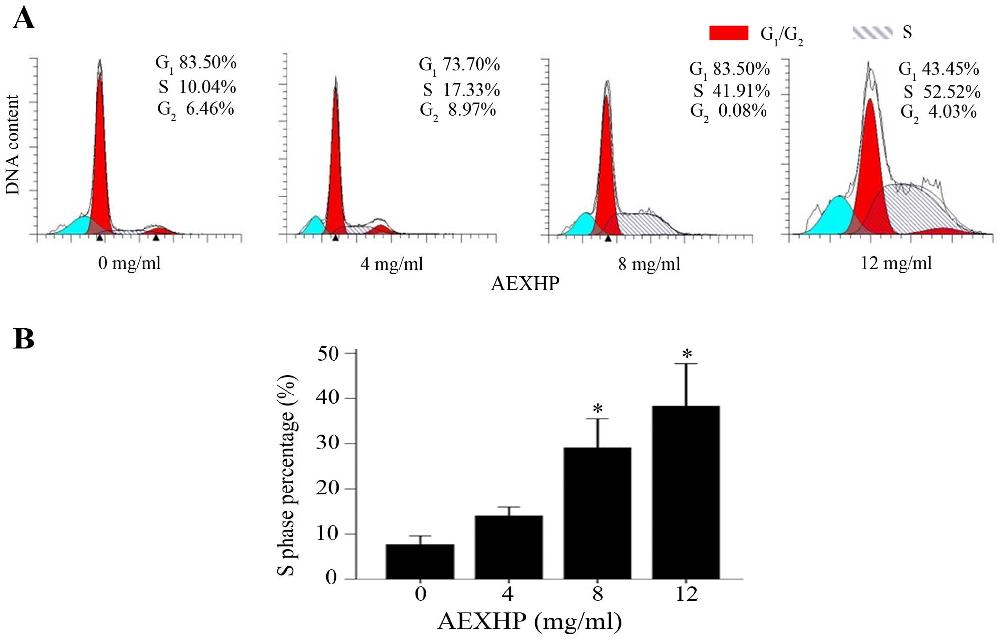
AEXHP induces cell cycle arrest in S
phase
Cell cycle regulation is important for cell
proliferation, and induction of cell cycle arrest is the major
mechanism via which numerous anti-tumor drugs exert their
anti-proliferative effects (15,16). To
examine whether the anti-proliferative effects of AEXHP on Hs578T
cells were associated with cell cycle arrest, their cell cycle
distribution was determined. The results showed that AEXHP
significantly affected the cell cycle distribution of Hs578T cells,
leading to cell cycle arrest at S phase in a dose-dependent manner
(Fig. 5B). While the S-phase
population was 10.04% in the untreated group, treatment with 4, 8
and 12 mg/ml AEXHP increased it to 17.33, 41.91 (P<0.05 vs.
control) and 52.52% (P<0.05 vs. control), respectively. Cell
populations in G1 or G2/M phase shifted
concomitantly to the changes detected in S phase (Fig. 5A), while there was no AEXHP
dose-dependent decrease or increase in the G1- or
G2/M-phase populations.
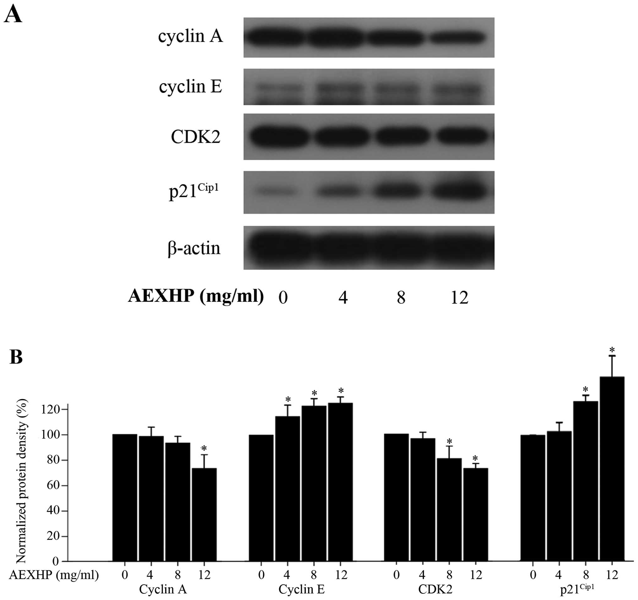
Effects of AEXHP on the expression of
cell cycle regulatory proteins
To explore the mechanisms by which AEXHP induced
cell cycle arrest of Hs578T cells at S phase, western blot analysis
was used to determine the expression of cell cycle regulatory
proteins. The results showed that treatment with various
concentrations of AEXHP (4, 8 or 12 mg/ml) for 48 h resulted in
decreased expression of cyclin A and CDK2, as well as increased
expression of cyclin E and p21Cip1, as compared to the
control group (Fig. 6A and B). The
decreased expression of cyclin A and CDK2, and the increased
expression of cyclin E and p21Cip1, may have contributed
to S-phase arrest of Hs578T cells induced by AEXHP.
Discussion
As mentioned above, the aim of the present study was
to assess the anti-proliferative effects of AEXHP on the Hs578T
TNBC cell line, and to investigate the underlying molecular
mechanisms, by performing MTT, apoptosis, cell cycle and western
blot assays.
The results of the MTT assay showed that AEXHP
inhibited the viability of Hs578T cells in a dose- and
time-dependent manner. The difference between the cytotoxic effects
of AEXHP on MCF-10A normal breast epithelial cells and Hs578T TNBCs
was depended on the cell density, which was mainly due to the cell
density-dependent sensitivity of the MCF-10A cells to AEXHP. When
the cell density was increased from 7,500 to 10,000 cells/well, the
viability of MCF-10A cells treated with 12 mg/ml AEXHP for 24 h
increased from 43.93±9.96 to 72.41±9.80%, while that of Hs578T
cells only increased marginally. The results of the present study
indicated that Hs578T was equally or less sensitive to AEXHP than
MCF-10A. However, due to differences in doubling times, it may be
impossible to directly compare the cytotoxic effects of AEXHP on
the MCF-10A normal breast epithelial cell line and Hs578T TNBCs at
identical seeding density, which represents a limitation of the MTT
assay performed in the present study. In fact, a previous study by
our group on another TNBC cell line, MDA-MB-231, demonstrated
cancer cell-specific toxicity in terms of MDA-MB-231 being more
sensitive to AEXHP than the MCF-10A cell line (17). It is therefore indicated that the
cytotoxic effects of AEXHP are cell type-specific, and whether they
are cancer-specific warrants further investigation in a panel of
cell lines.
In order to elucidate the mechanisms of the
anti-proliferative activity of AEXHP, the apoptotic rate and cell
cycle distribution of Hs578T treated with various concentrations of
AEXHP were assessed in the present study. The results showed that
AEXHP dose-dependently decreased the viability of Hs578T cells, and
simultaneously induced apoptosis and S-phase arrest. In order to
investigate via which pathways AEXHP induces apoptosis in Hs578T,
the protein expression of caspase-8 and caspase-3 were detected by
western blot analysis. While procaspase-3 was not significantly
affected, the levels of cleaved caspase-3 were significantly
increased following AEXHP treatment. However, the protein levels of
procaspase-8 and cleaved caspase-8 were not affected, leading to
the conclusion that AEXHP induces apoptosis via the intrinsic, but
not the extrinsic pathway.
Bax constitutes the ion channel of the mitochondrial
membrane in human cells, while Bcl-2 combines with Bax to inhibit
it, with the Bcl-2/Bax ratio therefore having a decisive role in
the induction of apoptosis (18). In
the present study, western blot analysis showed that Bax, Bcl-2 and
the Bcl-2/Bax ratio were not affected by AEXHP, leading to the
conclusion that the depletion of the Δψm induced by
AEXHP was not associated with the Bcl-2/Bax ratio, and was
therefore likely to be linked with factors directly destroying the
mitochondrial membrane, which requires to be further
elucidated.
Cell cycle progression is orchestrated by a complex
network of interactions between proteins, including cyclins, CDKs,
E3 ubiquitin ligase complexes, CDK activating kinase, CDC25
phosphatases and CDK inhibitors (19).
To explore the mechanisms by which AEXHP induces cell cycle arrest
in Hs578T cells at S phase, western blot analysis was used to
assess the modulation of the expression of cell cycle regulatory
proteins by AEXHP. The results showed that AEXHP treatment resulted
in decreased expression of cyclin A and CDK2, and increased
expression of cyclin E and p21Cip1, which explains for
the S-phase arrest observed. Cyclin A and CDK2 are specific S-phase
regulatory proteins (19), and the
increased cyclin E enhances G1-to-S-phase progression.
p21Cip1 not only forms complexes with cyclin D-, cyclin
E- or cyclin A-dependent kinase and inhibits their activities, but
also binds to and inhibits proliferating cell nuclear antigen to
curb the synthesis of DNA (20), which
is the main task during S phase. In addition, the expression of
cyclin B and CDK1 were assessed in the present study (data not
shown). The expression of these two proteins was not significantly
affected by AEXHP, which is in line with the observed S-phase
arrest, as cyclin B and CDK1 G2/M phase-specific cell
cycle regulatory proteins. The limitation of the present study was
that the effects of AEXHP were only assessed on one TNBC cell line,
Hs578T, in vitro. However, in a previous study by our group,
a xenograft tumor model using another TNBC cell line, MDA-MB-231,
was established, which proved the anti-tumor effects of AEXHP in
vivo (results not shown). Another limitation of the present
study was that the active components in the AEXHP may be different
from those in the serum of patients taking XHP for the treatment of
cancer. Furthermore, the cytotoxic effects of AEXHP on the Hs578T
cell line may not be equivalent to those of XHP in TNBC patients.
Further studies in the clinical setting are required to confirm the
antitumor effects of XHP on TNBC. Furthermore, the active
components of AEXHP should be isolated and their structure should
be identified in future studies.
In conclusion, the present study revealed that AEXHP
significantly inhibited the viability of Hs578T cells in
vitro in a dose- and time-dependent manner. The possible
underlying mechanisms include induction of apoptosis and cell cycle
arrest at S phase. AEXHP was indicated to induce apoptosis in
Hs578T cells via the intrinsic, Bcl-2/Bax-independent pathway.
S-phase arrest was likely to be due to the combined effects of
decreased expression of cyclin A and CDK2 as well as increased
expression of cyclin E and p21Cip1. The present study
therefore indicated that AEXHP is potent against the Hs578T TNBC
cell line, providing in vitro evidence to support the
clinical use of XHP as a prescription for TNBC.
Acknowledgements
The present study was supported by the key program
foundation of Beijing Administration of Traditional Chinese
Medicines (no. 2004-IV15).
References
|
1
|
Liedtke C, Bernemann C, Kiesel L and Rody
A: Genomic profiling in triple-negative breast cancer. Breast Care
(Basel). 8:408–413. 2013. View Article : Google Scholar : PubMed/NCBI
|
|
2
|
Bosch A, Eroles P, Zaragoza R, Viña JR and
Lluch A: Triple-negative breast cancer: Molecular features,
pathogenesis, treatment and current lines of research. Cancer Treat
Rev. 36:206–215. 2010. View Article : Google Scholar : PubMed/NCBI
|
|
3
|
Yadav BS, Sharma SC, Chanana P and Jhamb
S: Systemic treatment strategies for triple-negative breast cancer.
World J Clin Oncol. 5:125–133. 2014. View Article : Google Scholar : PubMed/NCBI
|
|
4
|
Jiao Q, Wu A, Shao G, Peng H, Wang M, Ji
S, Liu P and Zhang J: The latest progress in research on triple
negative breast cancer (TNBC): Risk factors, possible therapeutic
targets and prognostic markers. J Thorac Dis. 6:1329–1335.
2014.PubMed/NCBI
|
|
5
|
Meng Z, Garrett CR, Shen Y, Liu L, Yang P,
Huo Y, Zhao Q, Spelman AR, Ng CS, Chang DZ, et al: Prospective
randomised evaluation of traditional Chinese medicine combined with
chemotherapy: A randomised phase II study of wild toad extract plus
gemcitabine in patients with advanced pancreatic adenocarcinomas.
Br J Cancer. 107:411–416. 2012. View Article : Google Scholar : PubMed/NCBI
|
|
6
|
Guo Q, Lin J, Liu R, Gao Y, He S, Xu X,
Hua B, Li C, Hou W, Zheng H and Bao Y: Review on the applications
and molecular mechanisms of Xihuang pill in tumor treatment. Evid
Based Complement Alternat Med. 2015:8543072015. View Article : Google Scholar : PubMed/NCBI
|
|
7
|
Jin SR, Zhu BD and Qin XH: Comparative
study of anti-tumors effects of Xi Huang Pellet by Different
Processing Methods. Shi Zhen Guo Yi Guo Yao. 19:1735–1737.
2008.
|
|
8
|
Pan G, Wang W, Wang L, Zhang F, Yin X,
Wang J and Liang R: Anti-breast cancer effects and mechanisms of
Xihuang pill on human breast cancer cell lines. J Tradit Chin Med.
33:770–778. 2013. View Article : Google Scholar : PubMed/NCBI
|
|
9
|
Wang W, Li N, Luo M, Zu Y and Efferth T:
Antibacterial activity and anticancer activity of Rosmarinus
officinalis L. essential oil compared to that of its main
components. Molecules. 17:2704–2713. 2012. View Article : Google Scholar : PubMed/NCBI
|
|
10
|
Fulda S and Debatin KM: Extrinsic versus
intrinsic apoptosis pathways in anticancer chemotherapy. Oncogene.
25:4798–4811. 2006. View Article : Google Scholar : PubMed/NCBI
|
|
11
|
Budihardjo I, Oliver H, Lutter M, Luo X
and Wang X: Biochemical pathways of caspase activation during
apoptosis. Annu Rev Cell Dev Biol. 15:269–290. 1999. View Article : Google Scholar : PubMed/NCBI
|
|
12
|
Nicholson DW: Caspase structure,
proteolytic substrates, and function during apoptotic cell death.
Cell Death Differ. 6:1028–1042. 1999. View Article : Google Scholar : PubMed/NCBI
|
|
13
|
Strasser A, Huang DCS and Vaux DL: The
role of the bcl-2/ced-9 gene family in cancer and general
implications of defects in cell death control for tumourigenesis
and resistance to chemotherapy. Biochim Biophys Acta.
1333:F151–F178. 1997.PubMed/NCBI
|
|
14
|
Gao N, Budhraja A, Cheng S, Yao H, Zhang Z
and Shi X: Induction of apoptosis in human leukemia cells by grape
seed extract occurs via activation of c-Jun NH2-terminal kinase.
Clin Cancer Res. 15:140–149. 2009. View Article : Google Scholar : PubMed/NCBI
|
|
15
|
Shapiro GI and Harper JW: Anticancer drug
targets: Cell cycle and checkpoint control. J Clin Invest.
104:1645–1653. 1999. View
Article : Google Scholar : PubMed/NCBI
|
|
16
|
Choi EJ, Oh HM, Wee H, Choi CS, Choi SC,
Kim KH, Han WC, Oh TY, Kim SH and Jun CD: Eupatilin exhibits a
novel anti-tumor activity through the induction of cell cycle
arrest and differentiation of gastric carcinoma AGS cells.
Differentiation. 77:412–423. 2009. View Article : Google Scholar : PubMed/NCBI
|
|
17
|
Zheng W, Han S, Jiang S, He X, Li X, Ding
H, Cao M and Li P: The multi-effect of Xihuang Pill on MDA-MB-231
cell lines in vitro and in vivo. Mol Med Rep. (In press).
|
|
18
|
Wong RS: Apoptosis in cancer: From
pathogenesis to treatment. J Exp Clin Cancer Res. 30:872011.
View Article : Google Scholar : PubMed/NCBI
|
|
19
|
Diaz-Moralli S, Tarrado-Castellarnau M,
Miranda A and Cascante M: Targeting cell cycle regulation in cancer
therapy. Pharmacol Ther. 138:255–271. 2013. View Article : Google Scholar : PubMed/NCBI
|
|
20
|
Sherr CJ and Roberts JM: CDK inhibitors:
Positive and negative regulators of G1-phase progression. Genes
Dev. 13:1501–1512. 1999. View Article : Google Scholar : PubMed/NCBI
|