Spandidos Publications Logo
  • About
    • About Spandidos
    • Aims and Scopes
    • Abstracting and Indexing
    • Editorial Policies
    • Reprints and Permissions
    • Job Opportunities
    • Terms and Conditions
    • Contact
  • Journals
    • All Journals
    • Oncology Letters
      • Oncology Letters
      • Information for Authors
      • Editorial Policies
      • Editorial Board
      • Aims and Scope
      • Abstracting and Indexing
      • Bibliographic Information
      • Archive
    • International Journal of Oncology
      • International Journal of Oncology
      • Information for Authors
      • Editorial Policies
      • Editorial Board
      • Aims and Scope
      • Abstracting and Indexing
      • Bibliographic Information
      • Archive
    • Molecular and Clinical Oncology
      • Molecular and Clinical Oncology
      • Information for Authors
      • Editorial Policies
      • Editorial Board
      • Aims and Scope
      • Abstracting and Indexing
      • Bibliographic Information
      • Archive
    • Experimental and Therapeutic Medicine
      • Experimental and Therapeutic Medicine
      • Information for Authors
      • Editorial Policies
      • Editorial Board
      • Aims and Scope
      • Abstracting and Indexing
      • Bibliographic Information
      • Archive
    • International Journal of Molecular Medicine
      • International Journal of Molecular Medicine
      • Information for Authors
      • Editorial Policies
      • Editorial Board
      • Aims and Scope
      • Abstracting and Indexing
      • Bibliographic Information
      • Archive
    • Biomedical Reports
      • Biomedical Reports
      • Information for Authors
      • Editorial Policies
      • Editorial Board
      • Aims and Scope
      • Abstracting and Indexing
      • Bibliographic Information
      • Archive
    • Oncology Reports
      • Oncology Reports
      • Information for Authors
      • Editorial Policies
      • Editorial Board
      • Aims and Scope
      • Abstracting and Indexing
      • Bibliographic Information
      • Archive
    • Molecular Medicine Reports
      • Molecular Medicine Reports
      • Information for Authors
      • Editorial Policies
      • Editorial Board
      • Aims and Scope
      • Abstracting and Indexing
      • Bibliographic Information
      • Archive
    • World Academy of Sciences Journal
      • World Academy of Sciences Journal
      • Information for Authors
      • Editorial Policies
      • Editorial Board
      • Aims and Scope
      • Abstracting and Indexing
      • Bibliographic Information
      • Archive
    • International Journal of Functional Nutrition
      • International Journal of Functional Nutrition
      • Information for Authors
      • Editorial Policies
      • Editorial Board
      • Aims and Scope
      • Abstracting and Indexing
      • Bibliographic Information
      • Archive
    • International Journal of Epigenetics
      • International Journal of Epigenetics
      • Information for Authors
      • Editorial Policies
      • Editorial Board
      • Aims and Scope
      • Abstracting and Indexing
      • Bibliographic Information
      • Archive
    • Medicine International
      • Medicine International
      • Information for Authors
      • Editorial Policies
      • Editorial Board
      • Aims and Scope
      • Abstracting and Indexing
      • Bibliographic Information
      • Archive
  • Articles
  • Information
    • Information for Authors
    • Information for Reviewers
    • Information for Librarians
    • Information for Advertisers
    • Conferences
  • Language Editing
Spandidos Publications Logo
  • About
    • About Spandidos
    • Aims and Scopes
    • Abstracting and Indexing
    • Editorial Policies
    • Reprints and Permissions
    • Job Opportunities
    • Terms and Conditions
    • Contact
  • Journals
    • All Journals
    • Biomedical Reports
      • Information for Authors
      • Editorial Policies
      • Editorial Board
      • Aims and Scope
      • Abstracting and Indexing
      • Bibliographic Information
      • Archive
    • Experimental and Therapeutic Medicine
      • Information for Authors
      • Editorial Policies
      • Editorial Board
      • Aims and Scope
      • Abstracting and Indexing
      • Bibliographic Information
      • Archive
    • International Journal of Epigenetics
      • Information for Authors
      • Editorial Policies
      • Editorial Board
      • Aims and Scope
      • Abstracting and Indexing
      • Bibliographic Information
      • Archive
    • International Journal of Functional Nutrition
      • Information for Authors
      • Editorial Policies
      • Editorial Board
      • Aims and Scope
      • Abstracting and Indexing
      • Bibliographic Information
      • Archive
    • International Journal of Molecular Medicine
      • Information for Authors
      • Editorial Policies
      • Editorial Board
      • Aims and Scope
      • Abstracting and Indexing
      • Bibliographic Information
      • Archive
    • International Journal of Oncology
      • Information for Authors
      • Editorial Policies
      • Editorial Board
      • Aims and Scope
      • Abstracting and Indexing
      • Bibliographic Information
      • Archive
    • Medicine International
      • Information for Authors
      • Editorial Policies
      • Editorial Board
      • Aims and Scope
      • Abstracting and Indexing
      • Bibliographic Information
      • Archive
    • Molecular and Clinical Oncology
      • Information for Authors
      • Editorial Policies
      • Editorial Board
      • Aims and Scope
      • Abstracting and Indexing
      • Bibliographic Information
      • Archive
    • Molecular Medicine Reports
      • Information for Authors
      • Editorial Policies
      • Editorial Board
      • Aims and Scope
      • Abstracting and Indexing
      • Bibliographic Information
      • Archive
    • Oncology Letters
      • Information for Authors
      • Editorial Policies
      • Editorial Board
      • Aims and Scope
      • Abstracting and Indexing
      • Bibliographic Information
      • Archive
    • Oncology Reports
      • Information for Authors
      • Editorial Policies
      • Editorial Board
      • Aims and Scope
      • Abstracting and Indexing
      • Bibliographic Information
      • Archive
    • World Academy of Sciences Journal
      • Information for Authors
      • Editorial Policies
      • Editorial Board
      • Aims and Scope
      • Abstracting and Indexing
      • Bibliographic Information
      • Archive
  • Articles
  • Information
    • For Authors
    • For Reviewers
    • For Librarians
    • For Advertisers
    • Conferences
  • Language Editing
Login Register Submit
  • This site uses cookies
  • You can change your cookie settings at any time by following the instructions in our Cookie Policy. To find out more, you may read our Privacy Policy.

    I agree
Search articles by DOI, keyword, author or affiliation
Search
Advanced Search
presentation
Biomedical Reports
Join Editorial Board Propose a Special Issue
Print ISSN: 2049-9434 Online ISSN: 2049-9442
Journal Cover
December-2017 Volume 7 Issue 6

Full Size Image

Sign up for eToc alerts
Recommend to Library

Journals

International Journal of Molecular Medicine

International Journal of Molecular Medicine

International Journal of Molecular Medicine is an international journal devoted to molecular mechanisms of human disease.

International Journal of Oncology

International Journal of Oncology

International Journal of Oncology is an international journal devoted to oncology research and cancer treatment.

Molecular Medicine Reports

Molecular Medicine Reports

Covers molecular medicine topics such as pharmacology, pathology, genetics, neuroscience, infectious diseases, molecular cardiology, and molecular surgery.

Oncology Reports

Oncology Reports

Oncology Reports is an international journal devoted to fundamental and applied research in Oncology.

Experimental and Therapeutic Medicine

Experimental and Therapeutic Medicine

Experimental and Therapeutic Medicine is an international journal devoted to laboratory and clinical medicine.

Oncology Letters

Oncology Letters

Oncology Letters is an international journal devoted to Experimental and Clinical Oncology.

Biomedical Reports

Biomedical Reports

Explores a wide range of biological and medical fields, including pharmacology, genetics, microbiology, neuroscience, and molecular cardiology.

Molecular and Clinical Oncology

Molecular and Clinical Oncology

International journal addressing all aspects of oncology research, from tumorigenesis and oncogenes to chemotherapy and metastasis.

World Academy of Sciences Journal

World Academy of Sciences Journal

Multidisciplinary open-access journal spanning biochemistry, genetics, neuroscience, environmental health, and synthetic biology.

International Journal of Functional Nutrition

International Journal of Functional Nutrition

Open-access journal combining biochemistry, pharmacology, immunology, and genetics to advance health through functional nutrition.

International Journal of Epigenetics

International Journal of Epigenetics

Publishes open-access research on using epigenetics to advance understanding and treatment of human disease.

Medicine International

Medicine International

An International Open Access Journal Devoted to General Medicine.

Journal Cover
December-2017 Volume 7 Issue 6

Full Size Image

Sign up for eToc alerts
Recommend to Library

  • Article
  • Citations
    • Cite This Article
    • Download Citation
    • Create Citation Alert
    • Remove Citation Alert
    • Cited By
  • Similar Articles
    • Related Articles (in Spandidos Publications)
    • Similar Articles (Google Scholar)
    • Similar Articles (PubMed)
  • Download PDF
  • Download XML
  • View XML
Case Report Open Access

Solitary pulmonary capillary hemangioma presents as ground glass opacity on computed tomography indicating adenocarcinoma in situ/atypical adenomatous hyperplasia: A case report

  • Authors:
    • Yanmei Zhu
    • Ning Qu
    • Lili Sun
    • Xiao Meng
    • Xiaoyan Li
    • Yong Zhang
  • View Affiliations / Copyright

    Affiliations: Department of Pathology, Cancer Hospital of China Medical University, Liaoning Cancer Hospital and Institute, Shenyang, Liaoning 110042, P.R. China, Department of Radiology, Cancer Hospital of China Medical University, Liaoning Cancer Hospital and Institute, Shenyang, Liaoning 110042, P.R. China
    Copyright: © Zhu et al. This is an open access article distributed under the terms of Creative Commons Attribution License.
  • Pages: 515-519
    |
    Published online on: October 9, 2017
       https://doi.org/10.3892/br.2017.997
  • Expand metrics +
Metrics: Total Views: 0 (Spandidos Publications: | PMC Statistics: )
Metrics: Total PDF Downloads: 0 (Spandidos Publications: | PMC Statistics: )
Cited By (CrossRef): 0 citations Loading Articles...

This article is mentioned in:


Abstract

Solitary pulmonary capillary hemangioma (SPCH) is a rare type of benign lung tumor, which must be distinguished from early lung cancer and precancerous lesions of the lung that manifest in a similar way upon imaging. The current study describes a case of SPCH and a review of the literature is performed. The patient was a 40-year-old Chinese woman who was referred to the Liaoning Cancer Hospital and Institute (Shenyang, China) in October, 2015 with a cough without obvious inducement. Computed tomography (CT) demonstrated pure ground glass opacity (GGO) in the right upper lung. Following systemic anti-inflammatory therapy over 6 months, the lesion did not exhibit any change on CT and was suspected to be an adenocarcinoma in situ (AIS) or atypical adenomatous hyperplasia (AAH). Video‑assisted thoracic surgery wedge resection was subsequently performed. Frozen section diagnosis revealed a benign tumor without atypical epithelial cells. Subsequent to surgery, paraffin sections demonstrated that the tumor contained narrow alveolar cavities, thickened alveolar septa and a clear boundary separating it from healthy lung tissue. Furthermore, the proliferation lumens in the alveolar septa were lined with a single layer of flat cells. Immunohistochemical staining revealed that the flat cells were positive for cluster of differentiation CD31 and CD34, and negative for thyroid transcription factor-1 and cytokeratin. The proliferation of capillary vessels lead to the thickened alveolar septa and the tumor was diagnosed as SPCH. When imaging examination demonstrates a GGO in lung, SPCH must be considered in the differential diagnosis of AIS/AAH. As the prognosis of these lesions is entirely different, a pathological examination must be conducted to ensure a correct diagnosis.

Introduction

Solitary pulmonary capillary hemangioma (SPCH) is a rare type of benign lung tumor that is difficult to diagnose due to a lack of symptoms, small lesion size and inconspicuous histological features (1). According to our search of previous literature, only 12 SPCH cases have been reported (1–10). However, each of these studies recorded only 1 or 2 cases, and thus SPCH has not been studied in depth, which may explain the absence of a description of SPCH in the World Health Organization classification of lung tumors in 2015 (11).

SPCH is a benign lesion with an unclear etiology. In the previously reported cases, all patients were asymptomatic and the lesion was discovered incidentally rather than clinically (1–10). Additionally, tumor growth was universally slow and no patients succumbed from the disease (1–10). Improvements in understanding and recognition of SPCH by radiologists, surgeons and pathologists may aid to elucidate its epidemiology.

SPCH exhibits ground glass opacity (GGO) on computed tomography (CT), and the GGO lesion may be suspected to be an adenocarcinoma in situ (AIS)/atypical adenomatous hyperplasia (AAH) and/or to have focal inflammation (1). The prognosis and treatment of these lesions vary; SPCH is a benign lesion and may be treated by complete tumor resection, while AIS/AAH may be malignant or exhibit malignant potential and require chemotherapy and/or radiotherapy in addition to surgical resection. Meanwhile, inflammation is typically treated with anti-inflammatory therapy (1,11). Thus, a detailed pathological examination is key to ensure correct diagnosis and subsequent treatment.

In the present study, a clinicopathological study of an SPCH case has been reported alongside a review of the literature. The purpose of the study was to define the clinical manifestation, radiological, histological and immunohistochemical features, and diagnosis of SPCH, as a currently unrecognized lesion.

Case report

A 40-year-old Chinese woman was referred to the Liaoning Cancer Hospital and Institute (Shenyang, China) with a cough in October, 2015 without obvious inducement. The patient had 
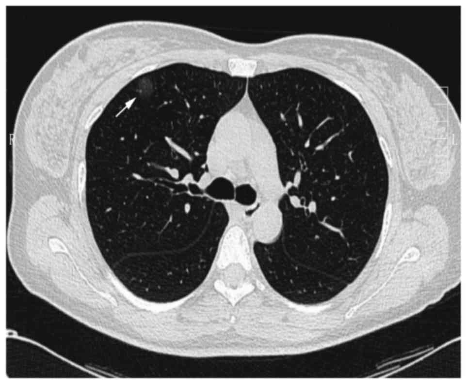
no phlegm or dyspnea chest discomfort. The chest CT indicated a pure GGO lesion (size, 19×17 mm) in the right upper lung (Fig. 1). Laboratory findings, including tumor markers, such as carcinoembryonic antigen and carbohydrate antigen 19-9 were within the normal ranges at 1.7 ng/ml (normal range 0–5 ng/ml) and 17.5 U/ml (normal range 0–37 U/ml), respectively (12). Following systemic anti-inflammatory therapy for 6 months, the size and density of the lesion had not changed according to CT. Although no abnormal uptake of fluorine-18-fluorodeoxyglucose in the lesion was observed during positron emission tomography-CT, it was suspected to be an AIS or AAH, and video-assisted thoracic surgery wedge resection was 
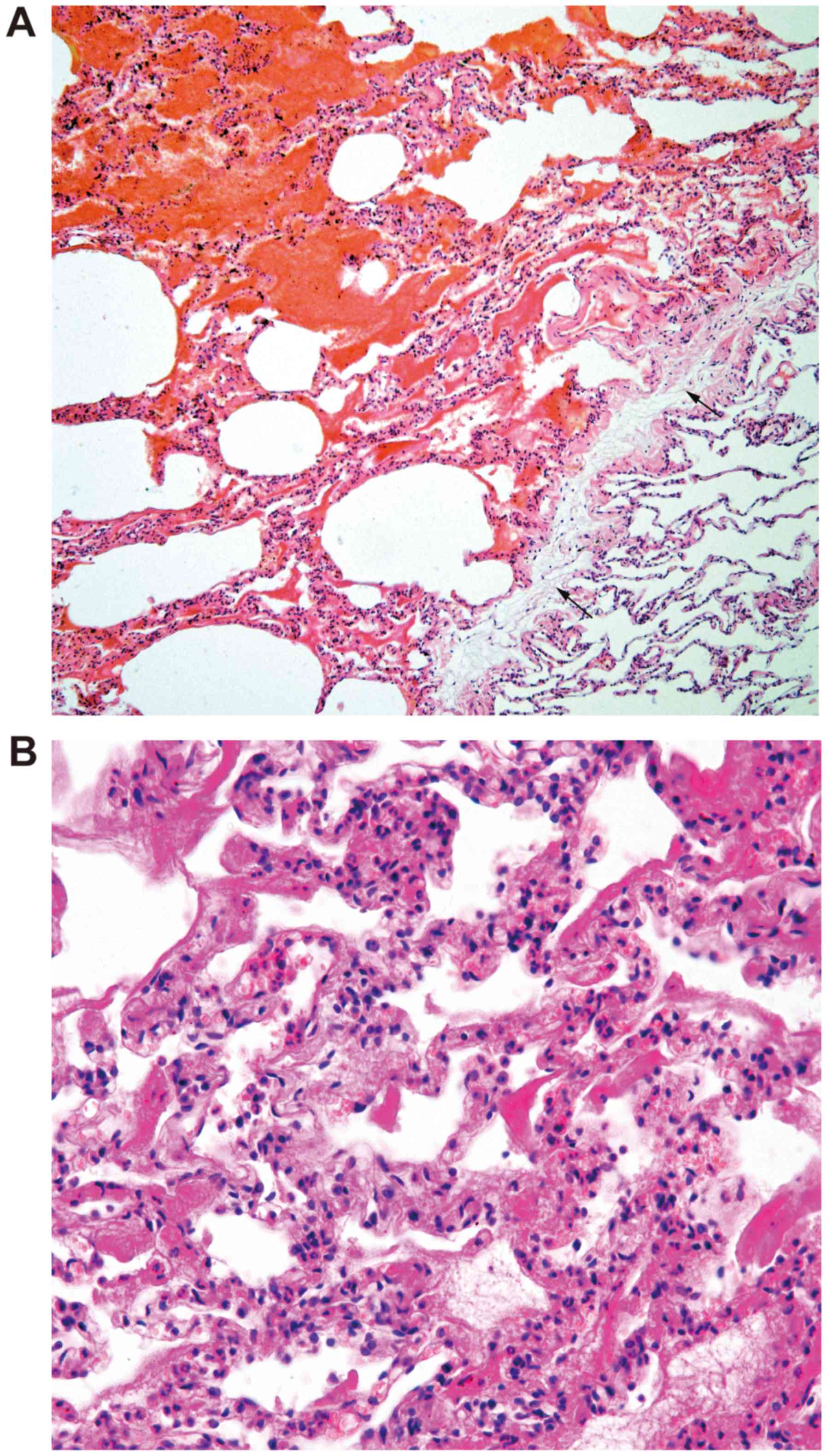
performed. Microscopically, a clear boundary between the tumor and normal lung tissue was observed in the low-power field (Fig. 2A). The tumor consisted of a narrow alveolar cavity, the alveolar septa was thickened with proliferated lumens that were various sizes and the lumens were lined with single-layer flat cells without atypical epithelial cells (Fig. 2B). Based on the above-mentioned findings, the intraoperative frozen sections demonstrated that the 
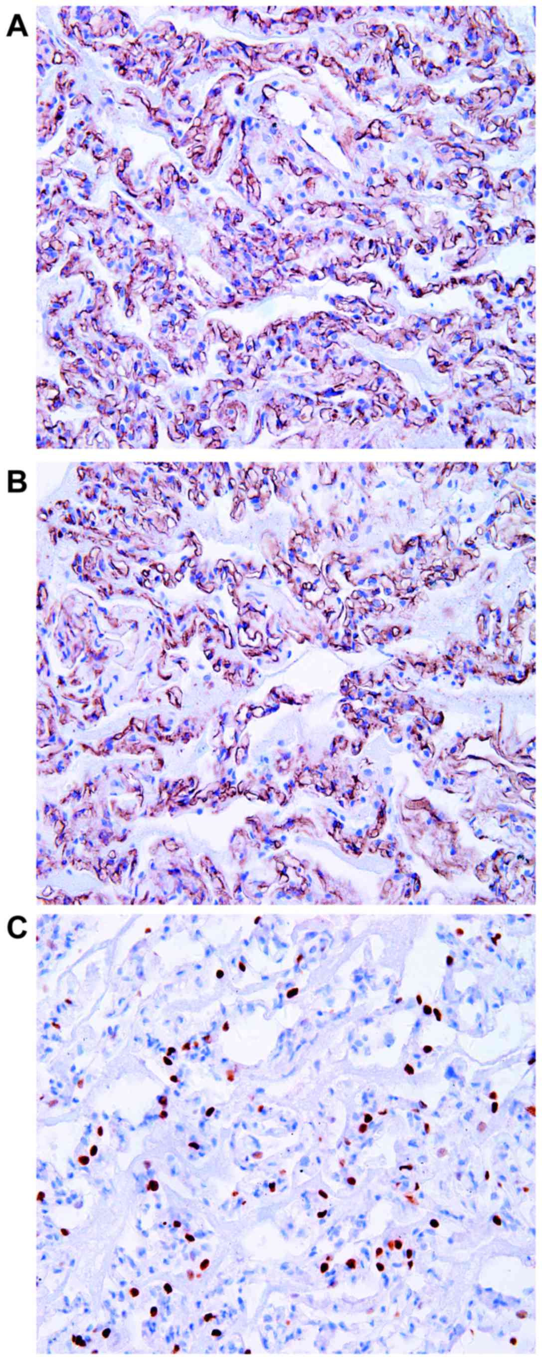
lesion was a benign tumor. Later, immunohistochemical staining of a paraffin section revealed the flat cells were positive for cluster of differentiation CD34 (Fig. 3A) and CD31 (Fig. 3B), and negative for D2-40, cytokeratin and thyroid transcription factor-1 (Fig. 3C). Therefore, the patient was diagnosed with SPCH and was followed up for 13 months after surgery with no recurrence.

Figure 1.

CT manifestation of the lung lesion. The chest CT exhibited a pure ground glass opacity lesion located in the subpleural area of the right upper lung, ~19×17 mm in size, with a clear boundary and no spiculations. CT, computed tomography.

Figure 2.

Morphological features of the lesion. (A) A clear boundary between the tumor and normal lung tissue is apparent (hematoxylin and eosin staining; magnification, ×40). (B) The lesion exhibited thickening of the alveolar septa caused by the proliferation of lumens that were varied in size. The lumens were lined with single-layer flat cells without cytological atypia. The alveolar cavity had become narrowed (hematoxylin and eosin staining; magnification, ×400).

Figure 3.

Immunohistochemical staining of the lesion. The flat cells were: (A) Positive for CD34 (DAB staining; magnification, ×400), (B) positive for CD31 (DAB staining; magnification, ×400) and (C) negative for thyroid transcription factor-1 (DAB staining; magnification, ×400). CD, cluster of differentiation; DAB, 3′-diaminobenzidine.

Discussion

SPCH is a particularly rare type of benign tumor, characterized by the proliferation of capillary vessels. It was initially identified by Fugo et al (1) in 2006. Since then, only 13 cases have been reported (including the present case; Table I). There were seven males and six females, and the age ranged from 40 to 61 years (mean age, 52.8 years). The lesions were located on the left side in six cases and on the right side in seven cases. Two were located in the upper lobe, two in the middle lobe and nine cases were located in the lower lobe. The tumor size ranged from 7 to 20 mm (mean, 12 mm). SPCH tends to present as a single small lesion, which is located in the lower lobe without any symptoms.

Table I.

Patient profile of solitary pulmonary capillary hemangioma.

Table I.

Patient profile of solitary pulmonary capillary hemangioma.

CaseAuthor, yearSexAge, yearsDetectedCT findingSize (mm)LocationSurgeryPathological finding(Refs)
  1Fugo et al 2006M59Medical check upMixed GGO19×11LL peripheralSegThe lesion showed  1
  2Fugo et al 2006F48Medical check upMixed GGO13×12RM peripheralWRthickening of the  1
  3Kato et al 2009M55Medical check upMixed GGO11RL peripheralSegalveolar septa caused  2
  4Hakiri et al 2010M45Medical check upMixed GGO12×11LL peripheralSegby the proliferation  3
  5Sakaguchi et al 2014F53Medical check upPure GGO20×20LU not peripheralLobof the capillary  4
  6Taniguchi et al 2010F59Medical check upPure GGO11×5RL peripheralWRvessels without  5
  7Yanagawa et al 2007M58Medical check upPure GGO  8RLWRcytological atypia or  6
  8Uegami et al 2008F54Medical check upSolid nodule12×11RM peripheralWRinflammatory  7
  9Shimada et al 2012M61Medical check upMixed GGO10LL peripheralLobbackground.  8
10Shimada et al 2012M42Medical check upPure GGO  7RL peripheralWR   8
11Matsushita et al 2012M58Medical check upMixed GGO  7LLN.D.   9
12Isaka et al 2013F55Medical check upMixed GGO7×4.6LL peripheralWR 10
13Yanmei et al 2017F40CoughPure GGO19×17RU peripheralWR Present case

[i] CT, computed tomography; GGO, ground-glass opacity; LL, left lower lobe; LU, left upper lobe; RL, right lower lobe; RM, right middle lobe; RU, right upper lobe; Seg, segmentectomy; WR, wedge resection; Lob, lobectomy; N.D., not defined; M, male; F, female.

Observation of GGO is typical upon imaging of cases of SPCH (1). In 13 cases, four were pure GGO, and eight were mixed GGO and one case exhibited a solid nodule. The pure GGO may represent a limited proliferation of capillary vessels in the alveolar septum, which is followed by a thickened alveolar septum and a narrowed alveolar space, although the space was preserved and still contained air. The solid aspect of the GGO may be the result of the capillary vessel congestion, which may cause alveolar collapse and a resulting lack of space to contain air.

When GGO in the peripheral lung is observed, AIS/AAH may be highly suspected. It is difficult to distinguish AIS/AAH from SPCH by imaging examination alone. During surgery, Shimada et al (8) observed that the SPCH became impalpable following reiterative palpation. This finding may be caused by squeezing the blood in capillary vessels followed by constriction of the thickened alveolar septum and the lesion feeling the same as normal lung tissue. However, in AIS/AAH, based upon the different histopathological manifestations, the nodule would not be impalpable, as there are collagenous fibers or elastic fibers in the alveolar septa rather than capillary vessels. Therefore, when tumors are located in the peripheral lung, present with a GGO image and cannot be palpated following reiterative palpation during surgery, a diagnosis of SPCH may be determined. SPCH is a benign tumor, whereas AIS/AAH are malignant or have malignant potential. Therefore, it is critical to differentiate these lesions in the clinical setting.

Histopathology is an important method, as the histological appearance between SPCH and AIS/AAH is completely different (11). AIS often occurs in the peripheral lung, very close to the pleura. AIS is a small (≤3 cm) adenocarcinoma with growth restricted to neoplastic cells along pre-existing alveolar structure (lepidic growth), lacking stroma, vascular, alveolar space or pleural invasion. Grossly, it is a poorly defined nodule measuring up to 3 cm in size, with a tan or pale cut surface. Microscopically, AIS typically exhibits type II pneumocytes and/or Clara cell differentiation. The cells demonstrate marked cytologic atypia, such as dark and big nucleolus, coarse granular chromatin and pathologic mitosis. Alveolar septa widening is common in AIS, which is caused by sclerosis/elastosis rather than proliferation of capillary vessels (11). Macroscopically, AAH is a millimeter-sized, poorly defined, tan-yellowed nodule. It is characterized by mildly to moderately atypical type II pneumocyte and/or Clara cell proliferation along the alveolar walls. Double nuclei are common, but mitoses are extremely rare. There are gaps along the surface of the basement membrane between the cells. Alveolar septa widening is not obvious and there is no capillary vessel proliferation (11).

Pulmonary capillary hemangiomatosis (PCH) is a multiple-lesion disease with numerous GGO nodules observed on CT (13). Although the histopathological characteristics are similar between PCH and SPCH, their clinical manifestations are completely different (14). PCH is a multiple-lesion disease with numerous GGO nodules on CT and is associated with pulmonary hypertension, whereas SPCH is a solitary nodular lesion with only solitary nodules observed on CT and the patient may present with mild or no symptoms (13). However, the underlying mechanisms have not been clarified (1). Evidence indicates that the increased expression levels of vascular endothelial growth factor and platelet-derived growth factor may be associated with PCH (1). However, the association between PCH and SPCH remains unknown. Based upon the similarities in histological manifestation, whether SPCH is the precursor lesion of PCH or whether they are independent of each other requires further investigation.

Pulmonary vein fibrosis is the main histological change observed in pulmonary veno-occlusive disease (PVOD) that leads to the complete blocking of pulmonary veins, venous stagnation and alveolar wall capillary dilatation (14). The histological features of PCH are the proliferation of capillaries within the alveolar walls, as well as muscularization of arterioles and medial hypertrophy of muscular pulmonary arteries. Therefore, PCH causes pulmonary hypertension (14). PCH and PVOD are similar in clinical manifestation, though are fundamentally different diseases due to their histopathological features.

Focal inflammation also exhibits a GGO on CT (15). Clinically, it may decrease or disappear following antibiotic therapy. Histopathologically, inflammation results in different types of inflammatory cell, such as lymphocytes, plasmocytes, neutrophil granulocytes and proliferation of fibroblasts, rather than the proliferation of capillary vessels in widened alveolar septa (11).

In conclusion, different types of lesion exhibit GGO on CT, and the GGO lesion may be SPCH rather than AIS/AAH. The prognosis of these types of lesions is entirely different, therefore, a pathological examination must be conducted to ensure the correct diagnosis.

Acknowledgements

The current study was supported by the Dr. Scientific Research Start Fund Project in Liaoning Province, China (grant no. 20170520046).

Glossary

Abbreviations

Abbreviations:

SPCH

solitary pulmonary capillary hemangioma

CT

computed tomography

GGO

ground glass opacity

AIS

adenocarcinoma in situ

AAH

atypical adenomatous hyperplasia

TTF1

thyroid transcription factor-1

CK

cytokeratin

References

1 

Fugo K, Matsuno Y, Okamoto K, Kusumoto M, Maeshima A, Kaji M, Takabatake H, Kondo H and Moriyama N: Solitary capillary hemangioma of the lung: Report of 2 resected cases detected by high-resolution CT. Am J Surg Pathol. 30:750–753. 2006. View Article : Google Scholar : PubMed/NCBI

2 

Kato H, Oizumi H, Kanauchi N and Sadahiro M: A case of pulmonary capillary hemangioma diagnosed by thoraco-scopic segmentectomy. J Jpn Assoc Chest Surg. 23:932–935. 2009. View Article : Google Scholar

3 

Hakiri S, Agatsuma H and Yoshioka H: A resected case of capillary hemangioma of the lung suspected to be lung cancer on chest computed tomography. Haigan. 50:841–845. 2010. View Article : Google Scholar

4 

Sakaguchi Y, Isowa N, Tokuyasu H and Miura H: A resected case of solitary pulmonary capillary hemangioma showing pure ground glass opacity. Ann Thorac Cardiovasc Surg. 20:578–581. 2014. View Article : Google Scholar : PubMed/NCBI

5 

Taniguchi D, Taniguchi H, Sano I, Tamura K, Shindou H, Shimizu K, Hamasaki K, Nakazaki T, Shigematsu K and Takahara O: Solitary capillary hemangioma in the lung: Report of a case. Kyobu Geka. 63:423–425. 2010.(In Chinese). PubMed/NCBI

6 

Yanagawa N, Kato H and Kanauchi N: Two cases of solitary peripheral small lung tumor needed to differentiate from small lung adenocarcinoma. Jpn J Diagn Pathol. 24:426–429. 2007.

7 

Uegami S, Hirai S, Mitsui N, Matsuura Y and Hamanaka Y: A case of solitary capillary hemangioma of the lung. J Jpn Assoc Chest Surg. 22:641–644. 2008. View Article : Google Scholar

8 

Shimada Y, Murakawa T, Sano A, Fukami T, Yoshida Y, Inoue Y, Morita S, Fukayama M and Nakajima J: Capillary hemangiomas of the lung presenting as ground glass opacities by high resolution computed tomography. Kyobu Geka. 65:1038–1043. 2012.(In Chinese). PubMed/NCBI

9 

Matsushita M, Kawakami S, Matsushita T, Sugiyama Y, Endo M, Shimojo H, Toishi M and Kadoya M: Changes in CT density of solitary capillary hemangioma of the lung upon varying patient position. Jpn J Radiol. 30:772–776. 2012. View Article : Google Scholar : PubMed/NCBI

10 

Isaka T, Yokose T, Ito H, Washimi K, Imamura N, Watanabe M, Imai K, Nishii T, Yamada K, Nakayama H, et al: Case of solitary pulmonary capillary hemangioma: Pathological features based on frozen section analysis. Pathol Int. 63:615–618. 2013. View Article : Google Scholar : PubMed/NCBI

11 

Travis WD, Brambilla E, Nicholson AG, Yatabe Y, Austin JHM, Beasley MB, Chirieac LR, Dacic S, Duhig E, Flieder DB, et al: WHO Panel: The 2015 World Health Organization Classification of Lung Tumors: Impact of Genetic, Clinical and Radiologic Advances Since the 2004 Classification. J Thorac Oncol. 10:1243–1260. 2015. View Article : Google Scholar : PubMed/NCBI

12 

Chen XL and Chen XY: The significance of serum tumor markers in lung cancer diagnosis. J Clin Pulm Med. 9:590–592. 2004.

13 

Li X, Jin ML, Wei P, Dai HP, Cui A, Zhang YG, Diao XL and Zhao HY: Pulmonary capillary hemangiomatosis: A clinicopathologic analysis of 2 cases with review of literature. Zhonghua Bing Li Xue Za Zhi. 41:16–19. 2012.(In Chinese). PubMed/NCBI

14 

Frazier AA, Franks TJ, Mohammed TL, Ozbudak IH and Galvin JR: From the archives of the AFIP: Pulmonary veno-occlusive disease and pulmonary capillary hemangiomatosis. Radiographics. 27:867–882. 2007. View Article : Google Scholar : PubMed/NCBI

15 

Lv YG, Bao JH, Xu DU, Yan QH, Li YJ, Yuan DL and Ma JH: Characteristic analysis of pulmonary ground-glass lesions with the help of 64-slice CT technology. Eur Rev Med Pharmacol Sci. 21:3212–3217. 2017.PubMed/NCBI

Related Articles

  • Abstract
  • View
  • Download
  • Twitter
Copy and paste a formatted citation
Spandidos Publications style
Zhu Y, Qu N, Sun L, Meng X, Li X and Zhang Y: Solitary pulmonary capillary hemangioma presents as ground glass opacity on computed tomography indicating adenocarcinoma in situ/atypical adenomatous hyperplasia: A case report. Biomed Rep 7: 515-519, 2017.
APA
Zhu, Y., Qu, N., Sun, L., Meng, X., Li, X., & Zhang, Y. (2017). Solitary pulmonary capillary hemangioma presents as ground glass opacity on computed tomography indicating adenocarcinoma in situ/atypical adenomatous hyperplasia: A case report. Biomedical Reports, 7, 515-519. https://doi.org/10.3892/br.2017.997
MLA
Zhu, Y., Qu, N., Sun, L., Meng, X., Li, X., Zhang, Y."Solitary pulmonary capillary hemangioma presents as ground glass opacity on computed tomography indicating adenocarcinoma in situ/atypical adenomatous hyperplasia: A case report". Biomedical Reports 7.6 (2017): 515-519.
Chicago
Zhu, Y., Qu, N., Sun, L., Meng, X., Li, X., Zhang, Y."Solitary pulmonary capillary hemangioma presents as ground glass opacity on computed tomography indicating adenocarcinoma in situ/atypical adenomatous hyperplasia: A case report". Biomedical Reports 7, no. 6 (2017): 515-519. https://doi.org/10.3892/br.2017.997
Copy and paste a formatted citation
x
Spandidos Publications style
Zhu Y, Qu N, Sun L, Meng X, Li X and Zhang Y: Solitary pulmonary capillary hemangioma presents as ground glass opacity on computed tomography indicating adenocarcinoma in situ/atypical adenomatous hyperplasia: A case report. Biomed Rep 7: 515-519, 2017.
APA
Zhu, Y., Qu, N., Sun, L., Meng, X., Li, X., & Zhang, Y. (2017). Solitary pulmonary capillary hemangioma presents as ground glass opacity on computed tomography indicating adenocarcinoma in situ/atypical adenomatous hyperplasia: A case report. Biomedical Reports, 7, 515-519. https://doi.org/10.3892/br.2017.997
MLA
Zhu, Y., Qu, N., Sun, L., Meng, X., Li, X., Zhang, Y."Solitary pulmonary capillary hemangioma presents as ground glass opacity on computed tomography indicating adenocarcinoma in situ/atypical adenomatous hyperplasia: A case report". Biomedical Reports 7.6 (2017): 515-519.
Chicago
Zhu, Y., Qu, N., Sun, L., Meng, X., Li, X., Zhang, Y."Solitary pulmonary capillary hemangioma presents as ground glass opacity on computed tomography indicating adenocarcinoma in situ/atypical adenomatous hyperplasia: A case report". Biomedical Reports 7, no. 6 (2017): 515-519. https://doi.org/10.3892/br.2017.997
Follow us
  • Twitter
  • LinkedIn
  • Facebook
About
  • Spandidos Publications
  • Careers
  • Cookie Policy
  • Privacy Policy
How can we help?
  • Help
  • Live Chat
  • Contact
  • Email to our Support Team