|
1
|
Michel O, Hess A, Bloch W, Stennert E, Su
J and Addicks K: Localization of the NO/cGMP-pathway in the cochlea
of guinea pigs. Hear Res. 133:1–9. 1999. View Article : Google Scholar : PubMed/NCBI
|
|
2
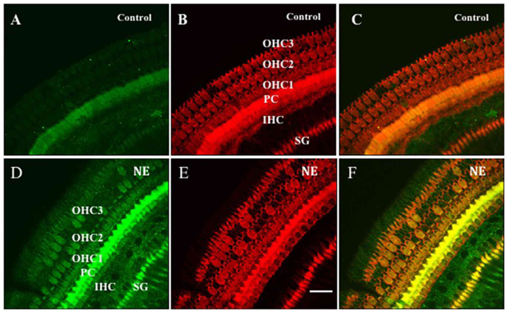
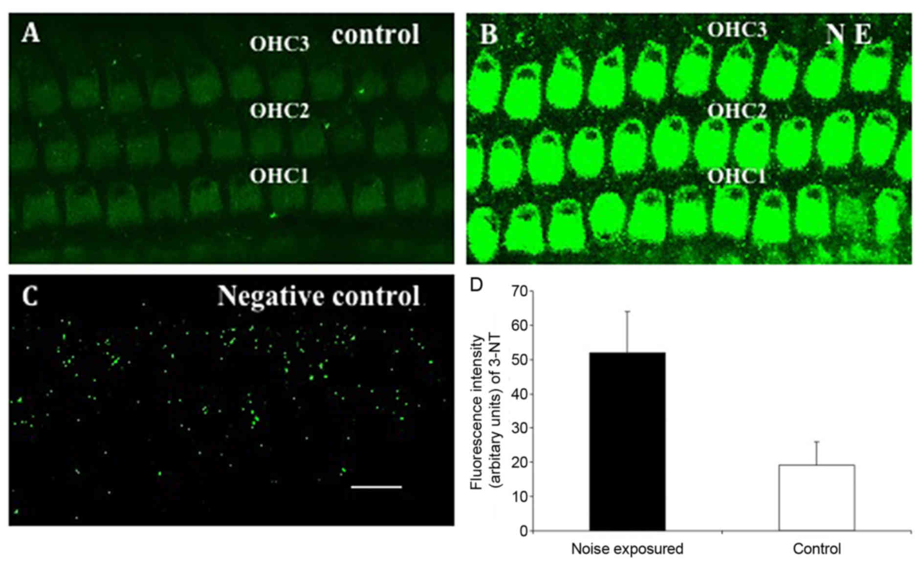
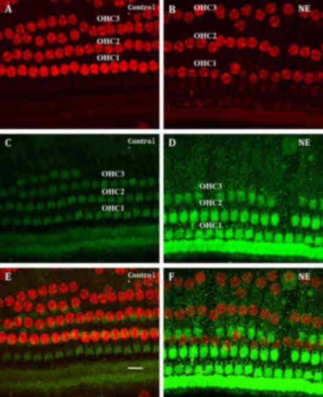
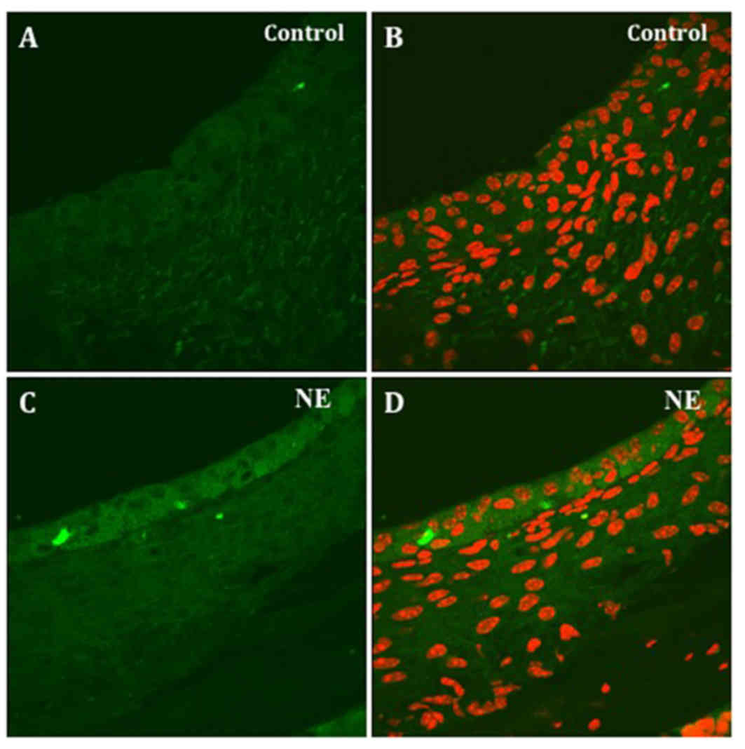
|
Radi R: Nitric oxide, oxidants, and
protein tyrosine nitration. Proc Natl Acad Sci USA. 101:4003–4008.
2004. View Article : Google Scholar : PubMed/NCBI
|
|
3
|
Gheddouchi S, Mokhtari-Soulimane N,
Merzouk H, Bekhti F, Soulimane F, Guermouche B, Meziane Tani A and
Narce M: Low SOD activity is associated with overproduction of
peroxynitrite and nitric oxide in patients with acute coronary
syndrome. Nitric Oxide. 49:40–46. 2015. View Article : Google Scholar : PubMed/NCBI
|
|
4
|
Koppenol WH, Moreno JJ, Pryor WA,
Ischiropoulos H and Beckman JS: Peroxynitrite, a cloaked oxidant
formed by nitric oxide and superoxide. Chem Res Toxicol. 5:834–842.
1992. View Article : Google Scholar : PubMed/NCBI
|
|
5
|
Huie RE and Padmaja S: The reaction of no
with superoxide. Free Radic Res Commun. 18:195–199. 1993.
View Article : Google Scholar : PubMed/NCBI
|
|
6
|
Beckman JS and Koppenol WH: Nitric oxide,
superoxide, and peroxynitrite: The good, the bad, and ugly. Am J
Physiol. 271:C1424–C1437. 1996. View Article : Google Scholar : PubMed/NCBI
|
|
7
|
Ischiropoulos H: Biological selectivity
and functional aspects of protein tyrosine nitration. Biochem
Biophys Res Commun. 305:776–783. 2003. View Article : Google Scholar : PubMed/NCBI
|
|
8
|
Crow JP and Ischiropoulos H: Detection and
quantitation of nitrotyrosine residues in proteins: In vivo marker
of peroxynitrite. Methods Enzymol. 269:185–194. 1996. View Article : Google Scholar : PubMed/NCBI
|
|
9
|
Han WJ, Shi XR and Nuttall A:
Noise-induced nitrotyrosine increase and outer hair cell death in
guinea pig cochlea. Chin Med J (Engl). 126:2923–2927.
2013.PubMed/NCBI
|
|
10
|
Zhang J, Chen X, Wu J and Han W: Detection
of guinea pigs' hearing functions before and after white noise
exposure. Chin J Otol. 31:141–144. 2015.
|
|
11
|
Konishi K, Yamane H, Iguchi H, Takayama M,
Nakagawa T, Sunami K and Nakai Y: Local substances regulating
cochlear blood flow. Acta Otolaryngol Suppl. 538:40–46.
1998.PubMed/NCBI
|
|
12
|
Hess A, Bloch W, Huverstuhl J, Su J,
Stennert E, Addicks K and Michel O: Expression of inducible nitric
oxide synthase (iNOS/NOS II) in the cochlea of guinea pigs after
intratympanical endotoxin-treatment. Brain Res. 830:113–122. 1999a.
View Article : Google Scholar
|
|
13
|
Kuhn DM, Sakowski SA, Sadidi M and Geddes
TJ: Nitrotyrosine as a marker for peroxynitrite-induced
neurotoxicity: The beginning or the end of the end of dopamine
neurons? J Neurochem. 89:529–536. 2004. View Article : Google Scholar : PubMed/NCBI
|
|
14
|
Shi X, Ren T and Nuttall AL: Nitric oxide
distribution and production in the guinea pig cochlea. Hear Res.
153:23–31. 2001. View Article : Google Scholar : PubMed/NCBI
|
|
15
|
Radi R, Denicola A, Alvarez B,
Ferrer-Sueta G and Rubbo H: Nitric Oxide. Ignarro L: Academic
Press; San Diego; pp. 57–82. 2000, View Article : Google Scholar : PubMed/NCBI
|
|
16
|
Yamashita D, Jiang HY, Schacht J and
Miller JM: Delayed production of free radicals following noise
exposure. Brain Res. 1019:201–209. 2004. View Article : Google Scholar : PubMed/NCBI
|
|
17
|
van Campen LE, Murphy WJ, Franks JR,
Mathias PI and Toraason MA: Oxidative DNA damage is associated with
intense noise exposure in the rat. Hear Res. 164:29–38. 2002.
View Article : Google Scholar : PubMed/NCBI
|
|
18
|
Brown GC: Nitric oxide as a competitive
inhibitor of oxygen consumption in the mitochondrial respiratory
chain. Acta Physiol Scand. 168:667–674. 2000. View Article : Google Scholar : PubMed/NCBI
|
|
19
|
Pisoschi AM and Pop A: The role of
antioxidants in the chemistry of oxidative stress: A review. Eur J
Med Chem. 97:55–74. 2015. View Article : Google Scholar : PubMed/NCBI
|
|
20
|
Yamane H, Nakai Y, Takayama M, Konishi K,
Iguchi H, Nakagawa T, Shibata S, Kato A, Sunami K and Kawakatsu C:
The emergence of free radicals after acoustic trauma and strial
blood flow. Acta Otolaryngol Suppl. 519:87–92. 1995. View Article : Google Scholar : PubMed/NCBI
|