Introduction
Systemic lupus erythematosus (SLE), a type of
immune-mediated destruction, is characterized by the breakdown of
self-tolerance in addition to the deposition of circulating immune
complexes throughout the body (1).
Lupus nephritis (LN) is one of the most common complications of
SLE. It is well established that SLE has been associated with
genetic, hormonal and environmental factors; specifically,
imbalances in these interactions determine the onset or progression
of SLE (2). Nevertheless, the
pathogenesis of SLE remains incompletely understood.
Toll-like receptor (TLR)/interleukin-1 receptor
(ILR) superfamily members are key regulators of immunity and
inflammation. Upon ligand binding, activation of multiple signaling
pathways occur, including ILR-associated kinases (IRAKs), myeloid
differentiation factor 88 (MyD88) and tumor necrosis factor
receptor-associated factor 6 (TRAF6), resulting in the activation
of activator protein-1 (AP-1), nuclear factor-κB (NF-κB) and c-Jun
N-terminal kinase (JNK). Subsequently, these active pathways induce
the secretion of interleukin-1 (IL-1), IL-6 and IL-23, which may
induce the differentiation of naive cluster of differentiation
(CD)4+ T cells to T helper cell 17 (Th17) cells
(3–5).
Th17 cells belong to a third lineage of CD4+ T cells,
and display an activated CD4+ T cell phenotype
characterized by the production of high quantities of IL-17, IL-21
and IL-22 (6,7). Upon T-cell receptor activation, a number
of cytokines including IL-6 and tumor growth factor-β are required
for Th17 cell differentiation through the induction of a set of
signature cytokines and cytokine receptors, including ILRs and
IL-23 receptors (4,8,9). Th17
cells, producing IL-17 amongst other cytokines, have been
demonstrated to serve a critical role in the pathogenesis of SLE
(10–12). Th17 cells and multiple cytokines are
involved in autoimmune diseases including SLE and LN (13).
Single immunoglobulin IL-1-related receptor (SIGIRR)
is a member of the TLR/ILR family and was first characterized as an
endogenous inhibitor of TLR/ILR signaling (14). Importantly, SIGIRR is expressed on T
cells and dendritic cells, exerting the fine-tuning modulation in
inflammatory responses (15). IL-17,
a proinflammatory cytokine, serves an important role in infections
and is involved in the pathogenesis of multiple autoimmune diseases
(16). One previous study has
suggested that the TLR/ILR family controls the differentiation of
Th17 cells and the secretion of IL-17 (4). SIGIRR, a negative regulator for TLR/ILR
signaling, was reported to regulate the differentiation of Th17
cells and secretion of IL-17 (17).
Altogether, the results of these studies demonstrate
that SIGIRR and Th17 cells are key elements in the control of
chronic autoimmune diseases, including SLE. However, relatively few
studies are available on the function of SIGIRR in patients with
SLE. Thus, in the present study, the frequency of Th17 cells and
SIGIRR+CD4+ T cells in patients with SLE and
their clinical associations were investigated to assess the
hypothesis that Th17 cells and SIGIRR may serve an important role
in the pathogenesis of human SLE.
Materials and methods
Patients and controls
Peripheral blood samples were recruited from 48
Chinese patients with SLE (46 women and 2 men, aged 28.9±9.03
years, with an age range of 15–57 years) and age and sex-matched 38
healthy volunteers (36 women and 2 men, aged 30.2±9.8 years, with
an age range of 18–53 years), who were admitted between May 2011
and December 2011. All samples were obtained from the Departments
of Rheumatology and Nephrology at the First and Second Affiliated
Hospital of Anhui Medical University (Anhui, China). Patients who
were diagnosed with SLE according to American College of
Rheumatology (ACR) criteria were included in the present study, and
healthy volunteers without a history of non-autoimmune diseases,
cancer or severe infectious diseases were included. Patients who
had suffered severe inflammation or any form of tumor type were
excluded from the present study. SLE diagnosis was established
according to the 1982 revised ACR criteria, and disease activity
was evaluated using the SLE disease activity index (SLEDAI) score
(18,19). Additionally, patients with SLE were
divided into 19 inactive SLE (ISLE; SLEDAI <10) and 29 active
SLE (ASLE; SLEDAI ≥10) cases (19).
Patients with LN were defined by persistent proteinuria (24-h
proteinuria >0.5 g/24 h) or the presence of cellular casts,
persistent hematuria or renal biopsy suggesting focal
proliferative, mesangial, diffuse proliferative or membranous
glomerulonephritis (19).
Demographic, clinical and laboratory data were collected from
hospital records in addition to questionnaires and reviewed by
experienced physicians from the Second Affiliated Hospital of Anhui
Medical University. The above protocol was ethically approved by
the Ethics Committee of Anhui Medical University, and written
informed consent was obtained from all participants.
Preparation of peripheral blood
mononuclear cells (PBMCs)
The anticoagulant blood samples were collected in
evacuated tubes containing ethylenediaminetetraacetic acid and were
recruited from healthy controls and patients with SLE from the
Departments of Rheumatology and Nephrology at the First and Second
Affiliated Hospital of Anhui Medical University. PBMCs were
purified by centrifugation at 4°C at 600 × g for 15 min, using a
Ficoll-Hypaque gradient (Tianjin HaoYang Biological Manufacture
Co., Ltd., Tianjin, China). Subsequently, PBMCs were adjusted to a
final concentration of 106/ml.
Flow cytometry for detecting Th17
cells
Th17 cells in PBMCs were quantified by flow
cytometry (Beckman Coulter, Inc., Brea, CA, USA). Cell staining was
analyzed using a CXP Cytometer system and analyzed using CXP 2.0
software (Beckman Coulter, Inc.).
CD3+CD8−IL-17+ T cells were
considered to be Th17 cells.
A total of 100 µlcells (1×10⁶ cells) were
transferred into a tube that contained 5 µl phycoerythrin (PE)-Cy7
anti-human CD3 diluted using 100 µl PBS containing 0.2% bovine
serum albumin (BSA; 1:20; cat no. 25-0037-42; eBioscience; Thermo
Fisher Scientific, Inc., Waltham, MA, USA) and 5 µl fluorescein
isothiocyanate-anti-human CD8 diluted by diluted using 100 µl PBS
containing 0.2% BSA (1:20; cat no. 11-0088; eBioscience; Thermo
Fisher Scientific, Inc.). Following incubation for 30 min at room
temperature, cells were then mixed with 100 µl FixA fixing agent
for 15 min at room temperature, and then washed in 3 ml PBS + 0.1%
NaN3 + 5% fetal bovine serum (FBS) once. Next, cells were mixed
with 100 µl PermB permeabilisation reagent for 5 min at room
temperature, then incubated with 5 µl PE-anti-human-IL-17 diluted
using 100 µl PBS containing 0.2% BSA (1:20; cat no. MHCD0412;
eBioscience; Thermo Fisher Scientific, Inc.) for 20 min at room
temperature. Cells were then washed in 3 ml PBS + 0.1% NaN3 + 5%
FBS for 1 min at room temperature once. Next, cells were
resuspended in 400 µl PBS for final flow cytometric analysis.
The frequency of SIGIRR+CD4+ T
cells detected by flow cytometry were determined as previously
described (20).
Statistical analysis
The differences in the frequency of Th17 cells
between different groups were analyzed using a Kruskal Wallis test
for non-parametric data and were presented as the mean ± standard
deviation. Spearman's rank correlation test was used to analyze the
correlation between the percentage of Th17 cells and clinical
laboratory data in addition to the correlation between the
percentage of Th17 cells and SIGIRR+CD4+ T
cells. All statistical analyses were performed using the
Statistical Package for the Social Sciences version 16.0 (SPSS,
Inc., Chicago, IL, USA). P<0.05 was considered to indicate a
statistically significant difference.
Results
Clinical laboratory data of patients
with SLE and controls
A total of 48 patients with SLE (28 of which had LN)
and 38 healthy volunteers were enrolled in the present study. The
clinical laboratory data of these patients were obtained from
hospital records and summarized in Table
I. There were no differences identified with regards to age,
sex, C3 or C4 between the SLE and LN group (P>0.05). Compared
with the SLE group, ESR and CRP were higher in the LN group
(P<0.001).
 | Table I.Clinical characteristics of patients
with SLE and control subjects. |
Table I.
Clinical characteristics of patients
with SLE and control subjects.
| Clinicopathological
characteristic | SLE (n=48) | Lupus nephritis
(n=28) | Controls
(n=38) |
|---|
| Age
(year)a | 28.9±9.03 | 30.7±7.08 | 30.2±9.8 |
| Sex
(male/female) | 2/46 | 0/28 | 2/36 |
| Duration of
diagnosis, monthsa | 23.3±12.5 | 20.1±15.7 | – |
| ESR
(mm/h)b | 39 (18.5,75) | 52 (20,102) | – |
| CRP
(mg/l)b | 6.49
(3.88,17.03) | 11.8
(5.0,20.35) | – |
| Anti-dsDNA
(−/+) | 16/32 | 7/21 | – |
| C3
(g/l)b | 0.51
(0.34,0.66) | 0.42
(0.22,0.86) | – |
| C4
(g/l)b | 0.09
(0.04,0.13) | 0.10
(0.02,0.17) | – |
| 24-h urinary
protein (g/24 h)b | 1.4 (0.73,2.8) | 2.4 (0.91,4.0) | – |
| SLEDAIb | 13 (8,17) | 15 (10,24) | – |
Frequency of Th17 cells in the PBMCs
of patients with SLE and controls
As presented in Fig.
1, flow cytometry revealed that compared with healthy controls
(median, 0.28%; range, 0.09–0.59%) a significantly higher
proportion of Th17 cells in patients with SLE (median, 0.75%;
range, 0.15–1.25%) were observed (Z=−5.82, P<0.001).
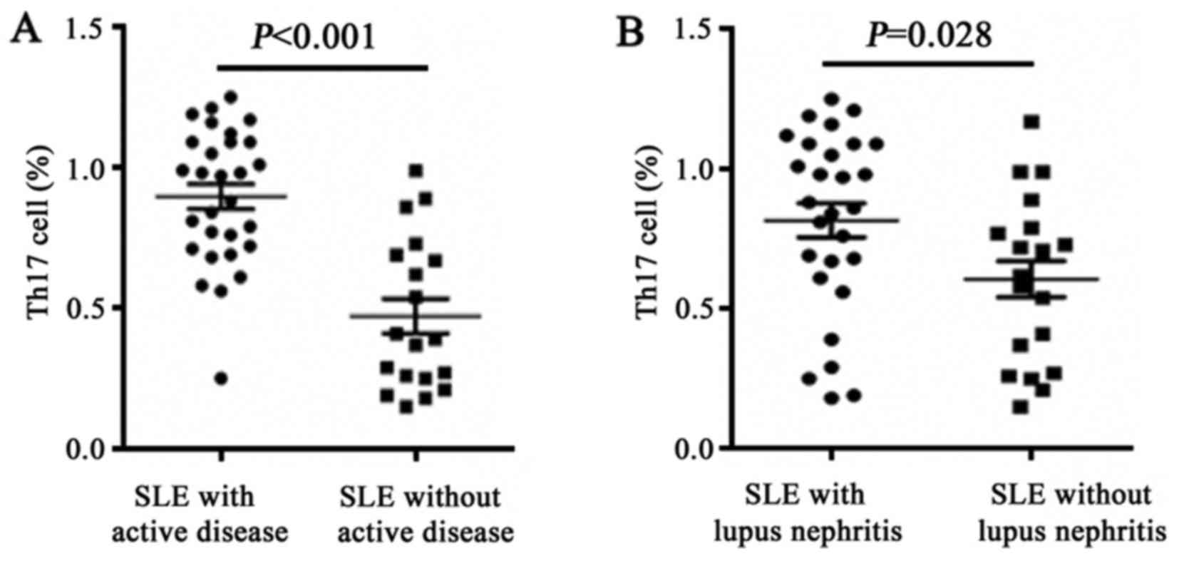
Frequency of Th17 cells in the PBMCs
of patients with ASLE and LN
As presented in Fig.
2, compared with patients with ISLE (median, 0.41%; range,
0.15–0.99%), a significantly higher proportion of Th17 cells in
patients with ASLE (median, 0.97%; range, 0.25–1.25%) was observed
(Z=−4.26, P<0.001).
Compared with non-LN patients (median, 0.65%; range,
0.15–1.17%), a significantly higher proportion of Th17 cells in
patients with LN (median, 0.87%; range, 0.18–1.25%) was observed
(Z=−2.20, P=0.028).
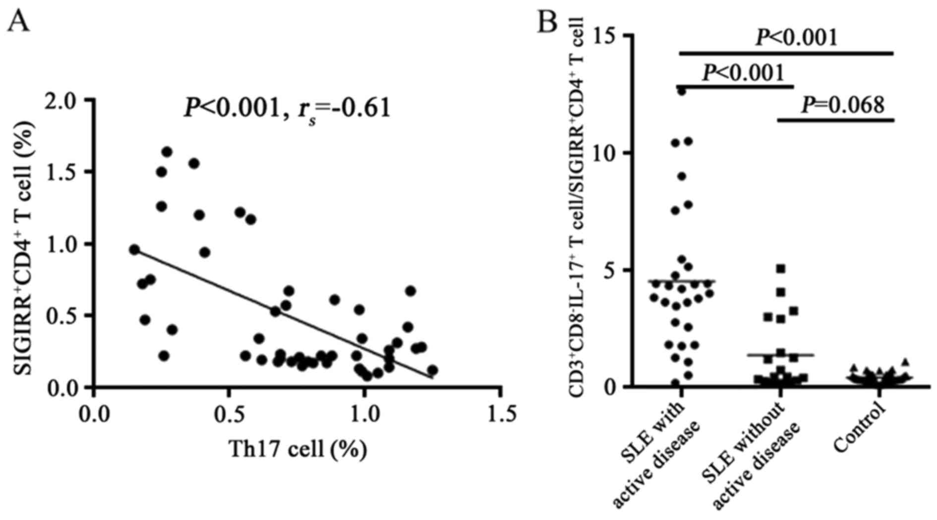
Associations of Th17 cells and
SIGIRR+CD4+ T cells and their ratio in
different groups
As presented in Fig.
3, the frequency of Th17 cells were inversely correlated with
the frequency of SIGIRR+CD4+ T cells
(r=−0.61, P<0.001). The ratio of Th17 cells to
SIGIRR+CD4+ T cells in patients with ASLE was
significantly increased compared with healthy controls or patients
with ISLE (P<0.001).
Association of the ratio of Th17
cells/SIGIRR+CD4+ T cells and clinical
data
As presented in Table
II, the ratio of Th17 cells to
SIGIRR+CD4+ T cells was significantly
correlated negatively with C3 (P<0.01) and C4 (P<0.05), and
was significantly correlated positively with SLEDAI (P<0.001)
and 24-h proteinuria (P<0.05). No significant correlations
between the ratio and erythrocyte sedimentation rate or C-reactive
protein were identified (P>0.05).
| Table II.Association between the ratio of T
helper cell 17 cells/single immunoglobulin IL-1-related
receptor+ cluster of differentiation 4+ T
cells and clinical data. |
Table II.
Association between the ratio of T
helper cell 17 cells/single immunoglobulin IL-1-related
receptor+ cluster of differentiation 4+ T
cells and clinical data.
| Clinicopathological
characteristic | r-value | P-value |
|---|
| ESR (mm/h) | 0.084 | 0.571 |
| CRP (mg/l) | −0.144 | 0.330 |
| C3 (g/l) | −0.377 | 0.008 |
| C4 (g/l) | −0.309 | 0.035 |
| SLEDAI | 0.609 | <0.001 |
| 24-h urinary
protein (g/24 h) | 0.345 | 0.016 |
Discussion
The TLR/ILR superfamily is a key regulator of
immunity and inflammation. Upon ligand binding, activation of a
signaling pathway occurs including IRAKs, MyD88 and TRAF6 resulting
in the activation of AP-1, NF-κβ and JNK (3,14,21). Subsequently this activated pathway
induced the secretion of IL-1, IL-6 and IL-23, which are able to
induce the differentiation of Th17 cells (6). It appears that Th17 cells serve crucial
roles in the development of a wide range of autoimmune disorders.
It has been suggested that the inappropriate regulation of Th17
cells may be a key event in the pathogenesis of rheumatoid
arthritis and SLE (22). A number of
studies have reported significantly higher serum levels of IL-17
and a higher frequency of IL-17-producing PBMCs in patients with
SLE compared with normal individuals (6,7,13).
In previous years, SIGIRR, a negative regulator of
the TLR/ILR superfamily, has been identified to be expressed in the
kidney, liver, lung and lymphoid tissue (23,24).
Additionally, SIGIRR was reported to regulate the differentiation
of Th17 cells in addition to the secretion of IL-17 (17,23).
Therefore, the present study estimated the immune response through
detecting the changes in Th17 cells and
SIGIRR+CD4+ T cells. Similarly, a previous
study revealed that the frequency of
SIGIRR+CD4+ T cells was decreased in patients
with SLE compared with controls (20). In the present study, the frequency of
Th17 cells in patients with SLE was investigated, and reanalysis of
the data (the frequency of SIGIRR+CD4+ T
cells) from the previous study was performed to investigate the
ratio of Th17 cells to SIGIRR+CD4+ T cells to
assess the hypothesis that Th17 cells and SIGIRR may serve an
important role in the pathogenesis of human SLE.
Studied have suggested that Th17 cells and IL-17 are
higher in PBMCs in patients with SLE compared with healthy
controls, and the levels of IL-17 were correlated positively with
SLEDAI (22,25,26). In
the present study, the percentage of Th17 cells was significantly
increased in the PBMCs of patients with SLE compared with healthy
controls. Compared with patients with ISLE, the percentage of Th17
cells were increased in patients with ASLE; and the frequency of
Th17 cells were also significantly increased in patients with LN
compared with non-LN patients. These results were consistent with
previous studies which suggested that Th17 cells may be involved in
the pathogenesis of SLE and LN (22,25).
SIGIRR, a negative regulator of the TLR/ILR pathway, has been
studied in patients with SLE (27).
Studies had revealed that SIGIRR-knockdown mice manifested with
diffuse membrane proliferative glomerulonephritis, macrophage
invasion, immune complex deposition and the formation of
autoantibodies including rheumatoid factor, anti-double stranded
DNA and anti-Sm (15,28,29).
Another study demonstrated that the SIGIRR rs7396562 polymorphism
was associated with SLE susceptibility in a Chinese population
(30). The mechanism of SIGIRR in SLE
remains unclear, and a previous study investigated
SIGIRR+CD4+ T cells in PBMCs in patients with
SLE through flow cytometry (20). The
results revealed that the percentage of
SIGIRR+CD4+ T cells was significantly
decreased compared with healthy controls. Compared with ISLE, the
percentage of SIGIRR+CD4+ T cells was
decreased in ASLE and the frequency of
SIGIRR+CD4+ T cells were also significantly
decreased in patients with LN compared with non-LN patients.
Theresults of the present study revealed that SIGIRR may serve an
important role in the development of SLE and LN.
IL-1-mediated signaling in T cells is essential for
Th17 cell differentiation. SIGIRR, a negative regulator of ILR and
TLR signaling, was induced during Th17 cell lineage commitment and
governed Th17 cell differentiation and expansion through its
inhibitory effects on IL-1 signaling (5,23). Gulen
et al (31) reported that
SIGIRR inhibited the differentiation of Th17 cells through the
TLR/ILR pathway. The absence of SIGIRR in T cells resulted in
increased Th17 cell polarization in vivo. Recombinant IL-1
promoted a marked increase in the proliferation of SIGIRR-deficient
T cells under in vitro Th17 cell-polarization conditions.
Compared with controls, induced experimental allergic
encephalomyelitis in SIGIRR-knockdown mice had a lower secretion of
IL-17 and IL-6. Gulen et al (31) reported that SIGIRR inhibited the
differentiation of Th17 cells through the TLR/ILR pathway.
Therefore, it may be inferred that SIGIRR and Th17 serve opposite
roles, as the following results revealed: Decreased
SIGIRR+CD4+ T cells in patients with SLE
compared with control subjects and the increased frequency of Th17
in patients with SLE compared with control subjects. Additionally,
the present study suggested that the frequency of Th17 cells were
correlated negatively with the frequency of
SIGIRR+CD4+ T cells. The ratio of Th17 cells
to SIGIRR+CD4+ T cells in ASLE was increased
compared with healthy controls or patients with ISLE. The ratio of
Th17 cells to SIGIRR+CD4+ T cells was
correlated negatively with C3 and C4, and was correlated positively
with SLEDAI and 24-h proteinuria. All results suggested that
interactions between Th17 cells and
SIGIRR+CD4+ T cells serve a crucial role in
the pathogenesis of SLE. However, the specific molecular mechanisms
of SIGIRR involved in SLE disease remain unclear. So, future
studies will examine SIGIRR overexpression to investigate the
mechanism of SIGIRR in IL-1β-induced epithelial-myofibroblast
transdifferentiation in human tubular cells.
Increased numbers of Th17 cells and decreased
numbers of SIGIRR+CD4+ T cells in patients
with SLE and their correlation with SLEDAI score in addition to the
clinical data suggested that SIGIRR+CD4+ T
and Th17 cells may be involved in the pathogenesis of SLE. In
summary, the negative regulation of TLR signaling may be required
to avoid inappropriate inflammatory responses. The results obtained
from the present study suggest that the ratio of Th17 cells to
SIGIRR+CD4+ T cells maybe a promising
therapeutic target for SLE. However, this is only a preliminary
descriptive study, and further mechanistic studies are required in
order to determine the exact role of SIGIRR in the pathogenesis of
SLE.
Acknowledgements
Preliminary results of the present study were
presented as a publication titled ‘The decreased frequency of
SIGIRR-positive CD4+ T cells in peripheral blood of
patients with SLE and its correlation with disease activity’ at the
Mol Biol Rep 42: 423–430, 2015.
Funding
The present study was supported the Natural Science
Foundation of Anhui Province (grant no. 1508085MH148), the China
Postdoctoral Science Foundation (grant no. 2012M511399) and the
Anhui Postdoctoral Science Foundation (grant no. 910101920).
Availability of data and materials
The datasets used and/or analysed during the current
study are available from the corresponding author on reasonable
request.
Authors' contributions
DeW and LH conceived and designed the experiments.
JX and DaW performed the experiments and wrote the manuscript. XW
and LY collected the clinical data.
Ethics approval and consent to
participate
The protocol was ethically approved by the Ethics
Committee of Anhui Medical University (Anhui, China), and written
informed consent was obtained from all participants.
Patient consent for publication
Informed consent was obtained from all patients for
the use of their tissues for research purposes.
Competing interests
The authors declare that they have no competing
interests.
References
|
1
|
Marrack P, Kappler J and Kotzin BL:
Autoimmune disease: Why and where it occurs. Nat Med. 7:899–905.
2001. View Article : Google Scholar : PubMed/NCBI
|
|
2
|
Lau CS, Yin G and Mok MY: Ethnic and
geographical differences in systemic lupus erythematosus: An
overview. Lupus. 15:715–719. 2006. View Article : Google Scholar : PubMed/NCBI
|
|
3
|
Adib-Conquy M, Adrie C, Fitting C,
Gattolliat O, Beyaert R and Cavaillon JM: Up-regulation of MyD88s
and SIGIRR, molecules inhibiting Toll-like receptor signaling, in
monocytes from septic patients. Crit Care Med. 34:2377–2385. 2006.
View Article : Google Scholar : PubMed/NCBI
|
|
4
|
Riva F, Bonavita E, Barbati E, Muzio M,
Mantovani A and Garlanda C: TIR8/SIGIRR is an interleukin-1
receptor/toll like receptor family member with regulatory functions
in inflammation and immunity. Front Immunol. 3:3222012. View Article : Google Scholar : PubMed/NCBI
|
|
5
|
Garlanda C, Riva F, Veliz T, Polentarutti
N, Pasqualini F, Radaelli E, Sironi M, Nebuloni M, Zorini EO,
Scanziani E, et al: Increased susceptibility to colitis-associated
cancer of mice lacking TIR8, an inhibitory member of the
interleukin-1 receptor family. Cancer Res. 67:6017–6021. 2007.
View Article : Google Scholar : PubMed/NCBI
|
|
6
|
Sha Y and Markovic-Plese S: A role of
IL-1R1 signaling in the differentiation of Th17 cells and the
development of autoimmune diseases. Self Nonself. 2:35–42. 2011.
View Article : Google Scholar : PubMed/NCBI
|
|
7
|
Bettelli E, Carrier Y, Gao W, Korn T,
Strom TB, Oukka M, Weiner HL and Kuchroo VK: Reciprocal
developmental pathways for the generation of pathogenic effector
TH17 and regulatory T cells. Nature. 441:235–238. 2006. View Article : Google Scholar : PubMed/NCBI
|
|
8
|
Yang XO, Pappu BP, Nurieva R, Akimzhanov
A, Kang HS, Chung Y, Ma L, Shah B, Panopoulos AD, Schluns KS, et
al: T helper 17 lineage differentiation is programmed by orphan
nuclear receptors ROR alpha and ROR gamma. Immunity. 28:29–39.
2008. View Article : Google Scholar : PubMed/NCBI
|
|
9
|
Dinarello CA: Immunological and
inflammatory functions of the interleukin-1 family. Annu Rev
Immunol. 27:519–550. 2009. View Article : Google Scholar : PubMed/NCBI
|
|
10
|
Poissonnier A, Sanséau D, Le Gallo M,
Malleter M, Levoin N, Viel R, Morere L, Penna A, Blanco P, Dupuy A,
et al: CD95-mediated calcium signaling promotes T Helper 17
trafficking to inflamed organs in lupus-prone mice. Immunity.
45:209–223. 2016. View Article : Google Scholar : PubMed/NCBI
|
|
11
|
Hammad A, Osman E, Mosaad Y and Wahba M:
Serum interleukin-17 in Egyptian children with systemic lupus
erythematosus: Is it related to pulmonary affection? Lupus.
26:388–395. 2017. View Article : Google Scholar : PubMed/NCBI
|
|
12
|
Wu H, Zhen Y, Ma Z, Li H, Yu J, Xu ZG,
Wang XY, Yi H and Yang YG: Arginase-1-dependent promotion of TH17
differentiation and disease progression by MDSCs in systemic lupus
erythematosus. Sci Transl Med. 8:331ra402016. View Article : Google Scholar : PubMed/NCBI
|
|
13
|
Sigdel KR, Duan L, Wang Y, Hu W, Wang N,
Sun Q, Liu Q, Liu X, Hou X, Cheng A, et al: Serum Cytokines Th1,
Th2, and Th17 expression profiling in active lupus nephritis-IV:
From a Southern Chinese Han Population. Mediators Inflamm.
2016:49275302016. View Article : Google Scholar : PubMed/NCBI
|
|
14
|
Qin J, Qian Y, Yao J, Grace C and Li X:
SIGIRR inhibits interleukin-1 receptor- and toll-like receptor
4-mediated signaling through different mechanisms. J Biol Chem.
280:25233–25241. 2005. View Article : Google Scholar : PubMed/NCBI
|
|
15
|
Lech M, Kulkarni OP, Pfeiffer S, Savarese
E, Krug A, Garlanda C, Mantovani A and Anders HJ: Tir8/Sigirr
prevents murine lupus by suppressing the immunostimulatory effects
of lupus autoantigens. J Exp Med. 205:1879–1888. 2008. View Article : Google Scholar : PubMed/NCBI
|
|
16
|
Datta SK, Zhang L and Xu L: T-helper cell
intrinsic defects in lupus that break peripheral tolerance to
nuclear autoantigens. J Mol Med (Berl). 83:267–278. 2005.
View Article : Google Scholar : PubMed/NCBI
|
|
17
|
Stahl M, Ries J, Vermeulen J, Yang H, Sham
HP, Crowley SM, Badayeva Y, Turvey SE, Gaynor EC, Li X, et al: A
novel mouse model of Campylobacter jejuni gastroenteritis
reveals key pro-inflammatory and tissue protective roles for
Toll-like receptor signaling during infection. PLoS Pathog.
10:e10042642014. View Article : Google Scholar : PubMed/NCBI
|
|
18
|
Tan EM, Cohen AS, Fries JF, Masi AT,
McShane DJ, Rothfield NF, Schaller JG, Talal N and Winchester RJ:
The 1982 revised criteria for the classification of systemic lupus
erythematosus. Arthritis Rheum. 25:1271–1277. 1982. View Article : Google Scholar : PubMed/NCBI
|
|
19
|
Bombardier C, Gladman DD, Urowitz MB,
Caron D, Chang CH, Austin A, Bell A, Bloch DA, Corey PN, Decker JL,
et al: The Committee on Prognosis Studies in SLE: Derivation of the
SLEDAI. A disease activity index for lupus patients. Arthritis
Rheum. 35:630–640. 1992. View Article : Google Scholar : PubMed/NCBI
|
|
20
|
Wang DY, Su C, Chen GM, Pan HF, Wang FM,
Liu GL, Hao L, Wang DG and Ye DQ: The decreased frequency of
SIGIRR-positive CD4+ T cells in peripheral blood of
patients with SLE and its correlation with disease activity. Mol
Biol Rep. 42:423–430. 2015. View Article : Google Scholar : PubMed/NCBI
|
|
21
|
Zhang C, Wu X, Zhao Y, Deng Z and Qian G:
SIGIRR inhibits toll-like receptor 4, 5, 9-mediated immune
responses in human airway epithelial cells. Mol Biol Rep.
38:601–609. 2011. View Article : Google Scholar : PubMed/NCBI
|
|
22
|
Yang J, Chu Y, Yang X, Gao D, Zhu L, Yang
X, Wan L and Li M: Th17 and natural Treg cell population dynamics
in systemic lupus erythematosus. Arthritis Rheum. 60:1472–1483.
2009. View Article : Google Scholar : PubMed/NCBI
|
|
23
|
Garlanda C, Anders HJ and Mantovani A:
TIR8/SIGIRR: An IL-1R/TLR family member with regulatory functions
in inflammation and T cell polarization. Trends Immunol.
30:439–446. 2009. View Article : Google Scholar : PubMed/NCBI
|
|
24
|
Xiao H, Gulen MF, Qin J, Yao J, Bulek K,
Kish D, Altuntas CZ, Wald D, Ma C, Zhou H, et al: The
Toll-interleukin-1 receptor member SIGIRR regulates colonic
epithelial homeostasis, inflammation, and tumorigenesis. Immunity.
26:461–475. 2007. View Article : Google Scholar : PubMed/NCBI
|
|
25
|
Zhao XF, Pan HF, Yuan H, Zhang WH, Li XP,
Wang GH, Wu GC, Su H, Pan FM, Li WX, et al: Increased serum
interleukin 17 in patients with systemic lupus erythematosus. Mol
Biol Rep. 37:81–85. 2010. View Article : Google Scholar : PubMed/NCBI
|
|
26
|
Chen DY, Chen YM, Wen MC, Hsieh TY, Hung
WT and Lan JL: The potential role of Th17 cells and Th17-related
cytokines in the pathogenesis of lupus nephritis. Lupus.
21:1385–1396. 2012. View Article : Google Scholar : PubMed/NCBI
|
|
27
|
Wang C, Feng CC, Pan HF, Wang DG and Ye
DQ: Therapeutic potential of SIGIRR in systemic lupus
erythematosus. Rheumatol Int. 33:1917–1921. 2013. View Article : Google Scholar : PubMed/NCBI
|
|
28
|
Lech M, Garlanda C, Mantovani A,
Kirschning CJ, Schlöndorff D and Anders HJ: Different roles of
TiR8/Sigirr on Τoll-like receptor signaling in intrarenal
antigen-presenting cells and tubular epithelial cells. Kidney Int.
72:182–192. 2007. View Article : Google Scholar : PubMed/NCBI
|
|
29
|
Zhu YY, Su Y, Li ZG and Zhang Y: The
largely normal response to Toll-like receptor 7 and 9 stimulation
and the enhanced expression of SIGIRR by B cells in systemic lupus
erythematosus. PLoS One. 7:e441312012. View Article : Google Scholar : PubMed/NCBI
|
|
30
|
Zhu Y, Wang DG, Yang XK, Tao SS, Huang Q,
Pan HF, Feng CC and Ye DQ: Emerging role of SIGIRR rs7396562(T/G)
polymorphism in systemic lupus erythematosus in a Chinese
population. Inflammation. 37:1847–1851. 2014. View Article : Google Scholar : PubMed/NCBI
|
|
31
|
Gulen MF, Kang Z, Bulek K, Youzhong W, Kim
TW, Chen Y, Altuntas CZ, Sass Bak-Jensen K, McGeachy MJ, Do JS, et
al: The receptor SIGIRR suppresses Th17 cell proliferation via
inhibition of the interleukin-1 receptor pathway and mTOR kinase
activation. Immunity. 32:54–66. 2010. View Article : Google Scholar : PubMed/NCBI
|