Introduction
Despite progress in prevention, diagnosis and
therapy, cardiovascular disease (CD) stands for the leading cause
of morbidity and mortality in the developed world (1). The majority of CD-related deaths are
due to acute thrombotic events, following the rupture of
atherosclerotic lesions, which are characterized by key
pathophysiologic features. The capability of positron emitting
tomography (PET) imaging to visualize and quantify these features
at the cellular and sub-cellular level provides the ground for the
employment of PET-radiopharmaceuticals, which target
rupture-related biochemical processes, in order to address the
challenge of detecting high-risk vulnerable atherosclerotic
lesions.
The pathophysiology of atherosclerosis is quite
complex, and is mainly characterized by an inflammatory cascade
triggered by the entrance of low-density lipoproteins (LDL) at
sites of endothelial injury, and the subsequent recruitment of
macrophages which take up the oxidized LDL remnants (2). An extensive description of the
atherosclerotic plaque pathophysiology is beyond the scope of the
current report. Our main interest is focused on the key role that
infiltration by macrophages plays in inflammatory processes
encountered in unstable atherosclerotic plaques.
Since the expression of somatostatin receptors
(SSTRs) subtype-2, has been detected on macrophages (3,4), these
cells can be effectively targeted with somatostatin analogues
radio-labelled with isotopes suitable for PET-imaging. Such
PET-tracers, which enable whole-body characterization of cell
surface SSTRs-expression, have become the imaging standard of
reference for the detections of neuroendocrine tumors (NETs) and
other SSTRs-positive lesions (5-12).
Furthermore, we have previously reported the increased uptake of
68Ga-DOTATATE, which is one of the commercially
available PET-tracers targeting cell surface
SSTRs-subtype-2-over-expression, at sites of reactive inflammatory
alterations (13,14).
Case report
A 82-year-old man presented with a constant
epigastric pain. The computed tomography (CT) scan of the abdomen,
showed a large (3 cm) pancreatic head mass. Subsequently,
endoscopic ultrasound (EUS) and biopsy of the tumor revealed a
low-grade NET. Therefore, a whole body 68Ga-DOTATATE
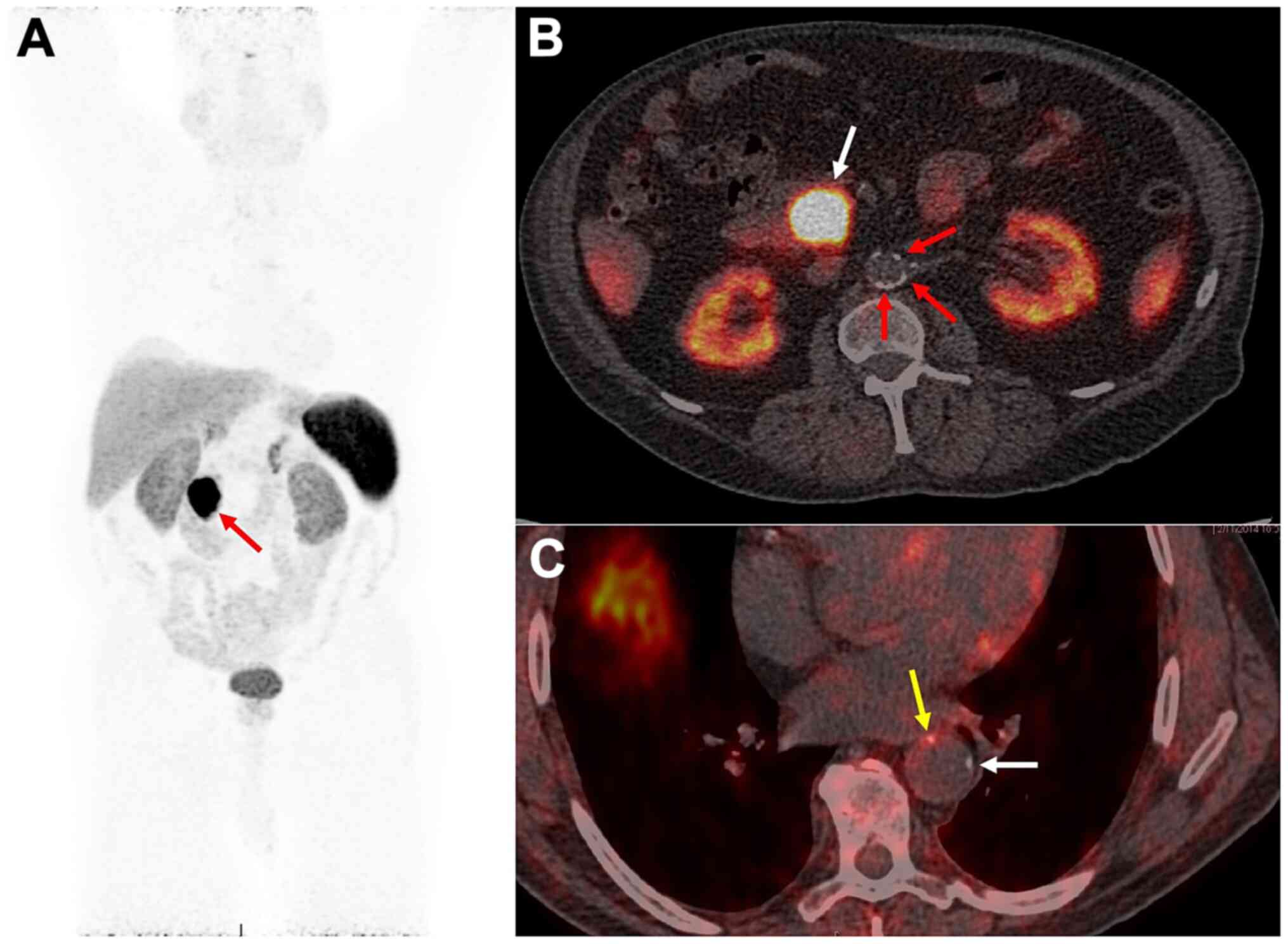
PET/CT scan was performed for staging purposes. The PET/CT study
showed (Fig. 1A) intense radiotracer
uptake (SUVmax: 85) by the pancreatic head tumor and
excluded the presence of metastatic disease.
Furthermore, there was extensive atherosclerosis
seen throughout medium- and large-sized arteries such as in the
abdominal aorta, most of which were
68Ga-DOTATATE-negative (Fig.
1B; red arrows). However, in the thoracic aorta a
radiotracer-positive plaque (SUVmax: 5.5) was
encountered (Fig. 1C; yellow arrow),
implying infiltration by macrophages, which are known to be
characterized by cell-surface over-expression of SSTRs-subtype-2,
leading to increased 68Ga-DOTATATE activity. At the same
level of the thoracic aorta, another
68Ga-DOTATATE-negative plaque was detected (Fig. 1C; white arrow), suggesting that not
all atherosclerotic lesions take up the administered PET-tracer.
Despite its small size, the 68Ga-DOTATATE avidity of the
plaque seen on the thoracic aorta, suggests an active inflammatory
cascade taking place in that specific lesion, raising suspicion for
a high-risk prone to rupture lesion.
Discussion
Early and accurate detection of high-risk, prone to
rupture atherosclerotic plaques, is the holy grail of CD research,
receiving great interest and extensive research efforts. Molecular
imaging of key rupture-related pathophysiological features of the
plaques, by means of PET-tracers, holds promise to address this
diagnostic challenge. Imaging the recruitment of macrophages at
sites of vessel wall inflammation (VWI), via PET-tracers aiming at
the cell-surface over-expression of STTRs subtype-2, also seen on
macrophages, is a promising molecular imaging strategy for the
detection of the unstable plaques.
In a series of 16 patients with NETS, Li X. et al.
reported association between coronary artery uptake of
68Ga-DOTATATE with known risk factors of CD (15). Furthermore, Pedersen SF et al, in a
cohort of 10 patients who underwent simultaneous PET/MRI scans
using 64Cu-DOTATATE, prior to carotid endarterectomy,
found increased tracer uptake in symptomatic plaques, while an
independent correlation with CD 163 gene expression (surrogate
marker of activated macrophages) was revealed (16). In a series of 42 patients with
atherosclerosis, Tarkin et al reported that
68Ga-DOTATATE correctly detected culprit arteries in
patients with acute coronary syndrome, predicted high-risk coronary
CT features and was positively associated with Framingham risk
score, implying the employment of the radiotracer as a novel
imaging probe for VWI (17).
Moreover, 68Ga-DOTATATE is superior to
18F-FDG which is the most widely used PET-tracer,
targeting metabolic activity, since the lack of physiologic uptake
by the myocardium, enables assessment of the coronary arteries.
In accordance to the existing literature, the
current report adds to the data that not all atherosclerotic
plaques exhibit elevated 68Ga-DOTATATE activity,
suggesting that only lesions harboring active inflammatory
processes and therefore are infiltrated by macrophages, take up the
tracer. A major limitation of our report is the lack of histologic
analysis of the 68Ga-DOTATATE-avid plaque in the
thoracic aorta, and the confirmation of macrophages accumulation.
However, our work enhances the need for further research efforts
being addressed towards employment of this specific molecular
imaging strategy for the detection of the vulnerable
atherosclerotic plaque.
Acknowledgements
Not applicable.
Funding
Funding
Part of this study was financially supported by the
Stavros Niarchos Foundation within the framework of the project
ARCHERS (Advancing Young Researchers' Human Capital in Cutting Edge
Technologies in the Preservation of Cultural Heritage and the
Tackling of Societal Challenges).
Availability of data and materials
All the information relevant to the present study is
available from the corresponding author on reasonable request.
Authors' contributions
GZP, AHK, and GK conceived and designed the study.
GZP, GL, KM, NK, FHS, GGI, TKN and SK researched the literature,
performed interpretation of data and drafted the manuscript. DAS,
and AHK critically revised the article for important intellectual
content, and assisted in the literature search for this case
report. All authors agree to be accountable for all aspects of the
work in ensuring that questions related to the accuracy or
integrity of any part of the work are appropriately investigated,
and finally approved the version of the manuscript to be
published.
Ethics approval and consent to
participate
The images were provided by esteemed physicians at
the NIH, with whom the first and corresponding author of the
article collaborates. All study participants at the NIH clinical
protocols provided all the extensive consent forms and strict
ethical approval documents that the NIH standards require.
Patient consent for publication
Not applicable.
Competing interests
DAS is the Editor-in-Chief for the journal, but had
no personal involvement in the reviewing process, or any influence
in terms of adjudicating on the final decision, for this article.
All the other authors declare that they have no competing
interests.
References
|
1
|
Bucerius J, Dijkgraaf I, Mottaghy FM and
Schurgers LJ: Target identification for the diagnosis and
intervention of vulnerable atherosclerotic plaques beyond
18F-fluorodeoxyglucose positron emission tomography imaging:
Promising tracers on the horizon. Eur J Nucl Med Mol Imaging.
46:251–265. 2019.PubMed/NCBI View Article : Google Scholar
|
|
2
|
Krishnan S, Otaki Y, Doris M, Slipczuk L,
Arnson Y, Rubeaux M, Dey D, Slomka P, Berman DS and Tamarappoo B:
Molecular imaging of vulnerable coronary plaque: A pathophysiologic
perspective. J Nucl Med. 58:359–364. 2017.PubMed/NCBI View Article : Google Scholar
|
|
3
|
Dalm VA, van Hagen PM, van Koetsveld PM,
Achilefu S, Houtsmuller AB, Pols DH, van der Lely AJ, Lamberts SW
and Hofland LJ: Expression of somatostatin, cortistatin, and
somatostatin receptors in human monocytes, macrophages, and
dendritic Cells. Am J Physiol Endocrinol Metab. 285:E344–E353.
2003.PubMed/NCBI View Article : Google Scholar
|
|
4
|
Armani C, Catalani E, Balbarini A, Bagnoli
P and Cervia D: Expression, pharmacology, and functional role of
somatostatin receptor subtypes 1 and 2 in human macrophages. J
Leukoc Biol. 81:845–855. 2007.PubMed/NCBI View Article : Google Scholar
|
|
5
|
Hofman MS, Lau WF and Hicks RJ:
Somatostatin receptor imaging with 68Ga DOTATATE PET/CT:
Clinical utility, normal patterns, pearls, and pitfalls in
interpretation. Radiographics. 35:500–516. 2015.PubMed/NCBI View Article : Google Scholar
|
|
6
|
Tirosh A, Papadakis GZ, Millo C, Hammoud
D, Sadowski SM, Herscovitch P, Pacak K, Marx SJ, Yang L, Nockel P,
et al: Prognostic utility of total 68Ga-DOTATATE-avid
tumor volume in patients with neuroendocrine tumors.
Gastroenterology. 154:998–1008.e1. 2018.PubMed/NCBI View Article : Google Scholar
|
|
7
|
Tirosh A, Papadakis GZ, Millo C, Sadowski
SM, Herscovitch P, Pacak K, Marx SJ, Yang L, Nockel P, Shell J, et
al: Association between neuroendocrine tumors biomarkers and
primary tumor site and disease type based on total
68Ga-DOTATATE-Avid tumor volume measurements. Eur J
Endocrinol. 176:575–582. 2017.PubMed/NCBI View Article : Google Scholar
|
|
8
|
Papadakis GZ, Millo C, Sadowski SM, Bagci
U and Patronas NJ: Kidney tumor in a von Hippel-Lindau (VHL)
patient with intensely increased activity on
68Ga-DOTA-TATE PET/CT. Clin Nucl Med. 41:970–971.
2016.PubMed/NCBI View Article : Google Scholar
|
|
9
|
El-Maouche D, Sadowski SM, Papadakis GZ,
Guthrie L, Cottle-Delisle C, Merkel R, Millo C, Chen CC, Kebebew E
and Collins MT: 68Ga-DOTATATE for tumor localization in
tumor-induced osteomalacia. J Clin Endocrinol Metab. 101:3575–3581.
2016.PubMed/NCBI View Article : Google Scholar
|
|
10
|
Papadakis GZ, Millo C, Sadowski SM, Bagci
U and Patronas NJ: Epididymal cystadenomas in von Hippel-Lindau
disease showing increased activity on 68Ga-DOTATATE
PET/CT. Clin Nucl Med. 41:781–782. 2016.PubMed/NCBI View Article : Google Scholar
|
|
11
|
Papadakis GZ, Millo C, Sadowski SM, Bagci
U and Patronas NJ: Endolymphatic sac tumor showing increased
activity on 68Ga-DOTATATE PET/CT. Clin Nucl Med.
41:783–784. 2016.PubMed/NCBI View Article : Google Scholar
|
|
12
|
Papadakis GZ, Bagci U, Sadowski SM,
Patronas NJ and Stratakis CA: Ectopic ACTH and CRH co-secreting
tumor localized by 68Ga-DOTA-TATE PET/CT. Clin Nucl Med.
40:576–578. 2015.PubMed/NCBI View Article : Google Scholar
|
|
13
|
Papadakis GZ, Millo C, Karantanas AH,
Bagci U and Patronas NJ: Avascular necrosis of the hips with
increased activity on 68Ga-DOTATATE PET/CT. Clin Nucl
Med. 42:214–215. 2017.PubMed/NCBI View Article : Google Scholar
|
|
14
|
Papadakis GZ, Millo C, Bagci U, Sadowski
SM and Stratakis CA: Schmorl nodes can cause increased
68Ga-DOTATATE activity on PET/CT, mimicking metastasis
in patients with neuroendocrine malignancy. Clin Nucl Med.
41:249–250. 2016.PubMed/NCBI View Article : Google Scholar
|
|
15
|
Li X, Samnick S, Lapa C, Israel I, Buck
AK, Kreissl MC and Bauer W: 68Ga-DOTATATE PET/CT for the
detection of inflammation of large arteries: Correlation with
18F-FDG, calcium burden and risk factors. xsEJNMMI Res.
2(52)2012.PubMed/NCBI View Article : Google Scholar
|
|
16
|
Pedersen SF, Sandholt BV, Keller SH,
Hansen AE, Clemmensen AE, Sillesen H, Højgaard L, Ripa RS and Kjær
A: 64Cu-DOTATATE PET/MRI for detection of activated
macrophages in carotid atherosclerotic plaques: Studies in patients
undergoing endarterectomy. Arterioscler Thromb Vasc Biol.
35:1696–1703. 2015.PubMed/NCBI View Article : Google Scholar
|
|
17
|
Tarkin JM, Joshi FR, Evans NR, Chowdhury
MM, Figg NL, Shah AV, Starks LT, Martin-Garrido A, Manavaki R, Yu
E, et al: Detection of atherosclerotic inflammation by
68Ga-DOTATATE PET compared to 18F-FDG PET imaging. J Am
Coll Cardiol. 69:1774–1791. 2017.PubMed/NCBI View Article : Google Scholar
|