Introduction
Diabetes mellitus is a metabolic disease
characterized by chronic hyperglycemia due to dysfunctional insulin
sensitivity. The incidence of diabetes mellitus is predicted to
double worldwide by 2030 compared to 2000(1). Type II diabetes mellitus (T2DM)
accounts ~90% of diabetic patients and is well-established to be
associated with low levels of physical activity, increased stress
and obesity (2,3). Inhibition of the activity of various
enzymes, such as α-glucosidase, protein tyrosine phosphatase 1B and
dipeptidyl peptidase 4 (DPP4), is involved in regulating blood
glucose, and analysis of molecular docking has been used as a
potential method for improving treatment of T2DM (4,5). In
general, α-glucosidase inhibitors delay the digestion and
absorption of carbohydrates in the small intestine, alleviating
postprandial hyperglycemia.
The family of DPP proteins comprises structurally
homologous enzymes, including DPP4, DPP8, DPP9 and fibroblast
activation protein (FAP) (6). Among
these, DPP4, also known as CD26, was first discovered in 1966 by
Hopsu-Havu and Glenner (7). The DPP4
enzyme is expressed in the epithelial cells of the liver, lung,
kidney and spleen (8). In addition
to its role in T-cell immune responses (9), DPP4 has been previously shown to be
involved in incretin hormone metabolism and has been used as a
target for the treatment of diabetes mellitus (10,11).
Effective inhibition of DPP4 is key for the development of natural
anti-diabetic agents.
Apios americana (A. americana) Medik
is a perennial vine of the Leguminosae family that is frequently
consumed by Native Americans (12).
Apios tubers contain isoflavonoids, such as genistein,
barpisoflavone A, 2'-hydroxygenistein, 5-methylgenistein and
2'-hydroxy genistein-7-O-gentibioside (13,14);
these have been shown to exhibit antioxidant, soluble epoxide
hydrolase inhibitory and tyrosinase inhibitory activities, which
are beneficial for the treatment of hypertension and diabetes
(14-18).
In our previous study, it was reported that lupinalbin A isolated
from A. americana exhibits anti-inflammatory effects in
LPS-induced RAW264.7 cells (19).
Lupinalbin A has also been shown to possess an inhibitory effect
against α-glucosidase (20).
Nonetheless, the mechanism underlying the inhibition of
α-glucosidase by lupinalbin A and the molecular interactions of
lupinalbin A with DPP4 are not fully understood.
The aim of the present study was to propose
determine the suitability of lupinalbin A (isolated from A.
americana) as a potentially novel compound for management of
diabetes mellitus, and its effects on inhibition of DPP4 and
α-glucosidase, by measuring the inhibitory activities and enzyme
kinetics, as well as performing molecular docking simulation
studies.
Materials and methods
General experimental procedures
Nuclear magnetic resonance experiments were
performed using an ECA500 (JEOL, Ltd.). Mass spectra were measured
using an Agilent LC-MS 6100 (Agilent Technologies, Inc.).
Thin-layer chromatography analysis was performed using Kieselgel 60
F254 plates (Merck KGaA). The compound was visualized by dipping
the plates into 10% (v/v) H2SO4
(Sigma-Aldrich; Merck KGaA) and then heating at 300˚C for 30 sec
using a 230-400 mesh silica gel (Merck KGaA), sephadex LH-20 (GE
Healthcare) and ODS-A silica gel (YMC, Co.) resins were used for
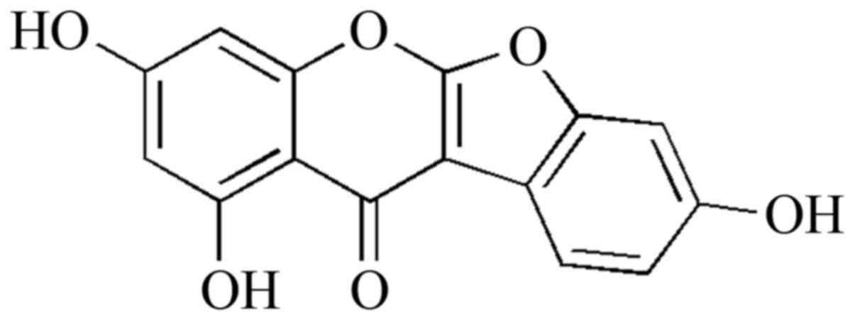
open column chromatography. Lupinalbin A was isolated from A.
americana tubers, as described previously (Fig. 1) (19).
DPP4 inhibitory assay and kinetic
analysis
DPP4 inhibitory activity was measured using a
DPP(IV) inhibitor screening assay kit (Cayman Chemical Company)
according to the manufacturer's protocol, with minor modifications.
Briefly, the enzyme solution (120 µl) was dissolved in 480 µl DPP
assay buffer [20 mM Tris-HCI (pH 8.0), 100 mM NaCl and 1 mM EDTA]
and was used as the enzyme solution. The substrate, 5 mM H-Gly-Pro
conjugated with aminomethylcoumarin (AMC), was prepared in the same
buffer. The assay procedure was performed according to the
manufacturer's protocols, and is briefly described as follows:
Diluted assay buffer (30 µl) and diluted enzyme solution (10 µl)
were added to the 96-well plates containing 10 µl solvent (blank)
or solvent-dissolved test compounds (0-75 µM). The reaction was
initiated by adding 50 µl diluted substrate solution, the reaction
was measured using fluorometric determination (excitation
wavelength, 350 nm; emission wavelength, 450 nm) using a plate
reader (Tecan Group, Ltd.) every 30 sec for 20 min at 37˚C.
Sitagliptin, which was included in the kit, was used as a positive
control. Various concentrations (0-75 µM) of the enzyme inhibitor
(tested compounds) and substrate were used in the reactions, the
initial rate of reaction (v) based on free AMC, and the release
rate were calculated using Lineweaver-Burk [1/v vs. 1/(substrate)]
or Dixon plots [1/v vs. (inhibitor)].
α-glucosidase inhibitory assay
The α-glucosidase inhibitory assay was performed as
described previously (21), with
minor modifications. A total of 130 µl enzyme (0.16 U/ml) in
phosphate buffer [0.1 mM phosphate (pH 6.8)], 20 μl methanol
(control) or ligand (0.062-1 mM) in methanol and 50 µl substrate
(2.5 mM p-nitrophenyl-α-D-glucopyranoside) were mixed in a 96-well
plate. After starting each reaction at 37˚C, p-nitrophenol was
quantified using a UV-vis spectrophotometer at 405 nm. Acarbose (20
µl) provided as ready to use in the kit was used as a positive
control.
Molecular docking
The molecular simulations for the interaction
between the inhibitors and enzyme were generated using AutoDock
software version 4.2 (The Scripps Research Institute). The 3D
structure of the ligand was built and minimized using MM2 in Chem
3D Pro (version 14.0). The flexible bonds of the ligand were
assigned with AutoDockTools. The 3D structure of DPP4 (PDB ID:
5T4E) was obtained from Research Collaboratory for Structural
Bioinformatics Protein Data Bank after substrates were removed from
the original enzyme. The grid for docking was formed using X, Y and
Z axes, all of which were 180 units in length. Default settings
were used for docking, except for the Lamarckian genetic algorithms
(run 50) and maximum number of evaluations (long). The results of
the molecular simulation were prepared using LigPlot and Chimera
software (22).
Statistical analysis
All inhibitory assays were performed in triplicate.
Data are presented as mean ± standard deviation, and were analyzed
using SigmaPlot (Systat Software Inc.) or GraphPad Prism version 6
(GraphPad Software Inc.). Differences were compared using a one-way
ANOVA followed by a post-hoc Dunnett's multiple comparison test.
P<0.05 was considered to indicate a statistically significant
difference.
Results
Inhibition of DPP4 and α-glucosidase
activities by lupinalbin A
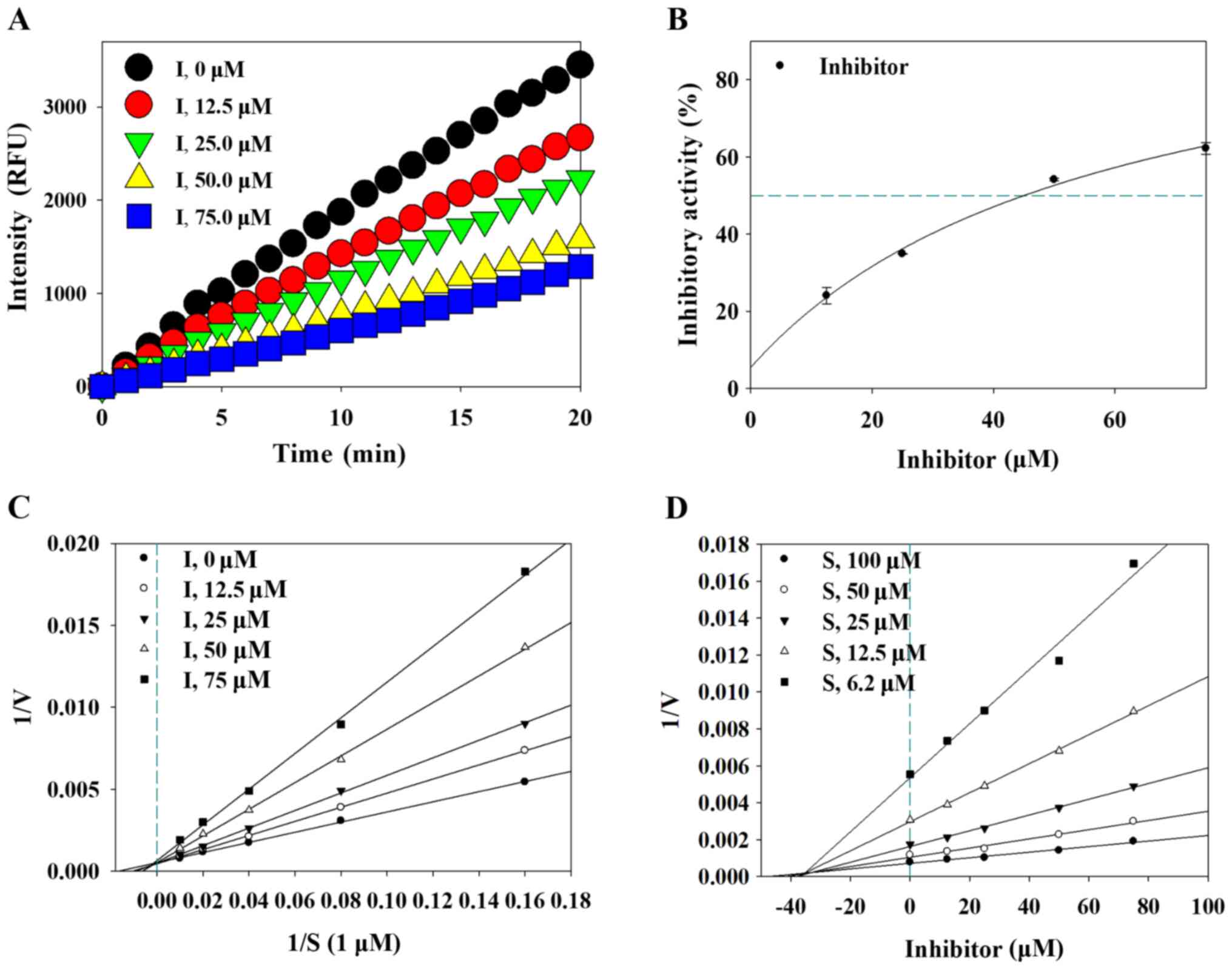
To investigate the inhibitory effect of lupinalbin
A, the inhibitory activity of lupinalbin A against DPP4 and
α-glucosidase were measured. Results showed that lupinalbin A
inhibited DPP4 activity in a time- and dose-dependent manner
(Fig. 2A and B). The IC50 values of lupinalbin
A against DPP4 and sitagliptin (positive control) were 45.2±0.8 µM
and 70.7±4.3 nM, respectively (Table
I). Furthermore, the IC50 values of lupinalbin A
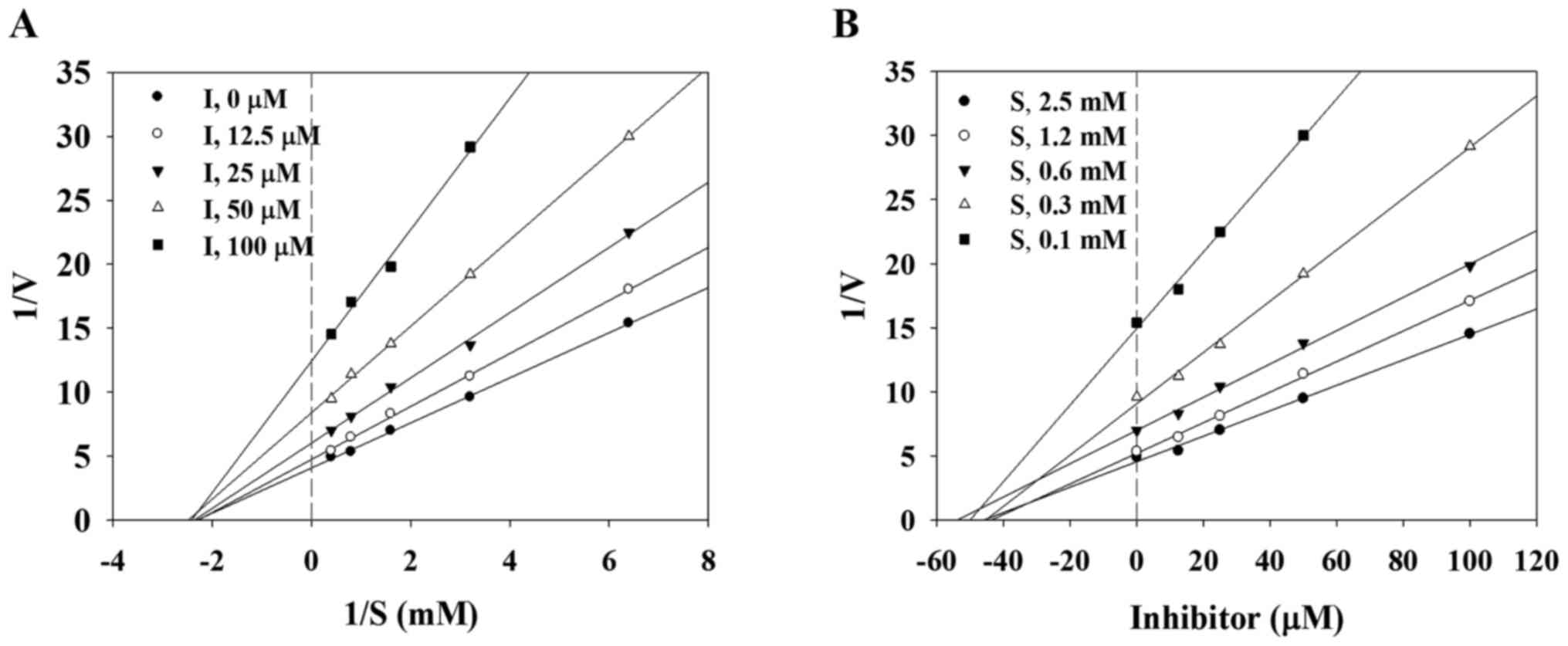
against α-glucosidase and acarbose (positive control) were 53.4±1.2
and 240.5±2.1 µM, respectively.
 | Table IInhibitory activities of lupinalbin A
on DPP4 and α-glucosidase. |
Table I
Inhibitory activities of lupinalbin A
on DPP4 and α-glucosidase.
| | DPP4 | α-glucosidase |
|---|
| Compound | IC50,
µMa | Binding mode
(Ki, µM)a | IC50,
µMa | Binding mode
(Ki, µM)a |
|---|
| Lupinalbin A | 45.2±0.8 | Competitive,
(35.1±2.0) | 53.4±1.2 | Non-competitive
(45.0±4.1) |
|
Sitagliptinb | 70.7±4.3 nM | | - | |
| Acarbosec | - | | 240.5±2.1 | |
Evaluation of enzyme kinetics
To determine whether lupinalbin A inhibits DPP4 and
α-glucosidase by interacting with the active site of these enzymes,
the enzyme kinetics of DPP4 and α-glucosidase were examined. An
association between lupinalbin A and the enzyme substrate was
confirmed using the Lineweaver-Burk plot, and the inhibition
constant (Ki) was determined using a Dixon plot. The slope
and X-axis of lupinalbin A changed in a dose-dependent manner,
whereas the Y-axis was intersected at only one point (Fig. 2C). This suggests that lupinalbin A
competitively suppressed the activity of DPP4. Analysis of
α-glucosidase kinetics using the Lineweaver-Burk plot showed that
the slope and Y-axis of lupinalbin A changed in a dose-dependent
manner, whereas the line only intersected the X-axis at one point,
indicating non-competitive inhibition (Fig. 3A). According to the Dixon plot, the
Ki values of lupinalbin A for DPP4 and α-glucosidase were
35.1 and 45.0 µM, respectively, indicating that lupinalbin A was a
competitive inhibitor of DPP4 and a non-competitive inhibitor of
α-glucosidase (Table I; Figs. 2D and 3B).
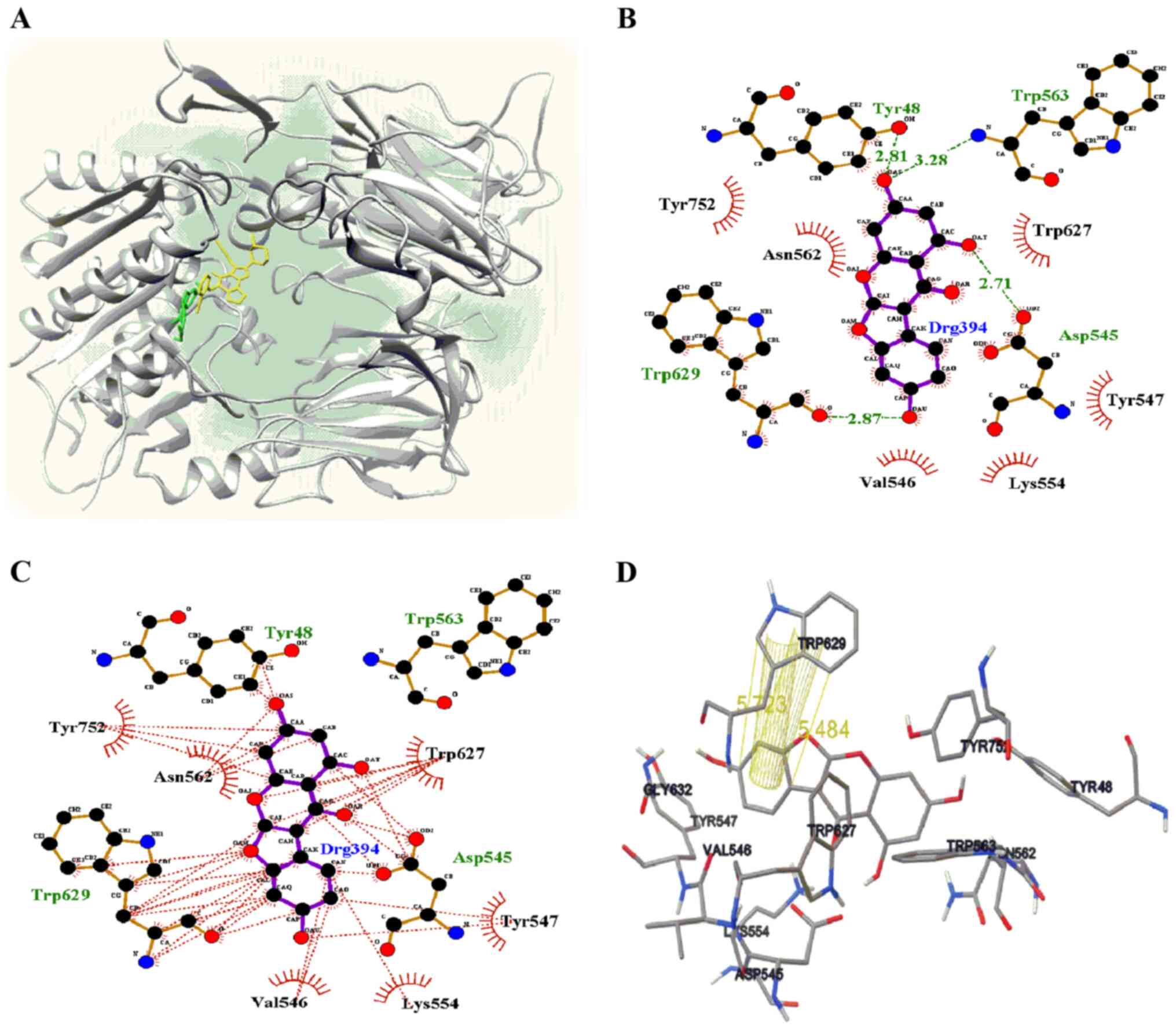
Molecular docking simulation of
lupinalbin A with DPP4
Molecular docking simulations are used to facilitate
the development of enzyme inhibitors (23,24),
determine the binding position of a ligand in an enzyme, and
identify key amino acids that participate in ligand-receptor
binding (25,26). Molecular docking was performed in the
present study to set up a grid containing the activity site based
on the results of the enzyme kinetic study. The binding pose of a
ligand with a receptor, as determined using AutoDock, was suggested
at the lowest energy values. Thus, lupinalbin A (1) occupied the right site next to the
active site and a small portion of the active site (Fig. 4A), with a binding energy of -7.32
kcal/mol. Lupinalbin A formed four hydrogen bonds with Tyr48 (2.81
Å), Asp545 (2.71 Å), Trp563 (3.28 Å) and Trp629 (2.87 Å) residues
surrounding the active site of DPP4 (Fig. 4B). A total of 9 amino acids of the
receptor participated in hydrophobic interactions to maintain a
stable binding pose with the ligand (Fig. 4C). Moreover, the B-ring of lupinalbin
A interacted two π-π stacking (5.48 and 5.72 Å) with the indole
ring of tryptophan (Fig. 4D).
Discussion
In the present study, the potential anti-diabetic
effects of lupinalbin A, which is isolated from A.
americana, were determined using enzyme assays. The
IC50 value of isoflavonoids isolated from the leaves of
Smilax china L. against DPP4 is greater than 100 µM (4); thus, the DPP4 inhibitory activity of
lupinalbin A (IC50, 45.2 µM) is stronger than these
isoflavonoids. Moreover, Bai et al (20) reported that lignans and flavonoids
isolated from mung beans exhibited inhibitory activity against
α-glucosidase, and lupinalbin A, a type of isoflavonoid was shown
to be more potent in their study compared with the present study,
with an IC50 value of 10.73±2.01 µM. Taken together,
lupinalbin A isolated from A. americana inhibited the
activity of DPP4 and α-glucosidase.
Through enzyme kinetics analysis, the ability of
lupinalbin A to inhibit the activity of different enzymes was
determined. Fan et al (27)
showed that phenolic compounds, including resveratrol and flavone,
competitively inhibited DPP4 activity, which is consistent with the
results of the present study. The binding force of flavone is
stronger than that of lupinalbin A; the Ki value of flavone
is 18.6 µM (23). Generally, the
lower the Ki value, the stronger the binding of the enzyme
and inhibitor; thus, an inhibitor with a lower Ki value is
more effective than an inhibitor with a higher Ki value. On
the basis of the Ki values of lupinalbin A for DPP4 and
α-glucosidase, lupinalbin A binds to DPP4 more strongly to
α-glucosidase. Thus, the DPP4 inhibitory activity of lupinalbin A
is stronger than its α-glucosidase inhibitory activity, which is
consistent with the results of the enzyme inhibition assay.
In conclusion Lupinalbin A, a compound isolated from
A. americana, exhibited anti-diabetic activity, and more
potently inhibited DPP4 compared with α-glucosidase. As a result,
it is suggested that lupinalbin A may be one of the active
components underlying the anti-diabetic effects of A.
americana.
Acknowledgements
Not applicable.
Funding
Funding: This work was supported by the National Research
Foundation of Korea (NRF) grant funded by the Korea government
(MSIP) (grant no. 2017M2A2A6A05018541).
Availability of data and materials
The datasets used and/or analyzed during the current
study are available from the corresponding author on reasonable
request.
Authors' contributions
CHJ designed the experiments. HYK performed the
experiments. HYK, JHK and CHJ analyzed the data. JHK isolated
lupinalbin A. HYK, JHK and CHJ wrote the manuscript. HGJ assisted
in designing the experiment and revising the manuscript. All
authors have read and approved the final manuscript. HYK, JHK and
CHJ confirm the authenticity of all the raw data.
Ethics approval and consent to
participate
Not applicable.
Patient consent for publication
Not applicable.
Competing interests
The authors declare that they have no competing
interests.
References
|
1
|
Wild S, Roglic G, Green A, Sicree R and
King H: Global prevalence of diabetes: estimates for the year 2000
and projections for 2030. Diabetes Care. 27:1047–1053.
2004.PubMed/NCBI View Article : Google Scholar
|
|
2
|
Kim SM, Lee JS, Lee J, Na JK, Han JH, Yoon
DK, Baik SH, Choi DS and Choi KM: Prevalence of diabetes and
impaired fasting glucose in Korea: Korean National Health and
Nutrition Survey 2001. Diabetes Care. 29:226–231. 2006.PubMed/NCBI View Article : Google Scholar
|
|
3
|
DeFronzo RA: Pathogenesis of type 2
diabetes mellitus. Med Clin North Am. 88:787–835, ix.
2004.PubMed/NCBI View Article : Google Scholar
|
|
4
|
Zhao BT, Le DD, Nguyen PH, Ali MY, Choi
JS, Min BS, Shin HM, Rhee HI and Woo MH: PTP1B, α-glucosidase, and
DPP-IV inhibitory effects for chromene derivatives from the leaves
of Smilax china L. Chem Biol Interact. 253:27–37. 2016.PubMed/NCBI View Article : Google Scholar
|
|
5
|
Li ZP, Song YH, Uddin Z, Wang Y and Park
KH: Inhibition of protein tyrosine phosphatase 1B (PTP1B) and
α-glucosidase by xanthones from Cratoxylum cochinchinense, and
their kinetic characterization. Bioorg Med Chem. 26:737–746.
2018.PubMed/NCBI View Article : Google Scholar
|
|
6
|
Yazbeck R, Howarth GS and Abbott CA:
Dipeptidyl peptidase inhibitors, an emerging drug class for
inflammatory disease? Trends Pharmacol Sci. 30:600–607.
2009.PubMed/NCBI View Article : Google Scholar
|
|
7
|
Hopsu-Havu VK and Glenner GG: A new
dipeptide naphthylamidase hydrolyzing
glycyl-prolyl-beta-naphthylamide. Histochemie. 7:197–201.
1966.PubMed/NCBI View Article : Google Scholar
|
|
8
|
Gorrell MD, Gysbers V and McCaughan GW:
CD26: A multifunctional integral membrane and secreted protein of
activated lymphocytes. Scand J Immunol. 54:249–264. 2001.PubMed/NCBI View Article : Google Scholar
|
|
9
|
Morimoto C and Schlossman SF: The
structure and function of CD26 in the T-cell immune response.
Immunol Rev. 161:55–70. 1998.PubMed/NCBI View Article : Google Scholar
|
|
10
|
Drucker DJ: Dipeptidyl peptidase-4
inhibition and the treatment of type 2 diabetes: Preclinical
biology and mechanisms of action. Diabetes Care. 30:1335–1343.
2007.PubMed/NCBI View Article : Google Scholar
|
|
11
|
Mulvihill EE and Drucker DJ: Pharmacology,
physiology, and mechanisms of action of dipeptidyl peptidase-4
inhibitors. Endocr Rev. 35:992–1019. 2014.PubMed/NCBI View Article : Google Scholar
|
|
12
|
Wilson PW, Pichardo FJ, Liuzzo JA,
Blackmon WJ and Reynolds BD: Amino acids in the american groundnut
(Apios americana). J Food Sci. 52:224–225. 1987.
|
|
13
|
Ichige M, Fukuda E, Miida S, Hattan J,
Misawa N, Saito S, Fujimaki T, Imoto M and Shindo K: Novel
isoflavone glucosides in groundnut (Apios americana Medik)
and their antiandrogenic activities. J Agric Food Chem.
61:2183–2187. 2013.PubMed/NCBI View Article : Google Scholar
|
|
14
|
Kaneta H, Koda M, Saito S, Imoto M, Kawada
M, Yamazaki Y, Momose I and Shindo K: Biological activities of
unique isoflavones prepared from Apios americana Medik.
Biosci Biotechnol Biochem. 80:774–778. 2016.PubMed/NCBI View Article : Google Scholar
|
|
15
|
Kim JH, Kim HY, Kang SY, Kim YH and Jin
CH: Soluble epoxide hydrolase inhibitory activity of components
isolated from Apios americana Medik. Molecules.
22(1432)2017.PubMed/NCBI View Article : Google Scholar
|
|
16
|
Kim JH, Kim HY, Kang SY, Kim JB, Kim YH
and Jin CH: Chemical constituents from Apios americana and
their inhibitory activity on tyrosinase. Molecules.
23(232)2018.PubMed/NCBI View Article : Google Scholar
|
|
17
|
Iwai K and Matsue H: Ingestion of Apios
americana Medikus tuber suppresses blood pressure and improves
plasma lipids in spontaneously hypertensive rats. Nutr Res.
27:218–224. 2007.
|
|
18
|
Gilbert ER and Liu D: Anti-diabetic
functions of soy isoflavone genistein: Mechanisms underlying its
effects on pancreatic β-cell function. Food Funct. 4:200–212.
2013.PubMed/NCBI View Article : Google Scholar
|
|
19
|
Kim HY, Kim JH, So Y, Kang SY, Jeong HG
and Jin CH: Anti-inflammatory effect of lupinalbin A isolated from
Apios americana on lipopolysaccharide-treated RAW264.7
cells. Molecules. 23(583)2018.PubMed/NCBI View Article : Google Scholar
|
|
20
|
Bai Y, Xu Y, Chang J, Wang X, Zhao Y and
Yu Z: Bioactives from stems and leaves of mung beans (Vigna
radiata L.). J Funct Foods. 25:314–322. 2016.
|
|
21
|
Yang D, Xie H, Jiang Y and Wei X:
Phenolics from strawberry cv. Falandi and their antioxidant and
α-glucosidase inhibitory activities. Food Chem. 194:857–863.
2016.PubMed/NCBI View Article : Google Scholar
|
|
22
|
Kim JH, Ryu YB, Lee WS and Kim YH:
Neuraminidase inhibitory activities of quaternary isoquinoline
alkaloids from Corydalis turtschaninovii rhizome. Bioorg Med Chem.
22:6047–6052. 2014.PubMed/NCBI View Article : Google Scholar
|
|
23
|
Meng XY, Zhang HX, Mezei M and Cui M:
Molecular docking: A powerful approach for structure-based drug
discovery. Curr Comput Aided Drug Des. 7:146–157. 2011.PubMed/NCBI View Article : Google Scholar
|
|
24
|
de Ruyck J, Brysbaert G, Blossey R and
Lensink MF: Molecular docking as a popular tool in drug design, an
in silico travel. Adv Appl Bioinform Chem. 9:1–11. 2016.PubMed/NCBI View Article : Google Scholar
|
|
25
|
Heitz MP and Rupp JW: Determining mushroom
tyrosinase inhibition by imidazolium ionic liquids: A spectroscopic
and molecular docking study. Int J Biol Macromol. 107 (Pt
B):1971–1981. 2018.PubMed/NCBI View Article : Google Scholar
|
|
26
|
Seong SH, Ali MY, Kim HR, Jung HA and Choi
JS: BACE1 inhibitory activity and molecular docking analysis of
meroterpenoids from Sargassum serratifolium. Bioorg Med Chem.
25:3964–3970. 2017.PubMed/NCBI View Article : Google Scholar
|
|
27
|
Fan J, Johnson MH, Lila MA, Yousef G and
de Mejia EG: Berry and citrus phenolic compounds inhibit dipeptidyl
peptidase IV: Implications in diabetes management. Evid Based
Complement Alternat Med. 2013(479505)2013.PubMed/NCBI View Article : Google Scholar
|