Introduction
Multiple sclerosis (MS) is a neurodegenerative
disease of the central nervous system with autoimmune and
inflammatory features. The etiology of MS remains unclear despite
extensive research over the past five decades. It is generally
hypothesized to be a multifactorial entity arising from complex
interactions between genetic predispositions, infectious exposures
and factors leading to pro-inflammatory conditions, such as
smoking, obesity and deficiency of vitamin D (1-4).
A growing body of evidence supports a possible role
of iron metabolism in a number of diseases with a neurodegenerative
component, including MS (5,6). Several post-mortem studies and studies
using conventional and the new iron-sensitive imaging techniques
have described changes in brain iron homeostasis, linking iron
deposition in different brain regions to the demyelinating process
in MS patients (7-11).
A recent study in MS patients found an association between the
serum iron concentration and evidence of increased iron deposition
in deep gray matter subcortical structures (12). In addition, in our previous study in
experimental autoimmune encephalomyelitis, the most commonly used
animal model of MS, it was found that chronic iron overload
influenced the clinical course of the disease in Dark Agouti rats;
it affected disease progression and mortality, with milder effects
on female rats (13). The human
hemochromatosis gene (HFE) is an important regulator of
cellular iron homeostasis and has the highest prevalence of
polymorphisms amongst the iron regulatory genes known to alter
brain iron levels and structure. In HFE, the single
nucleotide polymorphisms C282Y and H63D can result in phenotypes
with altered iron parameters. Homozygosity or compound
heterozygosity for the C282Y and H63D variants can lead to iron
overload and the disorder known as hereditary hemochromatosis.
Since HFE also acts as a signal peptide, it has two
domains (α1 and α2) that form an extracellular transferrin
receptor-binding region and an immunoglobulin-like domain (α3)
(14,15). Cysteine 282 is essential for the
binding of β-2-microglobulin (β2M) to the α3 domain and for the
extracellular presentation of HFE. When HFE binds to β2M, it forms
a heterodimer expressed at the cell surface (16). In the case of C282Y mutation, the
disulfide bond in the α3 domain is disrupted, thus β2M cannot bind
and HFE is not present at the cell surface, but instead aggregates
in the cytoplasm (17). It was
confirmed that when HFE C282Y is overexpressed, the binding
capacity of HFE and TFR is significantly reduced (16). Therefore, C282Y homozygosity results
in higher serum iron and ferritin levels and an increase in
transferrin saturation (18).
Histidine 63 forms a salt bridge in the transferrin
receptor-binding region (19). The
H63D mutation disrupts the salt bridge and alters the tertiary
structure of HFE and its function (17).
Animal models have also been used to improve our
understanding of the role of the C282Y and H63D polymorphisms. For
example, Hfe knockout mice, homozygous for the deletion
corresponding to the α1 and α2 domains of HFE, showed increased
iron absorption and plasma concentration, as well as increased
transferrin saturation and iron overload. Homozygosity for the
C282Y mutation also caused iron overload (20).
The few studies investigating the association
between the C282Y and H63D polymorphisms and the risk of MS have
reported inconclusive results (21-27).
As a single study typically has a relatively small number of
participants with low statistical power, a meta-analysis may be an
appropriate approach for obtaining a more definitive conclusion.
Therefore, a meta-analysis of 7 studies conducted in different
populations of Caucasian origin was performed. To the best of our
knowledge, this is the first study to integrate the association
between the C282Y and H63D polymorphisms in the HFE gene and
the risk of MS.
Materials and methods
PubMed, EMBASE and Web of Science were searched
independently by two investigators to identify all relevant studies
published before January 2021 that addressed the association
between HFE polymorphisms and MS. The key words used in the
search were: ‘hemochromatosis’ OR ‘HFE’ OR ‘C282Y’ OR ‘H63D’,
‘multiple sclerosis’ OR ‘MS’, ‘polymorphism’ OR ‘SNP’ OR ‘mutation’
OR ‘variant’, where ‘OR’ was used as a Boolean modifier. The
following information was collected from each study: First author,
year of publication, country, ethnicity of study population, study
design, number of cases and controls, and genotype and allele
frequencies of the C282Y and H63D polymorphisms. A meta-analysis
was performed in accordance with the PRISMA guidelines (28) using Comprehensive Meta-analysis 3.0
software (Biostatic Inc.).
The strength of the association between the C282Y
and H63D polymorphisms and MS risk was estimated by odds ratios
(ORs) with 95% confidence intervals (CIs). Due to the relatively
small sample sizes of each study and the low frequency of variant
alleles, as well as the practical clinical significance, a
meta-analysis of only two comparison models was performed: The
dominant model (YY + CY vs. CC or DD + HD vs. HH) and the allele
contrast (Y vs. C or D vs. H). The distribution of genotypes in the
control groups was tested for Hardy-Weinberg equilibrium using a
χ2 test.
A Cochran's Q statistic and I2 tests were
applied to quantify the heterogeneity between studies. Fixed/random
effects models were used to calculate pooled ORs. Funnel plots and
Egger's linear regression test were used to assess publication
bias.
Results
A total of 7 studies that examined the association
between HFE polymorphisms and MS were identified and
included in the present meta-analysis. The final meta-analysis
included 2,271 patients and 2,180 controls for the C282Y
polymorphism (21-27),
and 1,782 cases and 2,076 ontrols, for the H63D polymorphism
(22-27).
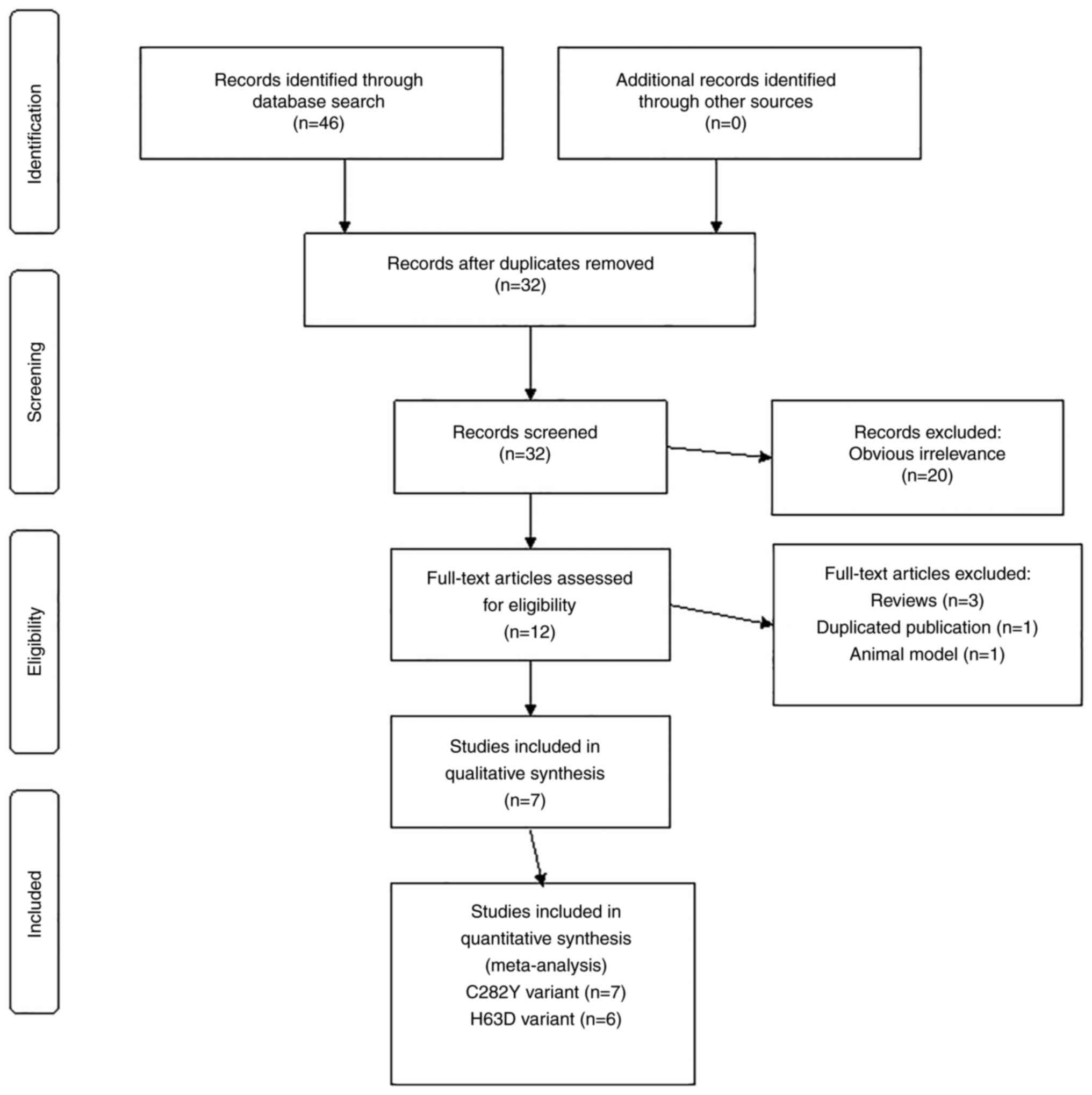
The flowchart of article selection and specific reasons for
exclusion/inclusion are shown in Fig.
1. The characteristics of the studies and the HFE
genotypes and allele distributions in MS patients and controls are
provided in Table I. To include the
data from the study by Ramagopalan et al (24) which lacked a control group, the
previously reported C282Y and H63D genotype and allele frequencies
from the Canadian population was used (29). All studies were performed in
populations of Caucasian origin. The HFE genotype
frequencies in the control groups of all studies were in
Hardy-Weinberg equilibrium.
 | Table ICharacteristics of studies included in
the meta-analysis. |
Table I
Characteristics of studies included in
the meta-analysis.
| First author,
year | Population
studied | Study group, n | HFE C/Y genotypes, n
(%) | HFE C/Y allele,
% | HFE H/D genotypes, n
(%) | HFE H/D allele,
% | (Refs.) |
|---|
| Rubio et al,
2004 | Australian
(Tasmanian, Victorian) | MS, 489 | CC, 395 (80.8); CY,
88 (18.0); YY, 6 (1.2) | C, 89.8; Y, 10.2 | - | - | (21) |
| | | Control, 104 | CC, 90 (86.5); CY, 14
(13.5); YY, 0 (0.0) | C, 93.3; Y, 6.7 | - | - | |
| Ristić et al,
2005 | Croatian and
Slovenian | MS, 314 | CC, 291 (92.7); CY,
23 (7.3); YY, 0 (0.0) | C, 96.3; Y, 3.7 | HH, 231 (73.6); HD,
80 (25.5); DD, 3 (0.9) | H, 86.3; D, 13.7 | (22) |
| | | Control, 400 | CC, 372 (93.0); CY,
27 (6.8); YY, 1 (0.2) | C, 96.4; Y, 3.6 | HH, 297 (74.3); HD,
90 (22.5); DD 13 (3.2) | H, 85.5; D, 14.5 | |
| Kotze et al,
2006 | Caucasian South
African | MS, 118 | CC, 95 (80.5); CY, 22
(18.6); YY, 1 (0.9) | C, 89.8; Y, 10.2 | HH, 100 (84.7); HD,
17 (14.4); DD, 1 (0.9) | H, 91.9; D, 8.1 | (23) |
| | | Control, 102 | CC, 85 (83.3); CY, 15
(14.7); YY, 2 (2.0) | C, 90.7; Y, 9.3 | HH, 77 (75.5); HD, 25
(24.5); DD, 0 (0.0) | H, 87.7; D, 12.3 | |
| Ramagopalan et
al, 2008 | Canadian | MS, 163 | CC, 151 (92.6); CY,
12 (7.4); YY, 0 (0.0) | C, 96.3; Y, 3.7 | HH, 119 (73.0); HD,
43 (26.4); DD, 1 (0.6) | H, 86.2; D, 13.8 | (24) |
| | | Control (C/Y, 881;
H/D, 870)a | CC, 805 (91.4); CY,
76 (8.6); YY, 0 (0.0) | C, 95.7; Y, 4.3 | HH, 578 (66.4); HD,
271 (31.1); DD, 21 (2.4) | H, 82.0; D, 18.0 | |
| Bettencourt et
al, 2011 | Northern
Portuguese | MS, 373 | CC, 341 (91.4); CY,
31 (8.3); YY, 1 (0.3) | C, 95.6; Y, 4.4 | HH, 251 (67.3); HD,
111 (29.8); DD, 11 (2.9) | H, 82.2; D, 17.8 | (25) |
| | | Control, 129 | CC, 115 (89.1); CY,
13 (10.1); YY, 1 (0.8) | C, 94.2; Y,
5.8 | HH, 87 (67.4); HD,
34 (26.4); DD, 8 (6.2) | H, 80.6; D,
19.4 | |
| Gemmati et
al, 2012 | Northern
Italian | MS, 414 | CC, 401 (96.6); CY,
13 (3.1); YY, 0 (0.0) | C, 98.4; Y,
1.6 | HH, 288 (69.6); HD,
113 (27.3); DD, 13 (3.1) | H, 83.2; D,
16.8 | (26) |
| | | Control, 414 | CC, 397 (95.9); CY,
17 (4.1); YY, 0 (0.0) | C, 97.9; Y,
2.1 | HH, 305 (73.7); HD,
101 (24.4); DD, 8 (1.9) | H, 85.9; D,
14.1 | |
| Hagemeier et
al, 2017 | USA | MS, 400 | CC, 353 (88.2); CY,
47 (11.8); YY, 0 (0.0) | C, 94.1; Y,
5.9 | HH, 285 (71.2); HD,
104 (26.0); DD, 11 (2.8) | H, 84.2; D,
15.8 | (27) |
| | | Control, 150 | CC, 137 (91.3); CY,
13 (8.7); YY, 0 (0.0) | C, 95.7; Y,
4.3 | HH, 116 (77.3); HD,
28 (18.7); DD, 6 (4.0) | H, 86.7; D,
13.3 | |
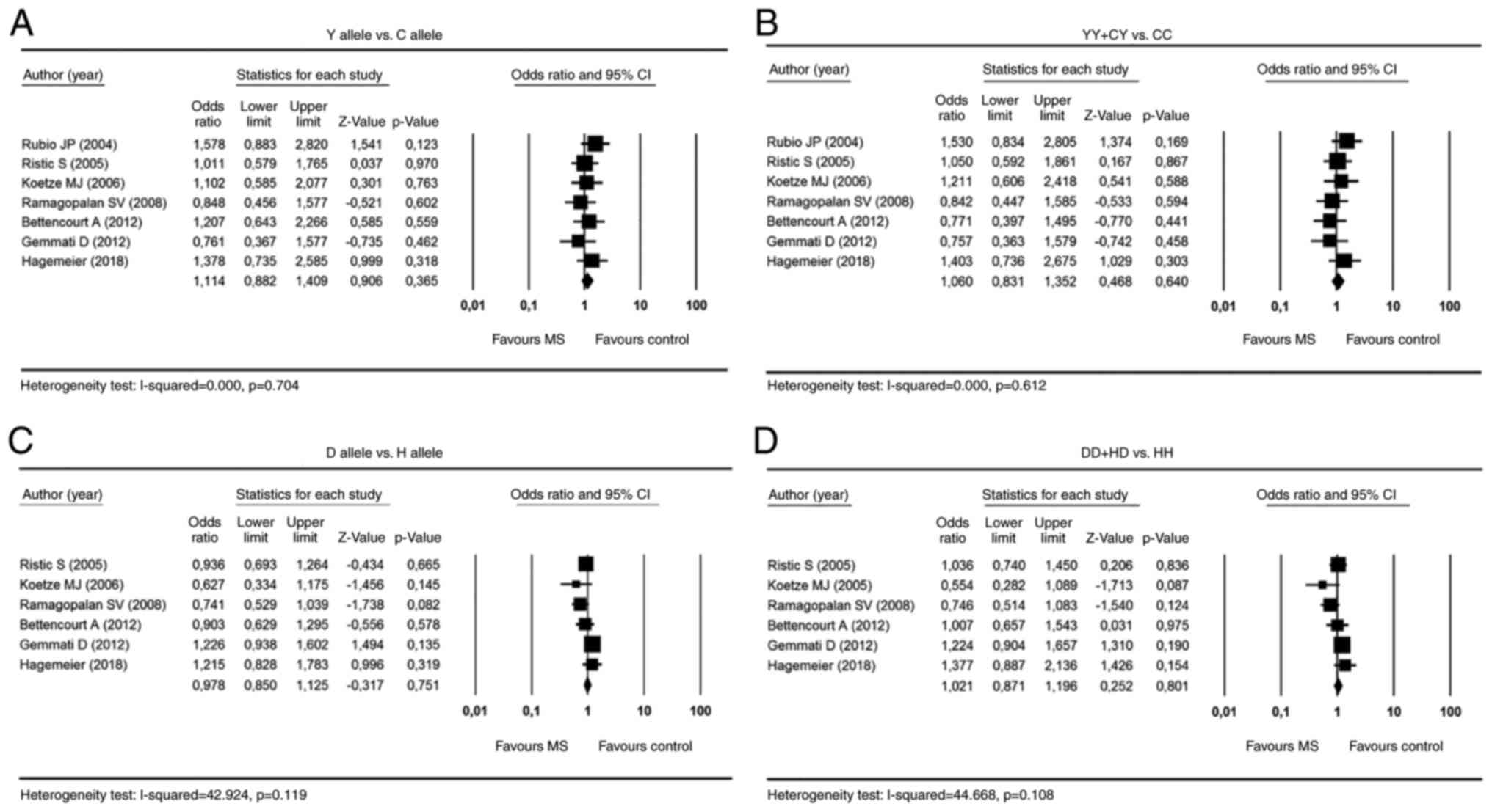
The results of the meta-analysis did not show a
significant association between HFE polymorphisms and
susceptibility to MS in any of the genetic comparison models (for Y
vs. C: OR=1.11, 95% CI 0.88-1.41, P=0.365; for YY + CY vs. CC:
OR=1.06, 95% CI 0.83-1.35, P=0.640; for D vs. H: OR=0.98, 95% CI
0.85-1.12, P=0.751; for DD + HD vs. HH: OR=1.02, 95% CI 0.87-1.20,
P=0.801; Fig. 2). No heterogeneity
was observed between studies in the meta-analysis (all P>0.05);
therefore, the fixed effects model was applied.
No publication bias was found amongst the studies
included in the meta-analysis. The shapes of the funnel plots for
all tested models were symmetrical, and no statistical evidence of
publication bias for any genetic model using Egger's linear
regression test was found (P=0.656 for the Y allele model, P=0.371
for the dominant C282Y model, P=0.147 for the D allele model, and
P=0.118 for the dominant H63D model).
Discussion
The present meta-analysis showed no evidence of a
significant association between the C282Y and H63D polymorphisms
and the risk of MS. Conversely, these mutations did influence
disease behavior rather than susceptibility, as some of the cited
studies reported their influence on the onset or severity of MS. A
higher MS disability score and disease progression have been shown
to correlate with C282Y carriers (25,27) or
H63D carriers (26). In our
previous study, it was reported an earlier onset of disease in
patients carrying the C282Y mutant allele (22). Recent research by Hagemeier et
al (27) using quantitative
susceptibility mapping MRI showed only a moderate association
between iron-linked genetic polymorphisms and susceptibility to
deep grey matter susceptibility. Furthermore, they found
differences by sex in MS patients, suggesting that iron-related
risk alleles are a potential risk factor for female MS patients.
Gemmati et al (26) also
reported high MS susceptibility associated with the H63 DD genotype
in progressive female patients. These findings are consistent with
the growing body of evidence showing that sex plays a significant
role in the development, progression and treatment of MS. Moreover,
our previous study in an animal model of MS showed that iron
overload accelerates disease onset in female rats, but accelerates
disease progression and increases mortality in male rats (13). However, due to the different study
designs and insufficient original data in the studies included in
the meta-analysis, analysis of the association between HFE
polymorphisms and clinical features of MS, including age of onset
and disease severity, could not be performed. As none of the
studies provided sex-specific subgroup values for the HFE
genotypes, it was not possible to perform a sex-based pooled
analysis. In addition, mean serum iron, transferrin saturation and
serum ferritin levels are known to be higher in individuals
homozygous or compound heterozygous for the C282Y and H63D
mutations compared with other HFE genotypes (16), but serum iron parameters were not
available in MS patients, which should be investigated in the
future.
Finally, it is well established that MS is a common
complex disease whose susceptibility most likely results from the
interplay of genes, environmental interactions and gene/environment
interactions (30). Considering
that ethnic confounding may be more problematic in disease
susceptibility than in disease behavior, possible ethnic
confounding effects were overcome in this meta-analysis as all
studies included consisted of Caucasian populations.
In summary, no evidence was found showing that the
C282Y and H63D polymorphisms contributed to MS susceptibility in
the Caucasian population. The low frequency of the C282Y mutation
and the compound C282Y/H63D genotype could possibly explain the
lack of association in both the individual studies and the
meta-analysis. Further studies with larger sample sizes addressing
the role of HFE and other genetic variants related to iron
regulation in the development and progression of MS are
warranted.
Acknowledgements
Not applicable.
Funding
Funding: This study was supported by funding from the University
of Rijeka (grant no. uniri-biomed-18-137).
Availability of data and materials
The datasets used and/or analyzed during the present
study are available from the corresponding author on reasonable
request.
Authors' contributions
NSČ designed the study, performed the data analysis
and wrote the manuscript. BĆC performed the data analysis and
drafted the manuscript. VBL and BP interpreted the data, and
drafted the manuscript. SR designed the study and drafted the
manuscript. NSČ, BP and SR confirm the authenticity of all the raw
data. All authors have read and approved the final manuscript.
Ethics approval and consent to
participate
Not applicable.
Patient consent for publication
Not applicable.
Competing interests
The authors declare that they have no competing
interests.
References
|
1
|
Dobson R and Giovannoni G: Multiple
sclerosis-a review. Eur J Neurol. 26:27–40. 2019.PubMed/NCBI View Article : Google Scholar
|
|
2
|
Tarlinton RE, Khaibullin T, Granatov E,
Martynova E, Rizvanov A and Khaiboullina S: The interaction between
viral and environmental risk factors in the pathogenesis of
multiple sclerosis. Int J Mol Sci. 20(303)2019.PubMed/NCBI View Article : Google Scholar
|
|
3
|
Arneth B: Multiple sclerosis and smoking.
Am J Med. 133:783–788. 2020.PubMed/NCBI View Article : Google Scholar
|
|
4
|
Pytel V, Matías-Guiu JA, Torre-Fuentes L,
Montero-Escribano P, Maietta P, Botet J, Álvarez S, Gómez-Pinedo U
and Matías-Guiu J: Exonic variants of genes related to the vitamin
D signaling pathway in the families of familial multiple sclerosis
using whole-exome next generation sequencing. Brain Behav.
9(e01272)2019.PubMed/NCBI View Article : Google Scholar
|
|
5
|
Kim Y and Connor JR: The roles of iron and
HFE genotype in neurological diseases. Mol Aspects Med.
75(100867)2020.PubMed/NCBI View Article : Google Scholar
|
|
6
|
Grubić Kezele T and Ćurko-Cofek B:
Age-related changes and sex-related differences in brain iron
metabolism. Nutrients. 12(2601)2020.PubMed/NCBI View Article : Google Scholar
|
|
7
|
Zecca L, Youdim MB, Riederer P, Connor JR
and Crichton RR: Iron, brain ageing and neurodegenerative
disorders. Nat Rev Neurosci. 5:863–873. 2004.PubMed/NCBI View
Article : Google Scholar
|
|
8
|
Zivadinov R, Heininen-Brown M, Schirda CV,
Poloni GU, Bergsland N, Magnano CR, Durfee J, Kennedy C, Carl E,
Hagemeier J, et al: Abnormal subcortical deep-gray matter
susceptibility-weighted imaging filtered phase measurements in
patients with multiple sclerosis: A case-control study. Neuroimage.
59:331–339. 2012.PubMed/NCBI View Article : Google Scholar
|
|
9
|
Hagemeier J, Weinstock-Guttman B,
Heininen-Brown M, Poloni GU, Bergsland N, Schirda C, Magnano CR,
Kennedy C, Carl E, Dwyer MG, et al: Gray matter SWI-filtered phase
and atrophy are linked to disability in MS. Front Biosci (Elite
Ed). 5:525–532. 2013.PubMed/NCBI View
Article : Google Scholar
|
|
10
|
Zivadinov R, Tavazzi E, Bergsland N,
Hagemeier J, Lin F, Dwyer MG, Carl E, Kolb C, Hojnacki D, Ramasamy
D, et al: Brain iron at quantitative MRI is associated with
disability in multiple sclerosis. Radiology. 289:487–496.
2018.PubMed/NCBI View Article : Google Scholar
|
|
11
|
Bergsland N, Tavazzi E, Schweser F,
Jakimovski D, Hagemeier J, Dwyer MG and Zivadinov R: Targeting iron
dyshomeostasis for treatment of neurodegenerative disorders. CNS
Drugs. 33:1073–1086. 2019.PubMed/NCBI View Article : Google Scholar
|
|
12
|
Bergsland N, Agostini S, Laganà MM,
Mancuso R, Mendozzi L, Tavazzi E, Cecconi P, Clerici M and Baglio
F: Serum iron concentration is associated with subcortical deep
gray matter iron levels in multiple sclerosis patients.
Neuroreport. 28:645–648. 2017.PubMed/NCBI View Article : Google Scholar
|
|
13
|
Ćurko-Cofek B, Kezele TG, Marinić J, Tota
M, Čizmarević NS, Milin Č, Ristić S, Radošević-Stašić B and
Barac-Latas V: Chronic iron overload induces gender-dependent
changes in iron homeostasis, lipid peroxidation and clinical course
of experimental autoimmune encephalomyelitis. Neurotoxicology.
57:1–12. 2016.PubMed/NCBI View Article : Google Scholar
|
|
14
|
Feder JN, Gnirke A, Thomas W, Tsuchihashi
Z, Ruddy DA, Basava A, Dormishian F, Domingo R Jr, Ellis MC, Fullan
A, et al: A novel MHC class I-like gene is mutated in patients with
hereditary haemochromatosis. Nat Genet. 13:399–408. 1996.PubMed/NCBI View Article : Google Scholar
|
|
15
|
Faraj SA and Al-Abedy NM: Serum hepcidin
hormone level and its genes polymorphism, Genetic Variation,
Trindade Maia R and de Araújo Campos M (eds). IntechOpen, 2021.
doi: 10.5772/intechopen.93622. Available from: https://www.intechopen.com/chapters/73214.
|
|
16
|
Barton JC, Edwards CQ and Acton RT: HFE
gene: Structure, function, mutations, and associated iron
abnormalities. Gene. 574:179–192. 2015.PubMed/NCBI View Article : Google Scholar
|
|
17
|
Hollerer I, Bachmann A and Muckenthaler
MU: Pathophysiological consequences and benefits of HFE mutations:
20 Years of research. Haematologica. 102:809–817. 2017.PubMed/NCBI View Article : Google Scholar
|
|
18
|
Adams PC, Reboussin DM, Barton JC, McLaren
CE, Eckfeldt JH, McLaren GD, Dawkins FW, Acton RT, Harris EL,
Gordeuk VR, et al: Hemochromatosis and iron-overload screening in a
racially diverse population. N Engl J Med. 352:1769–1778.
2005.PubMed/NCBI View Article : Google Scholar
|
|
19
|
Fleming RE and Sly WS: Mechanisms of iron
accumulation in hereditary hemochromatosis. Annu Rev Physiol.
64:663–680. 2002.PubMed/NCBI View Article : Google Scholar
|
|
20
|
Bahram S, Gilfillan S, Kühn LC, Moret R,
Schulze JB, Lebeau A and Schümann K: Experimental hemochromatosis
due to MHC class I HFE deficiency: Immune status and iron
metabolism. Proc Natl Acad Sci USA. 96:13312–13317. 1999.PubMed/NCBI View Article : Google Scholar
|
|
21
|
Rubio JP, Bahlo M, Tubridy N, Stankovich
J, Burfoot R, Butzkueven H, Chapman C, Johnson L, Marriott M, Mraz
G, et al: Extended haplotype analysis in the HLA complex reveals an
increased frequency of the HFE-C282Y mutation in individuals with
multiple sclerosis. Hum Genet. 114:573–580. 2004.PubMed/NCBI View Article : Google Scholar
|
|
22
|
Ristić S, Lovrecić L, Brajenović-Milić B,
Starcević-Cizmarević N, Jazbec SS, Sepcić J, Kapović M and Peterlin
B: Mutations in the hemochromatosis gene (HFE) and multiple
sclerosis. Neurosci Lett. 383:301–304. 2005.PubMed/NCBI View Article : Google Scholar
|
|
23
|
Kotze MJ, de Villiers JN, Warnich L,
Schmidt S, Carr J, Mansvelt E, Fourie E and van Rensburg SJ: Lack
of clinical manifestation of hereditary haemochromatosis in South
African patients with multiple sclerosis. Metab Brain Dis.
21:109–120. 2006.PubMed/NCBI View Article : Google Scholar
|
|
24
|
Ramagopalan SV, Cukjati M, Cernilec M,
DeLuca GC, Dyment DA, Degenhardt A, Sadovnick AD, Serbec VC, Ebers
GC and Duquette P: Mutations in the hemochromatosis gene and the
clinical outcome of multiple sclerosis. J Neuroimmunol.
203:104–107. 2008.PubMed/NCBI View Article : Google Scholar
|
|
25
|
Bettencourt A, Silva AM, Santos E, Gomes
S, Mendonça D, Costa PP, Faustino P and Silva BM: HFE gene
polymorphisms and severity in Portuguese patients with multiple
sclerosis. Eur J Neurol. 18:663–666. 2011.PubMed/NCBI View Article : Google Scholar
|
|
26
|
Gemmati D, Zeri G, Orioli E, De Gaetano
FE, Salvi F, Bartolomei I, D'Alfonso S, Dall'osso C, Leone MA,
Singh AV, et al: Polymorphisms in the genes coding for iron binding
and transporting proteins are associated with disability, severity,
and early progression in multiple sclerosis. BMC Med Genet.
13(70)2012.PubMed/NCBI View Article : Google Scholar
|
|
27
|
Hagemeier J, Ramanathan M, Schweser F,
Dwyer MG, Lin F, Bergsland N, Weinstock-Guttman B and Zivadinov R:
Iron-related gene variants and brain iron in multiple sclerosis and
healthy individuals. Neuroimage Clin. 17:530–540. 2017.PubMed/NCBI View Article : Google Scholar
|
|
28
|
Page MJ, McKenzie JE, Bossuyt PM, Boutron
I, Hoffmann TC, Mulrow CD, Shamseer L, Tetzlaff JM, Akl EA, Brennan
SE, et al: The PRISMA 2020 statement: An updated guideline for
reporting systematic reviews. BMJ. 372(n71)2021.PubMed/NCBI View
Article : Google Scholar
|
|
29
|
Girouard J, Giguère Y, Delage R and
Rousseau F: Prevalence of HFE gene C282Y and H63D mutations in a
French-Canadian population of neonates and in referred patients.
Hum Mol Genet. 11:185–189. 2002.PubMed/NCBI View Article : Google Scholar
|
|
30
|
Sadovnick AD: Genetic background of
multiple sclerosis. Autoimmun Rev. 11:163–166. 2012.PubMed/NCBI View Article : Google Scholar
|