Introduction
Physiological and morphological changes that occur
during aging impact the functional process related to an individual
quality of life. The brain irreversibly and progressively declines
in function, and is subject to several debilitating ailments such
as neurodegenerative diseases, cognitive impairment disorders,
neuroinflammatory disorders, or Alzheimer's disease (1-3).
The major risk factor concerning aging is an increase in free
radical levels, with a concurrent decrease in antioxidant levels.
These are the two major aspects that lead to oxidative stress, an
increase in reactive oxygen species (ROS) related to the
diminishing antioxidant defenses, and an increase in chronic
low-grade inflammation (4-6).
Previous studies have shown that a diet rich in antioxidants may
decrease the risk of developing various neurodegenerative diseases
(7,8). Prevention of brain deterioration
before aging is important. Curcumin and γ-oryzanol have been
reported to reduce ROS, oxidative stress, and inflammation.
Moreover, these two elements have been identified as compounds that
increase antioxidant levels such as those of superoxide dismutase
(SOD), catalase (CAT), and glutathione peroxidase (GPx) (9-12).
However, studies have found that the beneficial effects of curcumin
and γ-oryzanol are limited by their low water-solubility, leading
to low bioavailability (13-16).
Therefore, curcumin and γ-oryzanol were prepared in a solid
dispersion to enhance their water solubility. The aim of this study
was to evaluate the effects of γ-oryzanol solid dispersion (GOSD)
and curcumin solid dispersion (CURSD) on learning and memory of
middle-aged rats. In order to measure the level of improvement,
rats were tasked with performing a series of behavioral
evaluations: The Morris water maze (MWM) test and a novel object
recognition (NOR) test. Subsequently, the c-Fos activity, the
levels of malondialdehyde (MDA), the levels of tumor necrosis
factor-α (TNF-α), and antioxidant activities (including SOD, CAT,
and GPx) in the hippocampus and prefrontal cortex area of
middle-aged rats were determined.
Materials and methods
Experimental animals
A total of 30 Sprague-Dawley male rats were
obtained; 5 rats were aged 6-weeks old and 25 rats were aged
42-weeks old. All rats were obtained from Nomura Siam International
Co., Ltd. The animals were acclimatized for 1 week prior to
initiation of the experiments. Dependent on the size of the rat, 2
or 3 rats were housed per cage under control condition (12-h
light/dark cycle, room temperature of 22±1˚C, 55±10% humidity) and
provided ad libitum access to standard rat chow and tap
water. All animal welfare considerations were accounted for under
veterinary care. The animals were observed by well-trained
technicians at least once daily for clinical signs of illness. This
observation allowed prompt reporting and appropriate actions to be
performed as necessary by the animal veterinarian. Death or
20% weight loss were considered as endpoints. The experimental
protocols were in compliance with the standards of animal care and
use established under the ethical guidelines and policies of
Naresuan University, and were approved by the Ethics Committee of
the Centre for Animal Research of Naresuan University (Phitsanulok,
Thailand) (approval no. NU-AE620514).
Preparation of GOSD and CURSD
GOSD and CURSD were prepared using a melting solvent
method with PVPK30 as a carrier and lecithin as a co-carrier. GOSD
appeared as a white powder with γ-oryzanol content of 12%, while
CURSD appeared as a yellow powder with curcumin content of 10%.
Upon dilution in water, the water solubility of γ-oryzanol and
curcumin increased to 1.3 and 14 mg/ml, respectively.
Experimental design
In this experiment, the sample population was
divided into two clusters. One cluster with 5 rats aged 6-weeks old
that were assigned as the adult control group (n=5); and the second
cluster consisted of 25 rats aged 42-weeks old (middle-aged) that
were randomly divided into 5 groups (n=5/group). Those clusters
were then labeled as the control group, the GO group (GOSD 10
mg/kg·BW), the Cur group (CURSD 50 mg/kg·BW), the GO-LCur group
(GOSD 10 mg/kg·BW plus CURSD 25 mg/kg·BW), and the GO-HCur group
(GOSD 10 mg/kg·BW plus CURSD 50 mg/kg·BW). Rats were administered
the substance orally once daily for 42 consecutive days. On days
40-42, learning and memory performance was evaluated using NOR and
MWM tests. Once the behavioral assessments had been completed, all
rats were euthanized by intraperitoneal injection of thiopental
sodium overdose (100 mg/kg) and death was verified by cessation of
the heartbeat and respiration, and a lack of response to a noxious
stimulus (hind paw pinch). Rats were sacrificed and the brains were
removed. The right hippocampus and prefrontal cortex were stored at
-80˚C and subsequently used to analyze MDA, SOD, CAT, GPx, and
TNF-α, levels as well as being used for histopathological
analysis.
MWM test
The MWM test was used to assess spatial learning and
referent memory using a circular pool (diameter 130 cm, height 60
cm) filled with water (26±1˚C) with a depth of 40 cm, and was made
to appear opaque by mixing in additional powder. The platform
(diameter 10 cm) was submerged 1 cm below the surface and placed in
a stable motionless position. Animals were placed in the pool and
allowed 60 sec to swim to the hidden platform (17). During the trials, each animal
underwent three trials on 7 consecutive days. During the test, the
platform was removed from the pool and the time to swim in the
former target quadrant (retention time) was recorded. The videos
were analyzed using tracking software (Smart 3.0; Panlab).
NOR test
The NOR test was used to assess the recognition
memory of familiar and novel objects (18). The test used in this study was
performed using a contained open field (100x100x40 cm) enclosure
that included three objects. The object used were of different
shapes but of similar sizes. In the familiarization phase, the rat
was placed in an open field with two objects, a triangle and sphere
(object A and B, respectively) for 10 min. The objects were placed
in the center, ~20 cm apart. For the test phase, the rat was placed
in the open field again; however, one of the objects was changed,
the sphere was changed to a square (object C), to test the
recognition memory of the rat. Between each phase, the rat was
placed back in their cages for 10 min. The time spent exploring the
object was considered when the rat's nose pointed towards an object
at a distance of ≤1 cm. The percentage recognition index of each
rat was calculated based on the ratio
(TAx100)/(TA+TC), where
TA and TC are the time span each animal spent
at object A and C, respectively.
Histopathological study
The left hippocampus and prefrontal cortex were
fixed in 10% formaldehyde buffer at 22±1˚C for 3 days. Next the
tissue samples were transferred to a 30% sucrose solution in PBS
(0.1 M, pH 7.4) at 4˚C for 2-3 days (until submerged); transferred
to a tissue freezing medium (Leica GmbH, cat no. 3801480); and then
frozen in dry ice and stored at -80˚C until required for
sectioning. The brain coronal was sectioned at 30 µm on a cryostat
machine. The sections were stored in cryoprotectant solution at
-20˚C until used for immunohistochemical processing.
c-Fos immunohistochemistry
The immunohistochemical staining used to detect
c-Fos activity was performed using the free-floating section
technique. The sections were washed three times in PBS (pH 7.4),
immersed in a 3% solution of H2O2 in PBS for
5 min, washed three times in PBS, incubated in BSA (Capricorn
scientific, cat no. BSA-1S) for 1 h at 22±1˚C and then incubated
with a Rabbit polyclonal antibody (anti-c-Fos, hippocampus 1:400,
prefrontal cortex 1:500, Abcam, cat no. Ab190289) for 15 h at 4˚C.
The sections were washed three times in PBS, and then the sections
were incubated with goat anti-rabbit IgG H&L (HRP) (1:500,
Abcam, cat no. Ab205718) for 1 h at 22±1˚C. Subsequently, they were
washed three times in PBS, and next incubated with DAB (Abcam, cat
no. Ab64238) for 5 min and then they were washed three times in
PBS. Finally, the sections were mounted on slides, air dried,
dehydrated in ethanol solutions and xylene, and a cover slip placed
on top with Mounting Medium (Thermo Fisher Scientific, Inc., cat
no. SP15-500). c-Fos was detected using dark-brown staining. The
number of c-Fos positive cells were counted in three regions of the
dorsal hippocampus (CA1, CA3, and Dentate gyrus; DG) and the medial
prefrontal cortex (mPFC). Images of the two sections per animal
brains were counted using ImageJ (Version 1.53; National Institutes
of Health).
Tissue preparation
The hippocampus and prefrontal cortex were
homogenized in 0.1 M PBS (pH 7.4) and centrifuged at 9,000 x g for
20 min at 4˚C. The supernatants were collected and stored at -80˚C
until required for biochemical processing.
Estimation of glutathione and
TNF-α
The glutathione activity and the levels of the
inflammatory cytokine TNF-α in the brain were measured using a
glutathione peroxidase assay kit (Abcam, cat no. Ab102530) and rat
TNF-α ELISA kit (Abcam, cat no. Ab100785) according to the
manufacturer's protocol.
Measurement of lipid peroxide
levels
MDA levels were determined as a measure of lipid
peroxidation in the brain, based on the formation of thiobarbituric
acid reactive substances (TBARS) as described by Liu et al
(19). TBARS are formed as a result
of the reaction between one molecule of MDA with 2 molecules of
2-thiobarbituric (TBA) at high temperatures under acidic
conditions. The experiment consisted of 100 µl sample or standard
(1,1',3,3' tetramethoxy propane), 1.5 ml 20% acetic acid solution
(pH 3.5), 200 µl 8.1% SDS, and a 1.5 ml 0.8% sodium thiobarbiturate
solutions. The mixtures were incubated at 95˚C for 60 min in a
heat-box to induce initiation of the chemical reaction. Once the
incubation process had completed, the mixtures were cooled and
centrifuged at 2,000 x g for 20 min at 22±1˚C, and then the
absorbance was measured at 532 nm using a spectrophotometer. The
protein content was measured using a bicinchoninic acid protein
assay reagent kit (Thermo Fisher Scientific, cat no. 23225)
according to the manufacture's protocol.
Superoxide dismutase (SOD) activity
assay
SOD activity was determined by measuring the ability
of SOD to inhibit the pyrogallol autoxidation according as
described by Marklund and Marlund (20). The reaction mixture consisted of 50
mM Tris-EDTA (pH 8.2), 0.2 mM pyrogallol in 50 mM Tris-HCl (pH
7.4), and sample. The reaction kinetics were measured, and the
results showed that there was an optical density change in
absorbance at 420 nm, 25˚C for 5 min. The percentage of inhibition
was calculated by using a comparison with a blank assay system. One
unit of SOD activity was defined as the amount of SOD in the sample
needed to inhibit pyrogallol oxidation by 50%. Results are reported
as U/mg protein.
Catalase (CAT) activity assay
CAT activity was determined by measuring the
reaction of the decomposition of hydrogen peroxide
(H2O2) to water and oxygen following the
method established by Beers and Sizer (21). The reaction mixture consisted of
0.05 M sodium phosphate buffer (pH7), 0.059 M
H2O2 in a buffer and the sample. The reaction
kinetics were measured based on the changes in absorbance at 240
nm, 25˚C for 5 min. The CAT activity was calculated using the molar
absorbance coefficient of H2O2 (represented
as 43.6) and reported as U/mg protein.
Statistical analysis
The data were analyzed using GraphPad Prism version
9 (GraphPad Software, Inc.) and are presented as the mean ± SEM.
Data were compared using a Student's-test or a one-way ANOVA with a
post-hoc Bonferroni test. P<0.05 was considered to indicate a
statistically significant difference.
Results
Effect of age on performance and
biochemical parameters
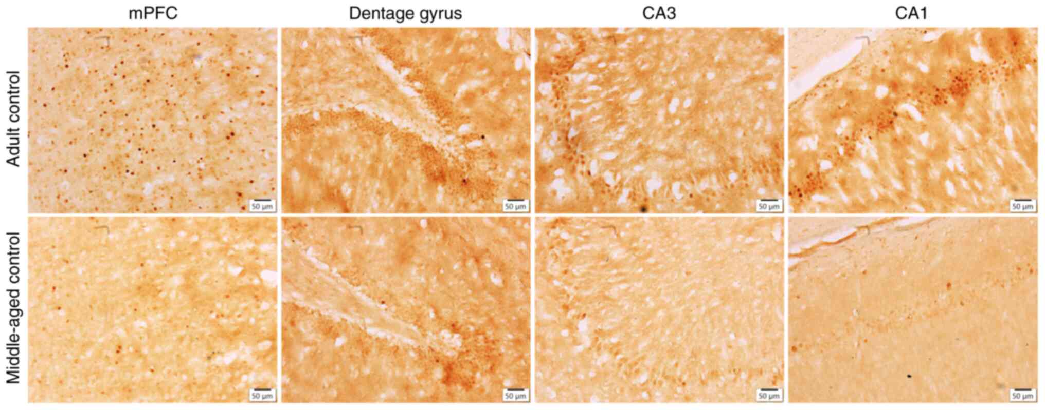
The middle-aged group (42-weeks old) showed a
significantly shorter retention time in the MWM test (P<0.05)
and a lower number of c-Fos positive cells in the mPFC (P<0.05),
DG (P<0.0001), CA3 (P<0.0001), and CA1 (P<0.0001) regions
of the brain compared with the adult group (Fig. 1). Moreover, the results found that
the levels of MDA (P<0.01) and TNF-α (P<0.05) in the
hippocampus of the middle-aged group were significantly higher than
the adult group (Table I).
 | Table IEffect of age on the assessed
variables in rats. |
Table I
Effect of age on the assessed
variables in rats.
| Variable | Adult control | Middle-aged
control | P-value |
|---|
| Retention time,
sec | 28.76±1.06 | 18.11±3.45 | 0.0185a |
| Percentage
recognition index | 57.72±1.75 | 55.05±2.78 | 0.4401 |
| MDA in hippocampus,
µmol/mg protein | 1.57±0.08 | 2.98±0.40 | 0.0092b |
| MDA in prefrontal
cortex, µmol/mg protein | 2.46±0.40 | 1.97±0.22 | 0.3206 |
| TNF-α in
hippocampus, pg/mg | 160.99±10.50 | 193.21±3.78 | 0.0203a |
| TNF-α in prefrontal
cortex, pg/mg | 168.38±3.46 | 172.11±4.46 | 0.5266 |
| c-Fos positive
cells in mPFC, cells/mm2 | 290.16±16.37 | 198.15±30.81 | 0.0167a |
| c-Fos positive
cells in dentate gyrus, cells/mm2 | 422.99±11.79 | 190.67±9.24 |
<0.0001c |
| c-Fos positive
cells in CA3, cells/mm2 | 186.50±8.23 | 105.00±3.49 |
<0.0001c |
| c-Fos positive
cells in CA1, cells/mm2 | 228.75±6.49 | 106.53±4.71 |
<0.0001c |
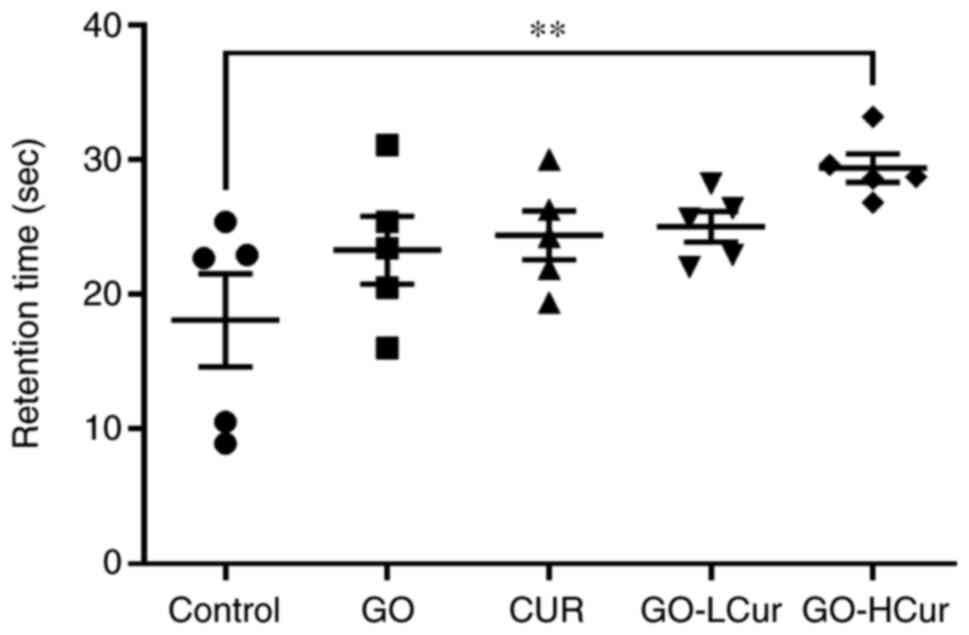
Effect of GOSD and CURSD on learning
and memory
Spatial learning and reference memory were
evaluated. The time that each rat spent in the target quadrant in
the testing period (retention time) was recorded in order to
observe the behavior of the rats. The data showed that GO-HCur
group significantly increased retention time when compared with the
control group (P<0.01). The results indicated that GOSD 10
mg/kg·BW plus CURSD 50 mg/kg·BW exhibited the largest improvement
in learning and memory performance in middle-aged rats (Fig. 2).
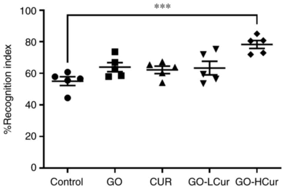
Effect of GOSD and CURSD on NOR
memory
The percentage recognition index was calculated from
the time spent exploring the novel object to the time whereby the
rats were able to distinguish familiar objects during the testing
period. GO-HCur group showed a significant increase in the
percentage recognition index when compared with the control group
(P<0.001). As the rats remembered the familiar objects, which
became mundane, they began to become more interested in exploring
the novel object. The results indicated that GOSD 10 mg/kg·BW plus
CURSD 50 mg/kg·BW exhibited the largest improvement in recognition
memory performance in the middle-aged rats (Fig. 3).
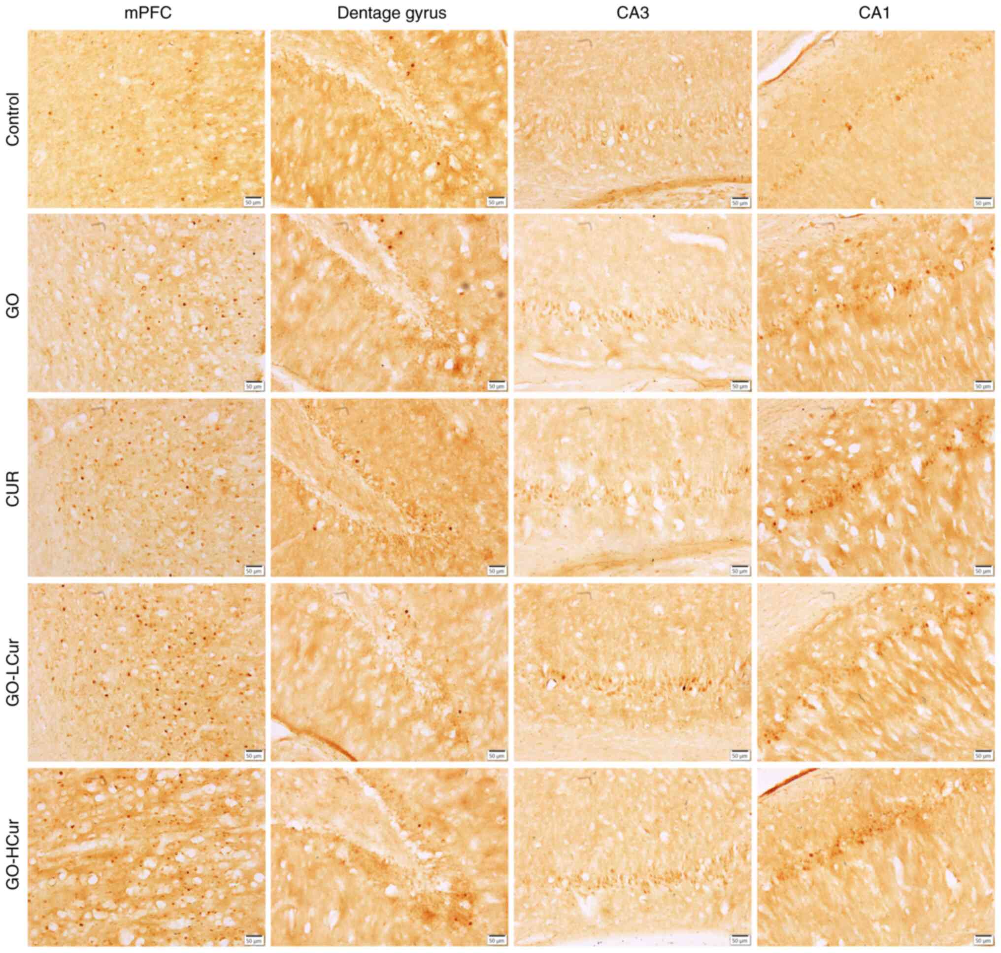
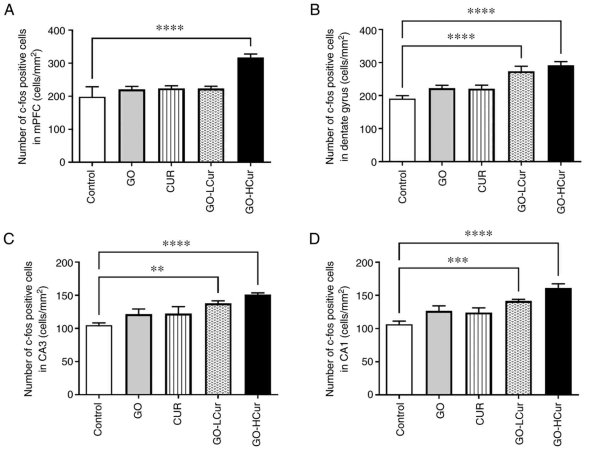
Effect of GOSD and CURSD on c-Fos
expression
c-Fos expression was evaluated using
immunohistochemistry to stain the prefrontal cortex and hippocampal
areas of the brain. The data from the prefrontal cortex showed that
the GO-HCur group exhibited significantly increased numbers of
c-Fos positive cells in the mPFC compared to the control group
(P<0.0001). This was especially apparent in the hippocampal
areas, which include the DG, CA3, and CA1 regions. The number of
c-Fos positive cells was significantly increased in GO-LCur (DG,
P<0.0001; CA3, P<0.01; and CA1, P<0.001, respectively),
and the GO-HCur (P<0.0001) compared to the control group. The
results indicated that GOSD 10 mg/kg·BW plus CURSD 50 mg/kg·BW
enhanced the c-Fos activity in both the prefrontal cortex and the
hippocampus in middle-aged rats (Figs.
4 and 5).
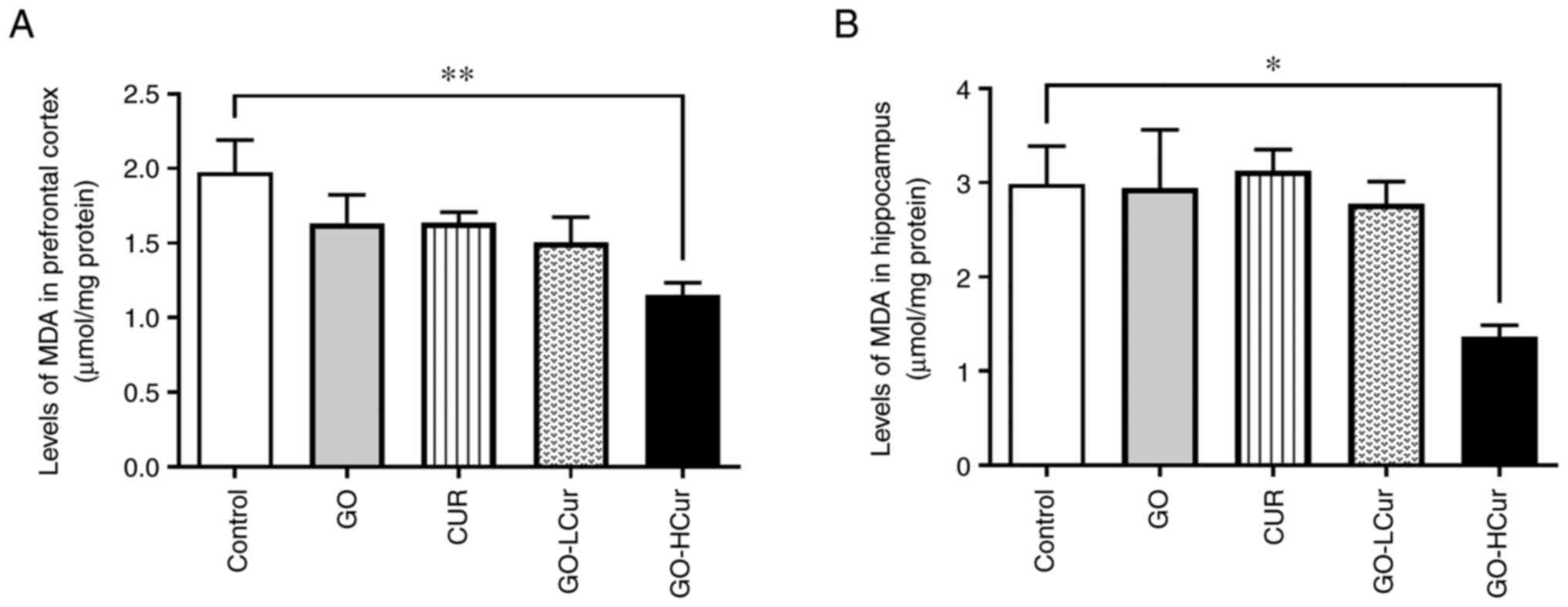
Determination of GOSD and CURSD on
oxidative stress and antioxidant status. Effect of GOSD and CURSD
on lipid peroxidation
Lipid peroxidation was used to evaluate the levels
of MDA in the prefrontal cortex and hippocampus using a TBARs
assay. The data showed that the GO-HCur group exhibited
significantly decreased levels of MDA in both the prefrontal cortex
and hippocampus compared with the control group (P<0.01 and
P<0.05, respectively). These results indicated that the
administration of GOSD 10 mg/kg·BW plus CURSD 50 mg/kg·BW reduced
lipid peroxidation in both the prefrontal cortex and the
hippocampus in middle-aged rats (Fig.
6).
Effect of GOSD and CURSD on SOD
activity
The data showed that the GO-HCur group exhibited
significantly increased SOD activity in the prefrontal cortex when
compared with the control group (P<0.001). Moreover, the data
demonstrated that the SOD activity in the hippocampus significantly
increased in middle-aged rats administered with GO (P<0.01), Cur
(P<0.05), GO-LCur (P<0.01), and GO-HCur (P<0.01) compared
with the control group. The results indicated that GOSD 10 mg/kg·BW
plus CURSD 50 mg/kg·BW increased the activity of SOD in both the
prefrontal cortex and the hippocampus in the middle-aged rats
(Fig. 7).
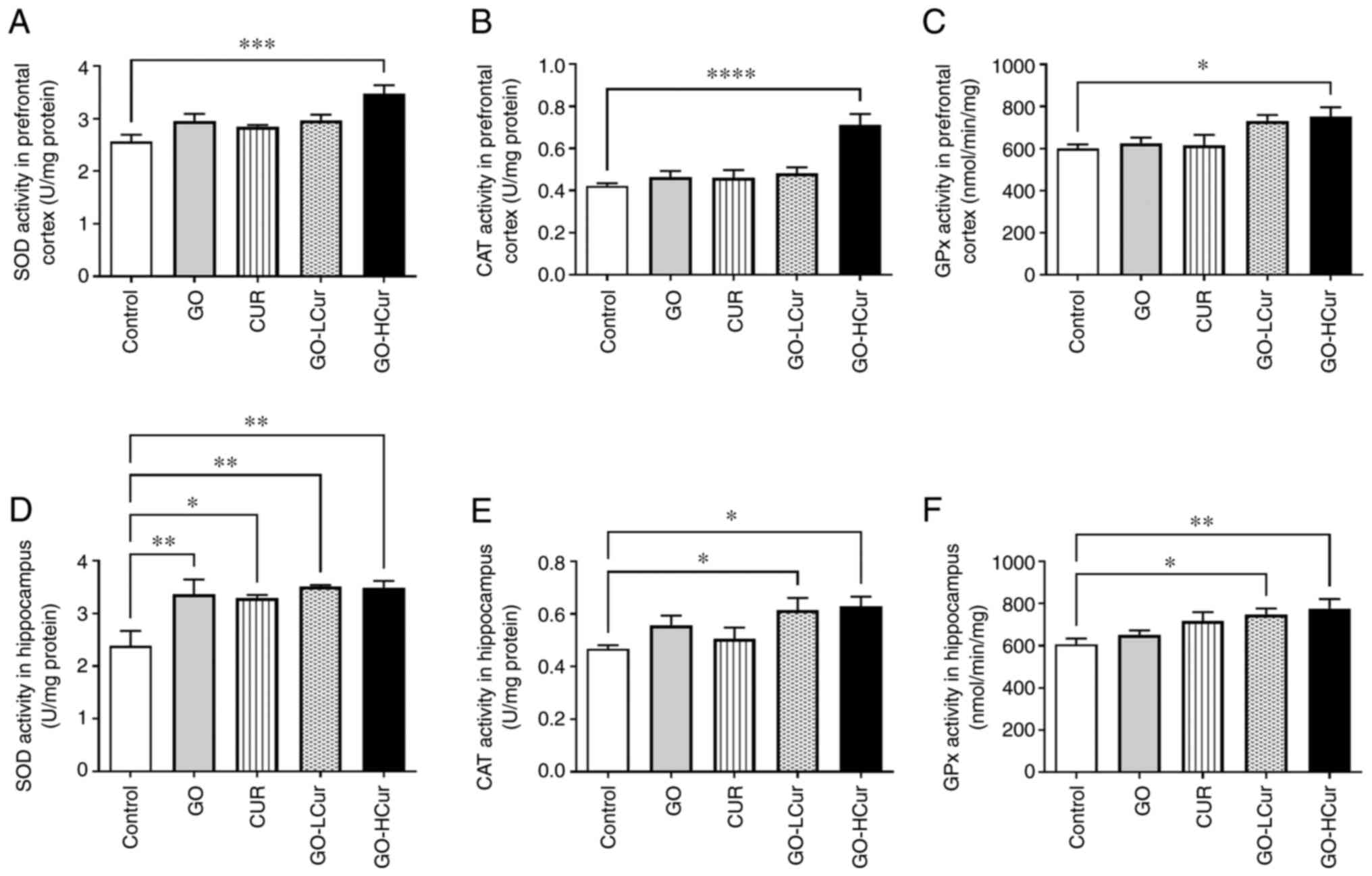 | Figure 7Effect of GOSD and CURSD on
antioxidant status. (A-C) Antioxidant status in the prefrontal
cortex and (D-F) in the hippocampus. (A and D) SOD activity, (B and
E) CAT activity, and (C and F) GPx activity. Data are presented as
the mean ± SEM. *P<0.05, **P<0.01,
***P<0.001, ****P<0.0001 vs. control.
SOD, superoxide dismutase; CAT, catalase; GPx, glutathione
peroxidase; GOSD, γ-oryzanol solid dispersion; CURSD, curcumin
solid dispersion. |
Effect of GOSD and CURSD on CAT
activity
The data showed that CAT activity in the prefrontal
cortex was significantly increased in the GO-HCur group when
compared with the control group (P<0.0001). In the hippocampus,
the activity of CAT was significantly increased in the GO-LCur and
GO-HCur groups when compared with the control group (both
P<0.05). The results indicated that GOSD 10 mg/kg·BW plus CURSD
50 mg/kg·BW enhanced CAT activity in both the prefrontal cortex and
the hippocampus in the middle-aged rats (Fig. 7).
Effect of GOSD and CURSD on GPx
activity
The data showed that GO-HCur group exhibited
significantly increased GPx activity in the prefrontal cortex when
compared with the control group (P<0.05). Moreover, the data
showed that GO-LCur and GO-HCur groups exhibited significantly
increased GPx activity in the hippocampus when compared with the
control group (P<0.05 and P<0.01, respectively). The results
indicated that GOSD 10 mg/kg·BW plus CURSD 50 mg/kg·BW enhanced the
activity of GPx in both the prefrontal cortex and the hippocampus
in the middle-aged rats (Fig.
7).
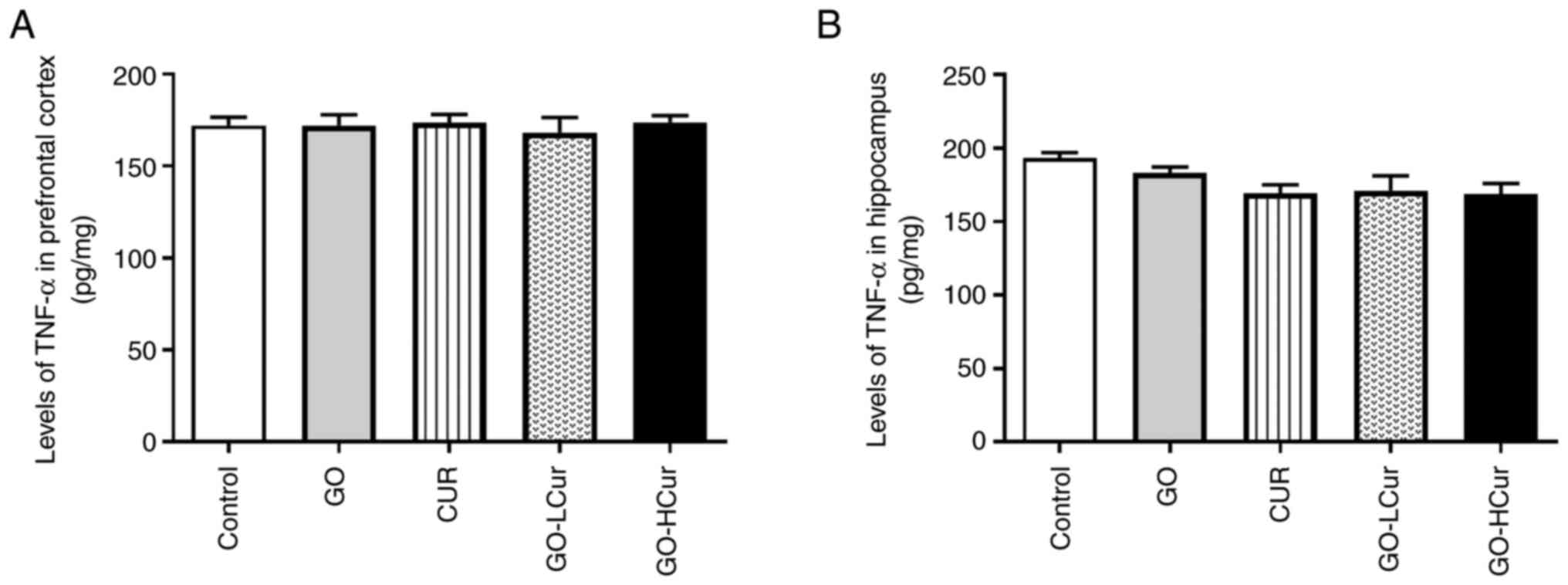
Effect of GOSD and CURSD on the levels
of TNF-α
The proinflammatory cytokine levels were assessed
based on the levels of TNF-α in the prefrontal cortex and
hippocampus. The data showed that levels of TNF-α did not
significantly differ among the five groups in both regions
(Fig. 8).
Discussion
The aim of this study was to evaluate the effects of
GOSD and CURSD on learning and memory in middle-aged rats. The
results showed an association between age with brain functions and
behavioral changes. Several studies reported that oxidative stress
and inflammation associated with age affect the brain's structure
and function, leading to a cognitive decline in an age-dependent
manner (22-24).
MDA and TNF-α are well-known indicators for the study of oxidative
stress and inflammation. The middle-aged rats used in this study
may not have shown the prominent alterations in the recognition
memory or the molecular changes of the prefrontal cortex yet.
However, some parameters such as the molecular changes in the
hippocampus including MDA, TNF-a, and c-Fos expression that are
associated with learning and memory, specifically in spatial
memory, were observed. The molecular changes in middle-aged
involved oxidative stress (6),
which is caused by the accumulation of free radicals and ROS, the
levels of which increase with age. Although free radicals and ROS
may be beneficial at optimal levels, excessive generation and
accumulation leads to oxidative stress (25). This can induce lipid peroxidation,
inflammation, and neurodegeneration in the brain (26). Furthermore, previous studies have
suggested that damage in the hippocampus leads to a reduction in
learning and memory functions in rats (27,28).
Immunohistochemical staining was used to detect c-Fos expression, a
marker of activated neurons in the brain. The expression of c-Fos
in the middle-aged rat group was lower than that in the adult rats
in both the hippocampus and prefrontal cortex. These results
indicated that middle-aged rats exhibited less neuronal activity
than the adult rats, which corresponds to the results of the MWM
test, which showed that the learning and memory performance
declined. These results also suggest that oxidative stress also
affected the structural alterations of neurons, which may have led
to a reduction in excitatory postsynaptic potential and neuronal
activity in the brain (2,29).
According to previous studies, the intake of dietary
antioxidants can prevent neurodegenerative diseases associated with
oxidative stress and aging (7,8).
Curcumin and γ-oryzanol compounds exhibit a wide range of
pharmacological properties, including antioxidant,
anti-inflammatory, anti-tumor, and anti-diabetic properties
(9-12,30).
Nevertheless, both compounds have been reported to be limited with
regard to their use as a nutritional product given their low
water-solubility, resulting in low absorption and bioavailability
(13-16).
The present study prepared curcumin and γ-oryzanol as a solid
dispersion to enhance their water solubility. Several studies have
reported that solid dispersion techniques enhance the absorption
and bioavailability by improving the water solubility and
dissolvability of poor water-soluble drugs (31,32).
In addition, previous studies have found that curcumin in solid
dispersion showed stronger pharmacological properties than native
curcumin (33). The present study
demonstrated GOSD and CURSD prevented the elevation of oxidative
stress in middle-aged rats by acting as an antioxidant, which
involved the elimination or neutralization of free radicals and/or
ROS. The increase in the levels of antioxidant enzymes, including
that of SOD, CAT, and GPx have been shown to associated with the
reduction of malondialdehyde in both the hippocampus and the
prefrontal cortex. Nevertheless, the results of the present study
did not show significantly reduced levels of TNF-α in any of the
treatment groups. Treatment with GOSD 10 mg/kg combined with CURSD
50 mg/kg was more effective than the GOSD, CURSD, or GOSD 10 mg/kg
combined with CURSD 25 mg/kg with regard to the defense against
oxidative stress in middle-aged rats. These results are in
agreement with previous studies that showed the ability of curcumin
and γ-oryzanol in reducing the oxidative stress and lipid
peroxidation by increasing the antioxidant activity (9,34). The
elevation in antioxidant levels played a key role in protecting
neurons from free radicals and ROS by decreasing lipid peroxidation
(35,36) and reducing any structural
alterations in neurons, in-turn maintaining neuronal activity and
improving learning and memory (34,37).
This was consistent with the results that administration of GOSD 10
mg/kg combined with CURSD 50 mg/kg significantly increased
expression the c-Fos, and this is associated with a significant
increase in reference memory and recognition memory in behavioral
tests (38).
In conclusion, the present study demonstrated that
GOSD and CURSD could protect against and slow down the aging of the
brain during the early stages of aging through attenuation of
oxidative stress by decreasing MDA levels and increasing
antioxidant enzyme activity, and this resulted in memory
enhancement. In addition, GOSD and CURSD did not exert any
noticeable neurotoxic effects on the central nervous system.
Acknowledgements
Not applicable.
Funding
Funding: This study was supported by Agricultural Research
Development Agency (Public Organization), the National Research
Council of Thailand (grant no. 2564/4, TP), and The Center of
Excellence for Innovation in Chemistry (PERCH-CIC), Ministry of
Higher Education, Science, Research and Innovation.
Availability of data and materials
The datasets used and/or analyzed during the present
study are available from the corresponding author on reasonable
request.
Authors' contributions
TP performed the experiments. TP and OK analyzed the
results and wrote the manuscript. WT, ST and OK designed the study,
and wrote and edited the manuscript. All authors read and approved
the final manuscript. TP and OK confirmed the authenticity of all
the raw data.
Ethics approval and consent to
participate
The present study was approved by the by the Ethics
Committee of the Centre for Animal Research of Naresuan University
(Phitsanulok, Thailand) (approval no. NU-AE620514).
Patient consent for publication
Not applicable.
Competing interests
The authors declare that they have no competing
interests.
References
|
1
|
Hou Y, Dan X, Babbar M, Wei Y, Hasselbalch
SG, Croteau DL and Bohr VA: Ageing as a risk factor for
neurodegenerative disease. Nat Rev Neurol. 15:565–581.
2019.PubMed/NCBI View Article : Google Scholar
|
|
2
|
Yankner BA, Lu T and Loerch P: The aging
brain. Annu Rev Pathol. 3:41–66. 2008.PubMed/NCBI View Article : Google Scholar
|
|
3
|
Gómez-Gonzalo M, Martin-Fernandez M,
Martínez-Murillo R, Mederos S, Hernández-Vivanco A, Jamison S,
Fernandez AP, Serrano J, Calero P, Futch HS, et al:
Neuron-astrocyte signaling is preserved in the aging brain. Glia.
65:569–580. 2017.PubMed/NCBI View Article : Google Scholar
|
|
4
|
Chandran R, Kumar M, Kesavan L, Jacob RS,
Gunasekaran S, Lakshmi S, Sadasivan C and Omkumar RV: Cellular
calcium signaling in the aging brain. J Chem Neuroanat. 95:95–114.
2019.PubMed/NCBI View Article : Google Scholar
|
|
5
|
Monti DM, Rigano MM, Monti SM and Peixoto
HS: Role of antioxidants in the protection from aging-related
diseases. Oxid Med Cell Longev. 2019(7450693)2019.PubMed/NCBI View Article : Google Scholar
|
|
6
|
Nikhra V: The aging brain: Recent research
and concepts. Gerontol Geriatr Stud. 1:1–11. 2017.
|
|
7
|
Vaiserman A, Koliada A, Zayachkivska A and
Lushchak O: Curcumin: A therapeutic potential in ageing-related
disorders. PharmaNutrition. 14(100226)2020.
|
|
8
|
Benameur T, Soleti R, Panaro MA, La Torre
ME, Monda V, Messina G and Porro C: Curcumin as prospective
anti-aging natural compound: Focus on brain. Molecules.
26(4794)2021.PubMed/NCBI View Article : Google Scholar
|
|
9
|
Samarghandian S, Azimi-Nezhad M,
Farkhondeh T and Samini F: Anti-oxidative effects of curcumin on
immobilization-induced oxidative stress in rat brain, liver and
kidney. Biomed Pharmacother. 87:223–229. 2017.PubMed/NCBI View Article : Google Scholar
|
|
10
|
Suryanarayana P, Satyanarayana A,
Balakrishna N, Kumar PU and Reddy GB: Effect of turmeric and
curcumin on oxidative stress and antioxidant enzymes in
streptozotocin-induced diabetic rat. Med Sci Monit. 13:BR286–BR292.
2007.PubMed/NCBI
|
|
11
|
Rungratanawanich W, Abate G, Serafini MM,
Guarienti M, Catanzaro M, Marziano M, Memo M, Lanni C and Uberti D:
Characterization of the antioxidant effects of γ-oryzanol:
Involvement of the Nrf2 pathway. Oxid Med Cell Longev: Mar 14, 2018
(Epub ahead of print).
|
|
12
|
Wang YX, Li Y, Sun AM, Wang FJ and Yu GP:
Hypolipidemic and antioxidative effects of aqueous enzymatic
extract from rice bran in rats fed a high-fat and -cholesterol
diet. Nutrients. 6:3696–3710. 2014.PubMed/NCBI View Article : Google Scholar
|
|
13
|
Islam A, Rebello L and Chepyala S: Review
on nanoformulations of curcumin (Curcuma longa Linn.): Special
emphasis on Nanocurcumin®. Int J Nat Life Sci. 3:1–12. 2019.
|
|
14
|
Hettiarachchi SS, Dunuweera SP, Dunuweera
AN and Rajapakse RMG: Synthesis of curcumin nanoparticles from raw
turmeric rhizome. ACS Omega. 6:8246–8252. 2021.PubMed/NCBI View Article : Google Scholar
|
|
15
|
Rawal T, Mishra N, Jha A, Bhatt A, Tyagi
RK, Panchal S and Butani S: Chitosan nanoparticles of
gamma-oryzanol: Formulation, optimization, and in vivo evaluation
of anti-hyperlipidemic activity. AAPS PharmSciTech. 19:1894–1907.
2018.PubMed/NCBI View Article : Google Scholar
|
|
16
|
Rodsuwan U, Pithanthanakul U, Thisayakorn
K, Uttapap D, Boonpisuttinant K, Vatanyoopaisarn S, Thumthanaruk B
and Rungsardthong V: Preparation and characterization of gamma
oryzanol loaded zein nanoparticles and its improved stability. Food
Sci Nutr. 9:616–624. 2020.PubMed/NCBI View Article : Google Scholar
|
|
17
|
Morris R: Developments of a water-maze
procedure for studying spatial learning in the rat. J Neurosci
Methods. 11:47–60. 1984.PubMed/NCBI View Article : Google Scholar
|
|
18
|
Dodart JC, Bales KR, Gannon KS, Greene SJ,
DeMattos RB, Mathis C, DeLong CA, Wu S, Wu X, Holtzman DM and Paul
SM: Immunization reverses memory deficits without reducing brain
Abeta burden in Alzheimer's disease model. Nat Neurosci. 5:452–457.
2002.PubMed/NCBI View
Article : Google Scholar
|
|
19
|
Liu J, Edamatsu R, Kabuto H and Mori A:
Antioxidant action of guilingji in the brain of rats with
FeCl3-induced epilepsy. Free Radic Biol Med. 9:451–454.
1990.PubMed/NCBI View Article : Google Scholar
|
|
20
|
Marklund S and Marklund G: Involvement of
the superoxide anion radical in the autoxidation of pyrogallol and
a convenient assay for superoxide dismutase. Eur J Biochem.
47:469–474. 1974.PubMed/NCBI View Article : Google Scholar
|
|
21
|
Beers RF Jr and Sizer IW: A
spectrophotometric method for measuring the breakdown of hydrogen
peroxide by catalase. J Biol Chem. 195:133–140. 1952.PubMed/NCBI
|
|
22
|
Hamezah HS, Durani LW, Ibrahim NF,
Yanagisawa D, Kato T, Shiino A, Tanaka S, Damanhuri HA, Ngah WZW
and Tooyama I: Volumetric changes in the aging rat brain and its
impact on cognitive and locomotor functions. Exp Gerontol.
99:69–79. 2017.PubMed/NCBI View Article : Google Scholar
|
|
23
|
Garg G, Singh S, Singh AK and Rizvi SI:
N-acetyl-l-cysteine attenuates oxidative damage and
neurodegeneration in rat brain during aging. Can J Physiol
Pharmacol. 96:1189–1196. 2018.PubMed/NCBI View Article : Google Scholar
|
|
24
|
Khairy EY and Attia MM: Protective effects
of vitamin D on neurophysiologic alterations in brain aging: Role
of brain-derived neurotrophic factor (BDNF). Nutr Neurosci.
24:650–659. 2021.PubMed/NCBI View Article : Google Scholar
|
|
25
|
Pyo IS, Yun S, Yoon YE, Choi JW and Lee
SJ: Mechanisms of aging and the preventive effects of resveratrol
on age-related diseases. Molecules. 25(4649)2020.PubMed/NCBI View Article : Google Scholar
|
|
26
|
Fischer R and Maier O: Interrelation of
oxidative stress and inflammation in neurodegenerative disease:
Role of TNF. Oxid Med Cell Longev. 2015(610813)2015.PubMed/NCBI View Article : Google Scholar
|
|
27
|
Anyanwu EC: Neurochemical changes in the
aging process: Implications in medication in the elderly.
ScientificWorldJournal. 7:1603–1610. 2007.PubMed/NCBI View Article : Google Scholar
|
|
28
|
Huang TT, Leu D and Zou Y: Oxidative
stress and redox regulation on hippocampal-dependent cognitive
functions. Arch Biochem Biophys. 576:2–7. 2015.PubMed/NCBI View Article : Google Scholar
|
|
29
|
Tönnies E and Trushina E: Oxidative
stress, synaptic dysfunction, and Alzheimer's disease. J Alzheimers
Dis. 57:1105–1121. 2017.PubMed/NCBI View Article : Google Scholar
|
|
30
|
Mastinu A, Bonini SA, Rungratanawanich W,
Aria F, Marziano M, Maccarinelli G, Abate G, Premoli M, Memo M and
Uberti D: Gamma-oryzanol prevents LPS-induced brain inflammation
and cognitive impairment in adult mice. Nutrients.
11(728)2019.PubMed/NCBI View Article : Google Scholar
|
|
31
|
Zhang X, Xing H, Zhao Y and Ma Z:
Pharmaceutical dispersion techniques for dissolution and
bioavailability enhancement of poorly water-soluble drugs.
Pharmaceutics. 10(74)2018.PubMed/NCBI View Article : Google Scholar
|
|
32
|
Jara MO, Warnken ZN and Williams RO III:
Amorphous solid dispersions and the contribution of nanoparticles
to in vitro dissolution and in vivo testing: Niclosamide as a case
study. Pharmaceutics. 13(97)2021.PubMed/NCBI View Article : Google Scholar
|
|
33
|
Oliveira VDS, de Almeida AS, Albuquerque
IDS, Duarte FÍC, Queiroz BCSH, Converti A and Lima ÁAND:
Therapeutic applications of solid dispersions for drugs and new
molecules: In vitro and in vivo activities. Pharmaceutics.
12(933)2020.PubMed/NCBI View Article : Google Scholar
|
|
34
|
Rungratanawanich W, Cenini G, Mastinu A,
Sylvester M, Wilkening A, Abate G, Bonini SA, Aria F, Marziano M,
Maccarinelli G, et al: γ-Oryzanol improves cognitive function and
modulates hippocampal proteome in mice. Nutrients.
11(753)2019.PubMed/NCBI View Article : Google Scholar
|
|
35
|
Birben E, Sahiner UM, Sackesen C, Erzurum
S and Kalayci O: Oxidative stress and antioxidant defense. World
Allergy Organ J. 5:9–19. 2012.PubMed/NCBI View Article : Google Scholar
|
|
36
|
Cui X, Song H and Su J: Curcumin
attenuates hypoxic-ischemic brain injury in neonatal rats through
induction of nuclear factor erythroid-2-related factor 2 and heme
oxygenase-1. Exp Ther Med. 14:1512–1518. 2017.PubMed/NCBI View Article : Google Scholar
|
|
37
|
Massaad CA and Klann E: Reactive oxygen
species in the regulation of synaptic plasticity and memory.
Antioxid Redox Signal. 14:2013–2054. 2011.PubMed/NCBI View Article : Google Scholar
|
|
38
|
Gallo FT, Katche C, Morici JF, Medina JH
and Weisstaub NV: Immediate early genes, memory and psychiatric
disorders: Focus on c-Fos, Egr1 and Arc. Front Behav Neurosci.
12(79)2018.PubMed/NCBI View Article : Google Scholar
|