Clinicopathological findings in refractory diabetic macular edema: A case report
- Authors:
- Takayuki Tanaka
- Satoru Kase
- Michiyuki Saito
- Ikuyo Hirose
- Miyuki Murata
- Emi Takakuwa
- Susumu Ishida
View Affiliations
Affiliations: Department of Ophthalmology, Faculty of Medicine and Graduate School of Medicine, Hokkaido University, Sapporo, Hokkaido 060‑8638, Japan, Department of Surgical Pathology, Hokkaido University Hospital, Sapporo, Hokkaido 060‑8648, Japan
- Published online on: November 29, 2023 https://doi.org/10.3892/br.2023.1701
-
Article Number:
13
-
Copyright: © Tanaka
et al. This is an open access article distributed under the
terms of Creative
Commons Attribution License.
Metrics: Total
Views: 0 (Spandidos Publications: | PMC Statistics: )
Metrics: Total PDF Downloads: 0 (Spandidos Publications: | PMC Statistics: )
This article is mentioned in:
Abstract
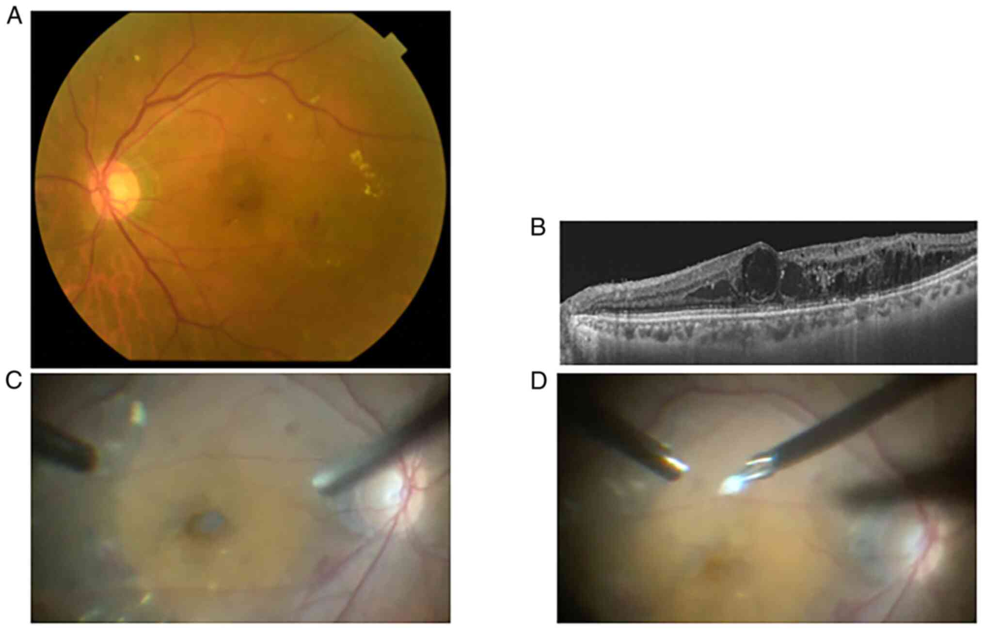
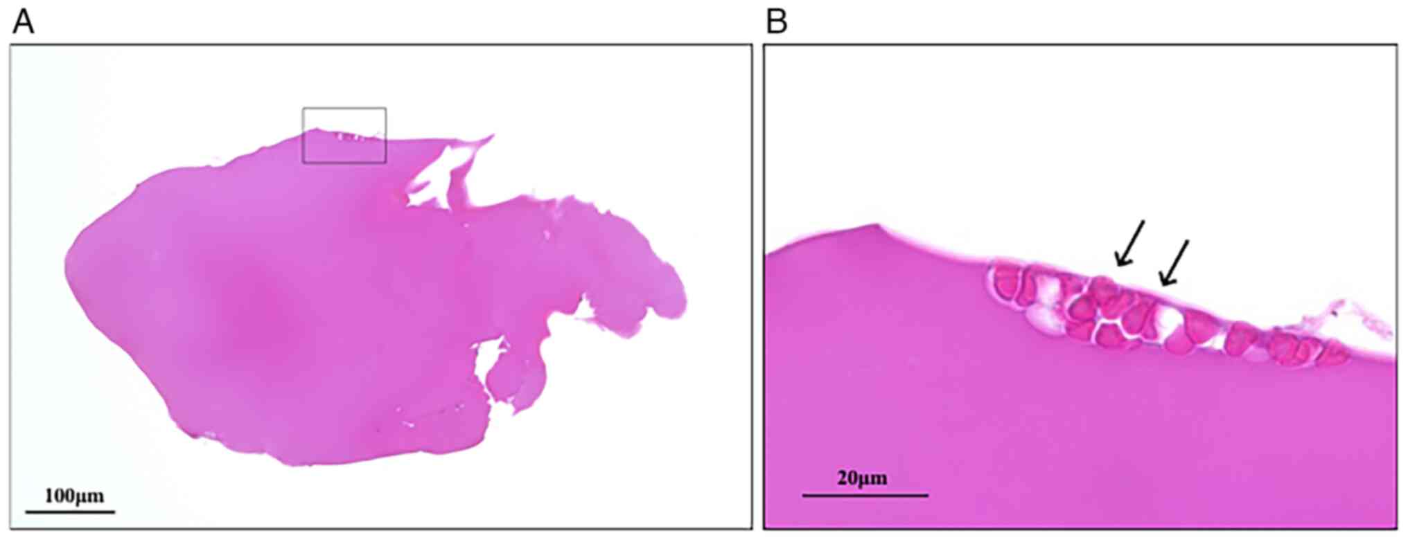
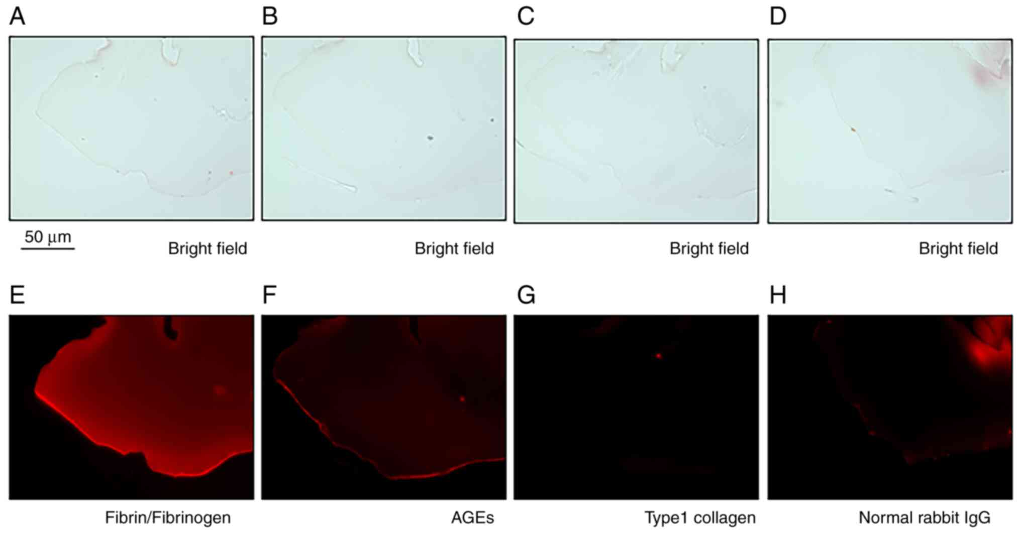
The present study describes the case of a patient with refractory diabetic cystoid macular edema who underwent vitrectomy with en bloc removal of the cystoid lesion component. The current study also performed histopathological and immunohistochemical analyses of the cystoid lesion component to assess fibrin/fibrinogen and advanced glycation end-products (AGEs) immunoreactivity. A 69‑year‑old Japanese man presented with visual loss in the left eye due to residual cystoid macular edema (CME) refractory to anti‑vascular endothelial growth factor therapy. Best‑corrected visual acuity was 1.2 in the right eye (OD) and 0.5 in the left eye (OS). Fundus examination showed dot hemorrhages and hard exudates in the peri‑macular region with pan‑retinal photocoagulation scars in both eye. Swept‑source optical coherence tomography revealed CME with slight hyperreflectivity in the cyst OS. A total of 3 months after the initial visit, pars plana vitrectomy was performed, and the translucent solidified component within the cystoid lesion was isolated. Histopathologically, the excised component was elliptical in shape, measuring 0.7x0.4 mm and exhibited homogeneous eosinophilic material without cellular components. No membranous structure was observed surrounding the component. Immunohistochemistry demonstrated that the tissue was positive for fibrin/fibrinogen and weakly positive for AGEs, but was negative for glial fibrillary acidic protein, type 1 collagen and receptor for AGEs. To the best of our knowledge, the present case report is the first to histopathologically examine the contents of refractory CME, and to immunohistochemically demonstrate that fibrin in diabetic CME may be post‑translationally modified by AGEs. These results suggested that fibrin in CME may escape degradation by plasmin due to post‑translational modifications.
View References
1
|
Tomkins-Netzer O, Ismetova F, Bar A,
Seguin-Greenstein S, Kramer M and Lightman S: Functional outcome of
macular edema in different retinal disorders. Prog Retin Eye Res.
48:119–136. 2015.PubMed/NCBI View Article : Google Scholar
|
2
|
Tachi N, Hashimoto Y and Ogino N:
Cystotomy for diabetic cystoid macular edema. Doc Ophthalmol.
97:459–463. 1999.PubMed/NCBI View Article : Google Scholar
|
3
|
Asahina Y, Tachi N, Asahina Y, Yoshimura
K, Ueta Y and Hashimoto Y: Six-month postoperative outcomes of
intraoperative OCT-guided surgical cystotomy for refractory cystoid
macular edema in diabetic eyes. Clin Ophthalmol. 11:2099–2105.
2017.PubMed/NCBI View Article : Google Scholar
|
4
|
Imai H, Tetsumoto A, Yamada H, Hayashida
M, Otsuka K, Miki A, Kusuhara S and Nakamura M: Long-term effect of
cystotomy with or without the fibrinogen clot removal for
refractory cystoid macular edema secondary to diabetic retinopathy.
Retina. 41:844–851. 2021.PubMed/NCBI View Article : Google Scholar
|
5
|
Imai H, Otsuka K, Tetsumoto A, Miki A and
Nakamura M: Effectiveness of en bloc removal of fibrinogen-rich
component of cystoid lesion for the treatment of cystoid macular
edema. Retina. 40:154–159. 2020.PubMed/NCBI View Article : Google Scholar
|
6
|
De Vries JJ, Snoek CJM, Rijken DC and De
Maat MPM: Effects of post-translational modifications of fibrinogen
on clot formation, clot structure, and fibrinolysis: A systematic
review. Arterioscler Thromb Vasc Biol. 40:554–569. 2020.PubMed/NCBI View Article : Google Scholar
|
7
|
Lund T, Svindland A, Pepaj M, Jensen AB,
Berg JP, Kilhovd B and Hanssen KF: Fibrin(ogen) may be an important
target for methylglyoxal-derived AGE modification in elastic
arteries of humans. Diab Vasc Dis Res. 8:284–294. 2011.PubMed/NCBI View Article : Google Scholar
|
8
|
Jennewein C, Tran N, Paulus P, Ellinghaus
P, Eble JA and Zacharowski K: Novel aspects of fibrin(ogen)
fragments during inflammation. Mol Med. 17:568–673. 2011.PubMed/NCBI View Article : Google Scholar
|
9
|
Stein TP, Leskiw MJ and Wallace HW:
Measurement of half-life human plasma fibrinogen. Am J Physiol.
234:D504–D510. 1978.PubMed/NCBI View Article : Google Scholar
|
10
|
Singh R, Barden A, Mori T and Beilin L:
Advanced glycation end-products: A review. Diabetologia.
44:129–146. 2001.PubMed/NCBI View Article : Google Scholar
|