Spandidos Publications Logo
  • About
    • About Spandidos
    • Aims and Scopes
    • Abstracting and Indexing
    • Editorial Policies
    • Reprints and Permissions
    • Job Opportunities
    • Terms and Conditions
    • Contact
  • Journals
    • All Journals
    • Oncology Letters
      • Oncology Letters
      • Information for Authors
      • Editorial Policies
      • Editorial Board
      • Aims and Scope
      • Abstracting and Indexing
      • Bibliographic Information
    • International Journal of Oncology
      • International Journal of Oncology
      • Information for Authors
      • Editorial Policies
      • Editorial Board
      • Aims and Scope
      • Abstracting and Indexing
      • Bibliographic Information
    • Molecular and Clinical Oncology
      • Molecular and Clinical Oncology
      • Information for Authors
      • Editorial Policies
      • Editorial Board
      • Aims and Scope
      • Abstracting and Indexing
      • Bibliographic Information
    • Experimental and Therapeutic Medicine
      • Experimental and Therapeutic Medicine
      • Information for Authors
      • Editorial Policies
      • Editorial Board
      • Aims and Scope
      • Abstracting and Indexing
      • Bibliographic Information
    • International Journal of Molecular Medicine
      • International Journal of Molecular Medicine
      • Information for Authors
      • Editorial Policies
      • Editorial Board
      • Aims and Scope
      • Abstracting and Indexing
      • Bibliographic Information
    • Biomedical Reports
      • Biomedical Reports
      • Information for Authors
      • Editorial Policies
      • Editorial Board
      • Aims and Scope
      • Abstracting and Indexing
      • Bibliographic Information
    • Oncology Reports
      • Oncology Reports
      • Information for Authors
      • Editorial Policies
      • Editorial Board
      • Aims and Scope
      • Abstracting and Indexing
      • Bibliographic Information
    • Molecular Medicine Reports
      • Molecular Medicine Reports
      • Information for Authors
      • Editorial Policies
      • Editorial Board
      • Aims and Scope
      • Abstracting and Indexing
      • Bibliographic Information
    • World Academy of Sciences Journal
      • World Academy of Sciences Journal
      • Information for Authors
      • Editorial Policies
      • Editorial Board
      • Aims and Scope
      • Abstracting and Indexing
      • Bibliographic Information
    • International Journal of Functional Nutrition
      • International Journal of Functional Nutrition
      • Information for Authors
      • Editorial Policies
      • Editorial Board
      • Aims and Scope
      • Abstracting and Indexing
      • Bibliographic Information
    • International Journal of Epigenetics
      • International Journal of Epigenetics
      • Information for Authors
      • Editorial Policies
      • Editorial Board
      • Aims and Scope
      • Abstracting and Indexing
      • Bibliographic Information
    • Medicine International
      • Medicine International
      • Information for Authors
      • Editorial Policies
      • Editorial Board
      • Aims and Scope
      • Abstracting and Indexing
      • Bibliographic Information
  • Articles
  • Information
    • Information for Authors
    • Information for Reviewers
    • Information for Librarians
    • Information for Advertisers
    • Conferences
  • Language Editing
Spandidos Publications Logo
  • About
    • About Spandidos
    • Aims and Scopes
    • Abstracting and Indexing
    • Editorial Policies
    • Reprints and Permissions
    • Job Opportunities
    • Terms and Conditions
    • Contact
  • Journals
    • All Journals
    • Biomedical Reports
      • Information for Authors
      • Editorial Policies
      • Editorial Board
      • Aims and Scope
      • Abstracting and Indexing
      • Bibliographic Information
    • Experimental and Therapeutic Medicine
      • Information for Authors
      • Editorial Policies
      • Editorial Board
      • Aims and Scope
      • Abstracting and Indexing
      • Bibliographic Information
    • International Journal of Epigenetics
      • Information for Authors
      • Editorial Policies
      • Editorial Board
      • Aims and Scope
      • Abstracting and Indexing
      • Bibliographic Information
    • International Journal of Functional Nutrition
      • Information for Authors
      • Editorial Policies
      • Editorial Board
      • Aims and Scope
      • Abstracting and Indexing
      • Bibliographic Information
    • International Journal of Molecular Medicine
      • Information for Authors
      • Editorial Policies
      • Editorial Board
      • Aims and Scope
      • Abstracting and Indexing
      • Bibliographic Information
    • International Journal of Oncology
      • Information for Authors
      • Editorial Policies
      • Editorial Board
      • Aims and Scope
      • Abstracting and Indexing
      • Bibliographic Information
    • Medicine International
      • Information for Authors
      • Editorial Policies
      • Editorial Board
      • Aims and Scope
      • Abstracting and Indexing
      • Bibliographic Information
    • Molecular and Clinical Oncology
      • Information for Authors
      • Editorial Policies
      • Editorial Board
      • Aims and Scope
      • Abstracting and Indexing
      • Bibliographic Information
    • Molecular Medicine Reports
      • Information for Authors
      • Editorial Policies
      • Editorial Board
      • Aims and Scope
      • Abstracting and Indexing
      • Bibliographic Information
    • Oncology Letters
      • Information for Authors
      • Editorial Policies
      • Editorial Board
      • Aims and Scope
      • Abstracting and Indexing
      • Bibliographic Information
    • Oncology Reports
      • Information for Authors
      • Editorial Policies
      • Editorial Board
      • Aims and Scope
      • Abstracting and Indexing
      • Bibliographic Information
    • World Academy of Sciences Journal
      • Information for Authors
      • Editorial Policies
      • Editorial Board
      • Aims and Scope
      • Abstracting and Indexing
      • Bibliographic Information
  • Articles
  • Information
    • For Authors
    • For Reviewers
    • For Librarians
    • For Advertisers
    • Conferences
  • Language Editing
Login Register Submit
  • This site uses cookies
  • You can change your cookie settings at any time by following the instructions in our Cookie Policy. To find out more, you may read our Privacy Policy.

    I agree
Search articles by DOI, keyword, author or affiliation
Search
Advanced Search
presentation
Biomedical Reports
Join Editorial Board Propose a Special Issue
Print ISSN: 2049-9434 Online ISSN: 2049-9442
Journal Cover
September-2025 Volume 23 Issue 3

Full Size Image

Sign up for eToc alerts
Recommend to Library

Journals

International Journal of Molecular Medicine

International Journal of Molecular Medicine

International Journal of Molecular Medicine is an international journal devoted to molecular mechanisms of human disease.

International Journal of Oncology

International Journal of Oncology

International Journal of Oncology is an international journal devoted to oncology research and cancer treatment.

Molecular Medicine Reports

Molecular Medicine Reports

Covers molecular medicine topics such as pharmacology, pathology, genetics, neuroscience, infectious diseases, molecular cardiology, and molecular surgery.

Oncology Reports

Oncology Reports

Oncology Reports is an international journal devoted to fundamental and applied research in Oncology.

Experimental and Therapeutic Medicine

Experimental and Therapeutic Medicine

Experimental and Therapeutic Medicine is an international journal devoted to laboratory and clinical medicine.

Oncology Letters

Oncology Letters

Oncology Letters is an international journal devoted to Experimental and Clinical Oncology.

Biomedical Reports

Biomedical Reports

Explores a wide range of biological and medical fields, including pharmacology, genetics, microbiology, neuroscience, and molecular cardiology.

Molecular and Clinical Oncology

Molecular and Clinical Oncology

International journal addressing all aspects of oncology research, from tumorigenesis and oncogenes to chemotherapy and metastasis.

World Academy of Sciences Journal

World Academy of Sciences Journal

Multidisciplinary open-access journal spanning biochemistry, genetics, neuroscience, environmental health, and synthetic biology.

International Journal of Functional Nutrition

International Journal of Functional Nutrition

Open-access journal combining biochemistry, pharmacology, immunology, and genetics to advance health through functional nutrition.

International Journal of Epigenetics

International Journal of Epigenetics

Publishes open-access research on using epigenetics to advance understanding and treatment of human disease.

Medicine International

Medicine International

An International Open Access Journal Devoted to General Medicine.

Journal Cover
September-2025 Volume 23 Issue 3

Full Size Image

Sign up for eToc alerts
Recommend to Library

  • Article
  • Citations
    • Cite This Article
    • Download Citation
    • Create Citation Alert
    • Remove Citation Alert
    • Cited By
  • Similar Articles
    • Related Articles (in Spandidos Publications)
    • Similar Articles (Google Scholar)
    • Similar Articles (PubMed)
  • Download PDF
  • Download XML
  • View XML
Article Open Access

Analysis of cell turnover in the macula densa through the normal aging process

  • Authors:
    • Marta Ortega‑Martínez
    • Yareth Gopar‑Cuevas
    • Karol García‑Aguilar
    • María-De-Lourdes Chávez‑Briones
    • Ivett Miranda‑Maldonado
    • Adriana Ancer‑Arellano
    • Jaime García‑Juárez
    • Jesús Ancer‑Rodríguez
    • Gilberto Jaramillo‑Rangel
  • View Affiliations

    Affiliations: Department of Pathology, School of Medicine, Autonomous University of Nuevo León, Monterrey, Nuevo León 64460, Mexico, Department of Histology, School of Medicine, Autonomous University of Nuevo León, Monterrey, Nuevo León 64460, Mexico
  • Published online on: July 21, 2025     https://doi.org/10.3892/br.2025.2033
  • Article Number: 155
  • Copyright: © Ortega‑Martínez et al. This is an open access article distributed under the terms of Creative Commons Attribution License.

Metrics: Total Views: 0 (Spandidos Publications: | PMC Statistics: )
Metrics: Total PDF Downloads: 0 (Spandidos Publications: | PMC Statistics: )
Cited By (CrossRef): 0 citations Loading Articles...

This article is mentioned in:


Abstract

The kidney undergoes changes during the lifespan of an individual, resulting in increased susceptibility to renal diseases in the elderly. The macula densa (MD) controls basic kidney functions, which deteriorate with aging. The present study for the first time, to the best of our knowledge, analyzed the relationship between cell proliferation and apoptosis (cell turnover) in the MD of mice through the normal aging process. Kidney specimens from CD1 mice aged 2, 6, 12, 18 or 24 months were fixed in neutral‑buffered formalin and embedded in paraffin. Tissue sections were immunostained to analyze cell proliferation, subjected to a TUNEL assay to evaluate apoptosis, or stained with hematoxylin and eosin to determine total cell number. A markedly dynamic cell turnover pattern was observed in the MD throughout the aging process, in which, when the number of proliferating cells increased, the number of apoptotic cells decreased and vice versa. However, there were no significant differences in the total cell number among the ages analyzed. Thus, other mechanisms may be involved in the deterioration of renal functions regulated by the MD in elderly people. Further research is required to determine these mechanisms, which could be the target of therapeutic strategies against age‑related kidney diseases in the future.
View Figures

Figure 1

Analysis of cell turnover in the MD
of 2-, 6-, 12-, 18- and 24-month-old mice. (A) Schematic of the MD
and adjacent structures in the kidney. (B) Tissue sections were
stained with hematoxylin and eosin to assess the total number of
cells. The arrow indicates a cell of the MD, while the arrowhead
indicates a cell of the thick ascending limb. (C) PCNA was detected
in proliferation analysis by immunohistochemistry. The arrow
indicates a proliferating cell (brown nucleus), while the arrowhead
indicates a non-proliferating cell (blue nucleus). (D) Apoptosis
was detected by in situ end-labeling of fragmented DNA using
the TUNEL assay. The arrow indicates an apoptotic cell (brown
nucleus), while the arrowhead indicates a non-apoptotic cell (blue
nucleus). (E) The number of MD cells undergoing proliferation in
mice at 24 months of age was scarce. The arrowheads indicate
non-proliferating MD cells (blue nuclei). The dotted arrow
indicates a proliferating tubular cell (brown nucleus). (F) The
number of MD cells undergoing apoptosis in mice at 24 months of age
was high. The arrows indicate apoptotic MD cells. Apoptosis was
also high in other tubular cells (dotted arrows, brown nuclei).
Mice in (B-D) belonged to the 6-month age group. Scale bar 20 µm.
MD, macula densa; PCNA, proliferating cell nuclear antigen; TUNEL,
terminal deoxynucleotidyl transferase-mediated dUTP nick end
labeling method.

Figure 2

Analysis of cell proliferation and
apoptosis in the macula densa of 2-, 6-, 12-, 18- and 24-month-old
mice. (A) The number of cells undergoing proliferation in mice at 2
months of age was significantly lower than in mice at 6, 12 and 18
months of age (*P<0.001). Mice at 6 months of age had
a significantly higher number of proliferating cells than mice at
18 and 24 months of age (both †P<0.001), but this
number was significantly lower than in mice at 12 months of age
(†P=0.043). Mice at 12 months of age had a significantly
higher number of proliferating cells than mice at 18 and 24 months
of age (both §P<0.001). Finally, mice at 18 months of
age had a significantly higher number of proliferating cells than
mice at 24 months of age (‡P<0.001). (B) The number
of cells undergoing apoptosis in mice at 2 months of age was
significantly higher than in mice at 12 months of age
(*P=0.002). Values are expressed as the means ± 1
standard error. Data were analyzed using ANOVA and the Bonferroni
post hoc test. Results were considered statistically significant at
P<0.05. PCNA, proliferating cell nuclear antigen; TUNEL,
terminal deoxynucleotidyl transferase-mediated dUTP nick end
labeling method.

Figure 3

Analysis of cell proliferation and
apoptosis in the macula densa-thick ascending limb of 2-, 6-, 12-,
18- and 24-month-old mice. (A) The number of cells undergoing
proliferation in mice at 2 months of age was significantly lower
than in mice at 6, 12 and 18 months of age *P<0.001).
Mice at 6 months of age had a significantly higher number of
proliferating cells than mice at 18 and 24 months of age (both
†P<0.001), but this number was significantly lower
than in mice at 12 months of age (†P=0.018). Mice at 12
months of age had a significantly higher number of proliferating
cells than mice at 18 and 24 months of age (both
§P<0.001). Finally, mice at 18 months of age had a
significantly higher number of proliferating cells than mice at 24
months of age (‡P<0.001). (B) The number of cells
undergoing apoptosis in mice at 2 months of age was significantly
higher than in mice at 12 months of age (*P=0.002).
Values are expressed as the means ± 1 standard error. Data were
analyzed using ANOVA and the Bonferroni post hoc test. Results were
considered statistically significant at P<0.05. PCNA,
proliferating cell nuclear antigen; TUNEL, terminal
deoxynucleotidyl transferase-mediated dUTP nick end labeling
method.

Figure 4

Analysis of total cell number in the
MD and MD-TAL of 2-, 6-, 12-, 18- and 24-month-old mice. There was
no significant difference in the number of total cells among the
analyzed ages in the (A) MD and in the (B) MD-TAL. Values are
expressed as the means ± 1 standard error. Data were analyzed using
ANOVA test. Results were considered statistically significant at
P<0.05. MD, macula densa; MD-TAL, macula densa-thick ascending
limb; BM, basement membrane.
View References

1 

United Nations and Departemtn of Economic and Social Affairs: World Population Ageing 2020 Highlights. https://www.un.org/development/desa/pd/sites/www.un.org.development.desa.pd/files/files/documents/2020/Sep/un_pop_2020_pf_ageing_10_key_messages.pdf. Accessed May 10, 2024.

2 

Shi C, de Wit S, Učambarlić E, Markousis-Mavrogenis G, Screever EM, Meijers WC, de Boer RA and Aboumsallem JP: Multifactorial diseases of the heart, kidneys, lungs, and liver and incident cancer: Epidemiology and shared mechanisms. Cancers (Basel). 15(729)2023.PubMed/NCBI View Article : Google Scholar

3 

He Y, Li Z, Niu Y, Duan Y, Wang Q, Liu X, Dong Z, Zheng Y, Chen Y, Wang Y, et al: Progress in the study of aging marker criteria in human populations. Front Public Health. 12(1305303)2024.PubMed/NCBI View Article : Google Scholar

4 

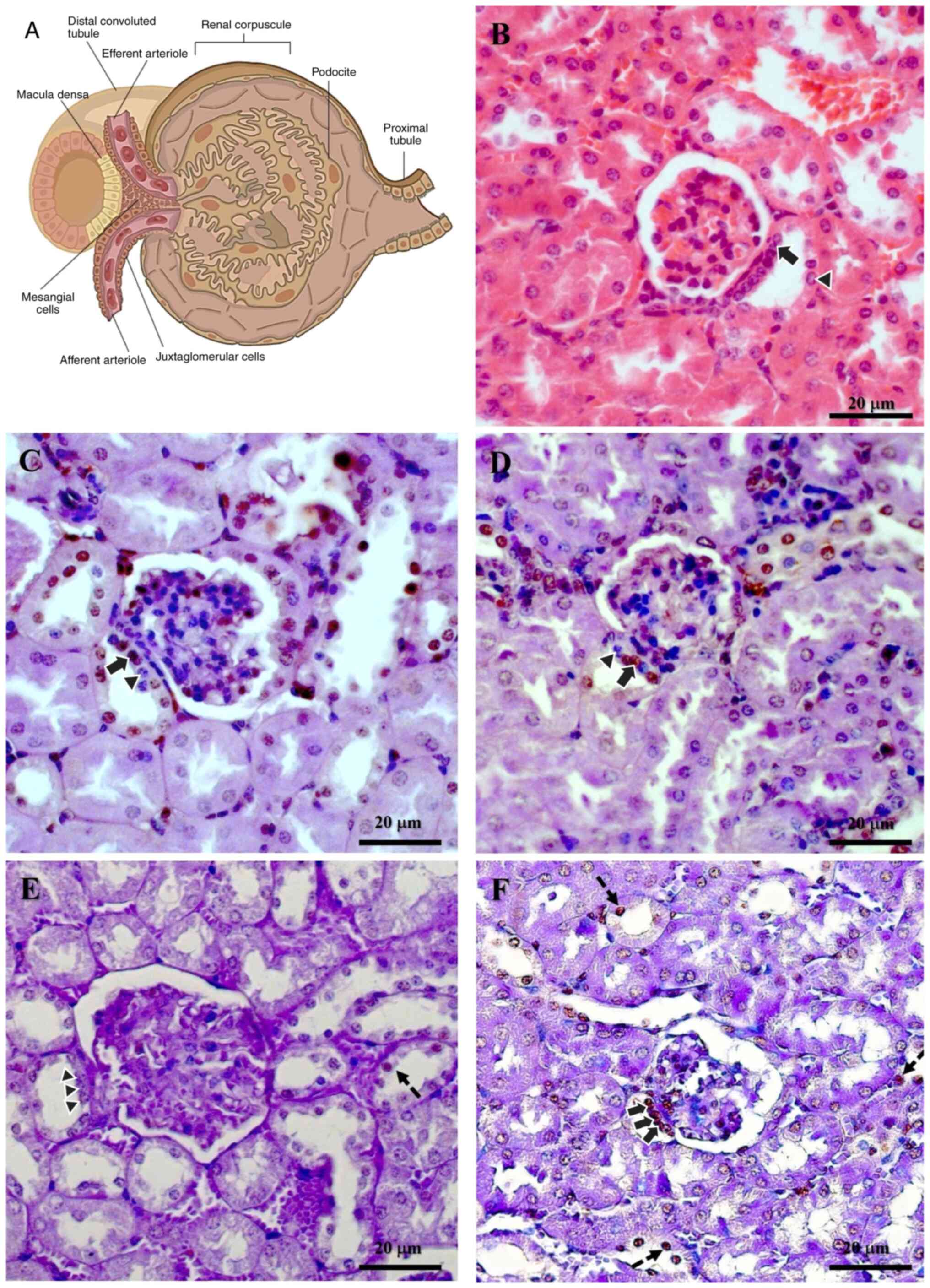
Jo MJ, Lee JK, Kim JE and Ko GJ: Molecular mechanisms associated with aging kidneys and future perspectives. Int J Mol Sci. 24(16912)2023.PubMed/NCBI View Article : Google Scholar

5 

Aging Biomarker Consortium. Bao H, Cao J, Chen M, Chen M, Chen W, Chen X, Chen Y, Chen Y, Chen Y, et al: Biomarkers of aging. Sci China Life Sci. 66:893–1066. 2023.PubMed/NCBI View Article : Google Scholar

6 

O'Sullivan ED, Hughes J and Ferenbach DA: Renal aging: Causes and consequences. J Am Soc Nephrol. 28:407–420. 2017.PubMed/NCBI View Article : Google Scholar

7 

Bolignano D, Mattace-Raso F, Sijbrands EJ and Zoccali C: The aging kidney revisited: A systematic review. Ageing Res Rev. 14:65–80. 2014.PubMed/NCBI View Article : Google Scholar

8 

Schmitt R and Melk A: Molecular mechanisms of renal aging. Kidney Int. 92:569–579. 2017.PubMed/NCBI View Article : Google Scholar

9 

Stocker SD, Kinsman BJ, Farquhar WB, Gyarmati G, Peti-Peterdi J and Sved AF: Physiological mechanisms of dietary salt sensing in the brain, kidney, and gastrointestinal tract. Hypertension. 81:447–455. 2024.PubMed/NCBI View Article : Google Scholar

10 

Peti-Peterdi J and Harris RC: Macula densa sensing and signaling mechanisms of renin release. J Am Soc Nephrol. 21:1093–1096. 2010.PubMed/NCBI View Article : Google Scholar

11 

Higashihara E, Harada T and Fukuhara H: Juxtaglomerular apparatus-mediated homeostatic mechanisms: Therapeutic implication for chronic kidney disease. Expert Opin Pharmacother. 25:819–832. 2024.PubMed/NCBI View Article : Google Scholar

12 

Saez F, Reverte V, Paliege A, Moreno JM, Llinás MT, Bachmann S and Salazar FJ: Sex-dependent hypertension and renal changes in aged rats with altered renal development. Am J Physiol Renal Physiol. 307:F461–F470. 2014.PubMed/NCBI View Article : Google Scholar

13 

Yabuki A, Miyazaki A, Ichii O, Kohyama M, Sawa M and Yamato O: Low expression of cyclooxygenase-2 in chronic kidney disease in young dogs. Res Vet Sci. 109:71–73. 2016.PubMed/NCBI View Article : Google Scholar

14 

Tower J: Programmed cell death in aging. Ageing Res Rev. 23 (Pt A):90–100. 2015.PubMed/NCBI View Article : Google Scholar

15 

Pellettieri J and Sánchez Alvarado A: Cell turnover and adult tissue homeostasis: From humans to planarians. Annu Rev Genet. 41:83–105. 2007.PubMed/NCBI View Article : Google Scholar

16 

Medh RD and Thompson EB: Hormonal regulation of physiological cell turnover and apoptosis. Cell Tissue Res. 301:101–124. 2000.PubMed/NCBI View Article : Google Scholar

17 

Yun MH: Changes in regenerative capacity through lifespan. Int J Mol Sci. 16:25392–25432. 2015.PubMed/NCBI View Article : Google Scholar

18 

Sousounis K, Baddour JA and Tsonis PA: Aging and regeneration in vertebrates. Curr Top Dev Biol. 108:217–246. 2014.PubMed/NCBI View Article : Google Scholar

19 

Ortega-Martínez M, Romero-Núñez E, Niderhauser-García A, de-la-Garza-González C, Ancer-Rodríguez J and Jaramillo-Rangel G: Evidence of chondrocyte turnover in lung cartilage, with the probable participation of nestin-positive cells. Cell Biol Int. 37:239–241. 2013.PubMed/NCBI View Article : Google Scholar

20 

Ortega-Martínez M, de-la-Garza-González C, Ancer-Rodríguez J and Jaramillo-Rangel G: Nestin-positive stem cells participate in chondrocyte renewal in healthy adult lung cartilage. Int J Morphol. 32:151–153. 2014.

21 

Ortega-Martínez M, Rodríguez-Flores LE, Ancer-Arellano A, Cerda-Flores RM, de-la-Garza-González C, Ancer-Rodríguez J and Jaramillo-Rangel G: Analysis of cell turnover in the bronchiolar epithelium through the normal aging process. Lung. 194:581–587. 2016.PubMed/NCBI View Article : Google Scholar

22 

Vanhooren V and Libert C: The mouse as a model organism in aging research: Usefulness, pitfalls and possibilities. Ageing Res Rev. 12:8–21. 2013.PubMed/NCBI View Article : Google Scholar

23 

Ackert-Bicknell CL, Anderson LC, Sheehan S, Hill WG, Chang B, Churchill GA, Chesler EJ, Korstanje R and Peters LL: Aging research using mouse models. Curr Protoc Mouse Biol. 5:95–133. 2015.PubMed/NCBI View Article : Google Scholar

24 

Messa GAM, Piasecki M, Hurst J, Hill C, Tallis J and Degens H: The impact of a high-fat diet in mice is dependent on duration and age, and differs between muscles. J Exp Biol. 223 (Pt 6)(jeb217117)2020.PubMed/NCBI View Article : Google Scholar

25 

Zhao B, Liu H, Wang J, Liu P, Tan X, Ren B, Liu Z and Liu X: Lycopene supplementation attenuates oxidative stress, neuroinflammation, and cognitive impairment in aged CD-1 mice. J Agric Food Chem. 66:3127–3136. 2018.PubMed/NCBI View Article : Google Scholar

26 

Izzotti A, Calin GA, Steele VE, Croce CM and De Flora S: Relationships of microRNA expression in mouse lung with age and exposure to cigarette smoke and light. FASEB J. 23:3243–3250. 2009.PubMed/NCBI View Article : Google Scholar

27 

Aldinger KA, Sokoloff G, Rosenberg DM, Palmer AA and Millen KJ: Genetic variation and population substructure in outbred CD-1 mice: Implications for genome-wide association studies. PLoS One. 4(e4729)2009.PubMed/NCBI View Article : Google Scholar

28 

Ortega-Martinez M, Gutierrez-Davila V, Gutierrez-Arenas E, Niderhauser-Garcia A, Cerda-Flores RM and Jaramillo-Rangel G: The convoluted tubules of the nephron must be considered elliptical, and not circular, in stereological studies of the kidney. Kidney Blood Press Res. 46:229–235. 2021.PubMed/NCBI View Article : Google Scholar

29 

National Research Council (US) Committee for the Update of the Guide for the Care and Use of Laboratory Animals: Guide for the Care and Use of Laboratory Animals. 8th edition. National Academies Press, Washington, DC, 2010.

30 

Ministry of the Interior. Official Mexican Standard NOM-062-ZOO-1999, Technical specifications for the production, care, and use of laboratory animals [Internet]. Mexico City: Official Gazette of the Federation, 2001 [cited 2025 Jul 7]. Available from: https://www.dof.gob.mx/nota_detalle.php?codigo=762506&fecha=22/08/2001#gsc.tab=0.

31 

Singal A, Sahni D, Gupta T, Aggarwal A and Gupta AK: Anatomic variability of oval window as pertaining to stapes surgery. Surg Radiol Anat. 42:329–335. 2020.PubMed/NCBI View Article : Google Scholar

32 

Murlimanju BV, Kumar BM, Kumar N, Prashanth KU, Rao CP, Guru A, Prabhu LV and Kumar CG: Morphometric parameters of the human adult kidney: An anatomical study. Int J Morphol. 32:656–659. 2014.

33 

Li XM, Yang L, Reng J, Xu GH and Zhou P: Non-invasive evaluation of renal structure and function of healthy individuals with multiparametric MRI: Effects of sex and age. Sci Rep. 9(10661)2019.PubMed/NCBI View Article : Google Scholar

34 

Onyeanusi BI, Adeniyi AA, Onyeanusi CG, Ayo JO and Ibe CS: A study of the kidney of the wistar rat in Northern Guinea savannah zone: The morphometric aspect. Pak J Nutr. 8:1040–1042. 2009.

35 

Mobini B and Abdollahi M: Effect of sex on histological and histochemical structures of different parts of the kidney in Japanese quail. Poult Sci. 95:2145–2150. 2016.PubMed/NCBI View Article : Google Scholar

36 

Bertram JF: Analyzing renal glomeruli with the new stereology. Int Rev Cytol. 161:111–172. 1995.PubMed/NCBI View Article : Google Scholar

37 

Ortega-Martínez M, Gutiérrez-Dávila V, Niderhauser-García A, Cerda-Flores RM, García-Juárez J, de-la-Garza-González C and Jaramillo-Rangel G: Morphometric analysis of the non-epithelial areas of mouse bronchioles through the normal aging process. Am J Transl Res. 11:3637–3644. 2019.PubMed/NCBI

38 

Elliott JE, Mantilla CB, Pabelick CM, Roden AC and Sieck GC: Aging-related changes in respiratory system mechanics and morphometry in mice. Am J Physiol Lung Cell Mol Physiol. 311:L167–L176. 2016.PubMed/NCBI View Article : Google Scholar

39 

Prophet EB, Mills B, Arrington JB and Sobin LH: Laboratory Methods in Histotechnology (Armed Forces Institute of Phatology). American Registry of Pathology, Washington, DC, 1992.

40 

Gutiérrez-Dávila VL: Evaluation of cell turnover in renal tubular structures throughout the aging process (Master's thesis). Nuevo León (Mexico): Autonomous University of Nuevo León, 2021.

41 

Romen W, Heine WD and Hollenz M: The regeneration of the cells of the macula densa after subtotal nephrectomy in the rat. Virchows Arch B Cell Pathol. 27:249–253. 1978.PubMed/NCBI View Article : Google Scholar

42 

Razga Z and Nyengaard JR: The effect of angiotensin II on the number of macula densa cells through the AT1 receptor. Nephron Physiol. 112:37–43. 2009.PubMed/NCBI View Article : Google Scholar

43 

Lorenzi T, Graciotti L, Sagrati A, Reguzzoni M, Protasoni M, Minardi D, Milanese G, Cremona O, Fabri M and Morroni M: Normal human macula densa morphology and cell turnover: A histological, ultrastructural, and immunohistochemical investigation. Anat Rec (Hoboken). 303:2904–2916. 2020.PubMed/NCBI View Article : Google Scholar

44 

Majumdar AP, Du J, Yu Y, Xu H, Levi E, Patel BB and Rishi AK: Cell cycle and apoptosis regulatory protein-1: A novel regulator of apoptosis in the colonic mucosa during aging. Am J Physiol Gastrointest Liver Physiol. 293:G1215–G1222. 2007.PubMed/NCBI View Article : Google Scholar

45 

Vazquez-Padron RI, Lasko D, Li S, Louis L, Pestana IA, Pang M, Liotta C, Fornoni A, Aitouche A and Pham SM: Aging exacerbates neointimal formation, and increases proliferation and reduces susceptibility to apoptosis of vascular smooth muscle cells in mice. J Vasc Surg. 40:1199–1207. 2004.PubMed/NCBI View Article : Google Scholar

46 

Robinson AM, Conley DB, Shinners MJ and Kern RC: Apoptosis in the aging olfactory epithelium. Laryngoscope. 112 (8 Pt 1):1431–1435. 2002.PubMed/NCBI View Article : Google Scholar

47 

Vinter-Jensen L: Pharmacological effects of epidermal growth factor (EGF) with focus on the urinary and gastrointestinal tracts. APMIS Suppl. 93:1–42. 1999.PubMed/NCBI

48 

Chou JS, Reiser IW and Porush JG: Aging and urinary excretion of epidermal growth factor. Ann Clin Lab Sci. 27:116–122. 1997.PubMed/NCBI

49 

Shurin GV, Yurkovetsky ZR, Chatta GS, Tourkova IL, Shurin MR and Lokshin AE: Dynamic alteration of soluble serum biomarkers in healthy aging. Cytokine. 39:123–129. 2007.PubMed/NCBI View Article : Google Scholar

50 

Zhang Z, Hou L, Liu D, Luan S, Huang M and Zhao L: Directly targeting BAX for drug discovery: Therapeutic opportunities and challenges. Acta Pharm Sin B. 14:2378–2401. 2024.PubMed/NCBI View Article : Google Scholar

51 

Palominos C, Fuentes-Retamal S, Salazar JP, Guzmán-Rivera D, Correa P, Mellado M, Araya-Maturana R and Urra FA: Mitochondrial bioenergetics as a cell fate rheostat for responsive to Bcl-2 drugs: New cues for cancer chemotherapy. Cancer Lett. 594(216965)2024.PubMed/NCBI View Article : Google Scholar

52 

Brown GC and Borutaite V: Regulation of apoptosis by the redox state of cytochrome c. Biochim Biophys Acta. 1777:877–881. 2008.PubMed/NCBI View Article : Google Scholar

53 

Yuan J and Ofengeim D: A guide to cell death pathways. Nat Rev Mol Cell Biol. 25:379–395. 2024.PubMed/NCBI View Article : Google Scholar

54 

Lee JH, Jung KJ, Kim JW, Kim HJ, Yu BP and Chung HY: Suppression of apoptosis by calorie restriction in aged kidney. Exp Gerontol. 39:1361–1368. 2004.PubMed/NCBI View Article : Google Scholar

55 

Razga Z: Functional relevancies of trans-differentiation in the juxtaglomerular apparatus of rat kidney. Int J Nephrol Renovasc Dis. 13:147–156. 2020.PubMed/NCBI View Article : Google Scholar

56 

Jaszewski R, Ehrinpreis MN and Majumdar AP: Aging and cancer of the stomach and colon. Front Biosci. 4:D322–D328. 1999.PubMed/NCBI View Article : Google Scholar

57 

Rex N, Melk A and Schmitt R: Cellular senescence and kidney aging. Clin Sci (Lond). 137:1805–1821. 2023.PubMed/NCBI View Article : Google Scholar

58 

Schmitt R and Cantley LG: The impact of aging on kidney repair. Am J Physiol Renal Physiol. 294:F1265–F1272. 2008.PubMed/NCBI View Article : Google Scholar

59 

Gandolfo MT, Verzola D, Salvatore F, Gianiorio G, Procopio V, Romagnoli A, Giannoni M and Garibotto G: Gender and the progression of chronic renal diseases: does apoptosis make the difference? Minerva Urol Nefrol. 56:1–14. 2004.PubMed/NCBI

60 

Bard JB: Growth and death in the developing mammalian kidney: Signals, receptors and conversations. Bioessays. 24:72–82. 2002.PubMed/NCBI View Article : Google Scholar

61 

Coles HS, Burne JF and Raff MC: Large-scale normal cell death in the developing rat kidney and its reduction by epidermal growth factor. Development. 118:777–784. 1993.PubMed/NCBI View Article : Google Scholar

62 

Koseki C, Herzlinger D and al-Awqati Q: Apoptosis in metanephric development. J Cell Biol. 119:1327–1333. 1992.PubMed/NCBI View Article : Google Scholar

Related Articles

  • Abstract
  • View
  • Download
  • Twitter
Copy and paste a formatted citation
Spandidos Publications style
Ortega‑Martínez M, Gopar‑Cuevas Y, García‑Aguilar K, Chávez‑Briones M, Miranda‑Maldonado I, Ancer‑Arellano A, García‑Juárez J, Ancer‑Rodríguez J and Jaramillo‑Rangel G: Analysis of cell turnover in the macula densa through the normal aging process. Biomed Rep 23: 155, 2025.
APA
Ortega‑Martínez, M., Gopar‑Cuevas, Y., García‑Aguilar, K., Chávez‑Briones, M., Miranda‑Maldonado, I., Ancer‑Arellano, A. ... Jaramillo‑Rangel, G. (2025). Analysis of cell turnover in the macula densa through the normal aging process. Biomedical Reports, 23, 155. https://doi.org/10.3892/br.2025.2033
MLA
Ortega‑Martínez, M., Gopar‑Cuevas, Y., García‑Aguilar, K., Chávez‑Briones, M., Miranda‑Maldonado, I., Ancer‑Arellano, A., García‑Juárez, J., Ancer‑Rodríguez, J., Jaramillo‑Rangel, G."Analysis of cell turnover in the macula densa through the normal aging process". Biomedical Reports 23.3 (2025): 155.
Chicago
Ortega‑Martínez, M., Gopar‑Cuevas, Y., García‑Aguilar, K., Chávez‑Briones, M., Miranda‑Maldonado, I., Ancer‑Arellano, A., García‑Juárez, J., Ancer‑Rodríguez, J., Jaramillo‑Rangel, G."Analysis of cell turnover in the macula densa through the normal aging process". Biomedical Reports 23, no. 3 (2025): 155. https://doi.org/10.3892/br.2025.2033
Copy and paste a formatted citation
x
Spandidos Publications style
Ortega‑Martínez M, Gopar‑Cuevas Y, García‑Aguilar K, Chávez‑Briones M, Miranda‑Maldonado I, Ancer‑Arellano A, García‑Juárez J, Ancer‑Rodríguez J and Jaramillo‑Rangel G: Analysis of cell turnover in the macula densa through the normal aging process. Biomed Rep 23: 155, 2025.
APA
Ortega‑Martínez, M., Gopar‑Cuevas, Y., García‑Aguilar, K., Chávez‑Briones, M., Miranda‑Maldonado, I., Ancer‑Arellano, A. ... Jaramillo‑Rangel, G. (2025). Analysis of cell turnover in the macula densa through the normal aging process. Biomedical Reports, 23, 155. https://doi.org/10.3892/br.2025.2033
MLA
Ortega‑Martínez, M., Gopar‑Cuevas, Y., García‑Aguilar, K., Chávez‑Briones, M., Miranda‑Maldonado, I., Ancer‑Arellano, A., García‑Juárez, J., Ancer‑Rodríguez, J., Jaramillo‑Rangel, G."Analysis of cell turnover in the macula densa through the normal aging process". Biomedical Reports 23.3 (2025): 155.
Chicago
Ortega‑Martínez, M., Gopar‑Cuevas, Y., García‑Aguilar, K., Chávez‑Briones, M., Miranda‑Maldonado, I., Ancer‑Arellano, A., García‑Juárez, J., Ancer‑Rodríguez, J., Jaramillo‑Rangel, G."Analysis of cell turnover in the macula densa through the normal aging process". Biomedical Reports 23, no. 3 (2025): 155. https://doi.org/10.3892/br.2025.2033
Follow us
  • Twitter
  • LinkedIn
  • Facebook
About
  • Spandidos Publications
  • Careers
  • Cookie Policy
  • Privacy Policy
How can we help?
  • Help
  • Live Chat
  • Contact
  • Email to our Support Team