Introduction
Eczema is a prevalent chronic inflammatory skin
condition characterized by recurrent episodes of erythema, papules
and exudation, often accompanied by intense itching (1,2). Its
etiology is multifactorial, involving complex interactions between
neuro-immune-endocrine regulatory mechanisms in the skin and
external triggers such as air pollution, house dust mites, water
hardness and psychological stressors such as anxiety and depression
(3-6).
These factors collectively modulate local and systemic immune
responses, with cytokines produced by the skin further influencing
endocrine regulation. Epidemiological data indicate a prevalence of
10-25% in children and 5-10% in adults, with rates steadily
increasing worldwide (7,8). Furthermore, most patients with eczema
also have comorbid respiratory allergic conditions, such as asthma
and hay fever (9). Genetic
susceptibility is another significant factor, with heritability
estimates reaching up to 80% in predisposed populations (10).
Current clinical management of eczema relies heavily
on antibiotics, antihistamines and glucocorticoids, such as
compound dexamethasone acetate cream (11). However, corticosteroid-based
therapies are associated with multiple adverse effects, including
local irritation, secondary infections and rebound symptoms upon
abrupt discontinuation. Prolonged use may also lead to
hyperpigmentation, skin atrophy and systemic reactions such as
hypertension and hyperglycemia (12,13).
Other drug classes, such as cyclosporine, are frequently linked to
renal dysfunction, further limiting treatment options.
Given these limitations, Cnidium monnieri
(Shechuangzi) has attracted increasing attention. Historically
documented in the Divine Farmer's Classic of Materia Medica, this
herb has been traditionally used to dispel wind and dampness (Zao
Shi Qu Feng; anti-inflammatory and antipruritic, it regulates the
immune system and is mainly used for skin-related disorders and
arthritis, etc.), relieve itching and treat parasitic skin
conditions (14). Modern
pharmacological studies have expanded its therapeutic profile,
revealing antitumor, anti-inflammatory and anti-osteoporotic
effects (15). The essential oil of
C. monnieri is of particular interest due to its rich
composition of bioactive sesquiterpenes and monoterpenes (16-20).
Key components, including β-pinene and cnidicin, have been shown to
antagonize histamine-induced responses, stabilize mast cells and
inhibit mast cell degranulation, contributing to their
anti-inflammatory and antibacterial effects (21-23).
Moreover, C. monnieri volatile oil (CMVO) exhibits strong
transdermal penetration, enhancing its suitability for topical
treatment for eczema (24,25).
Despite its therapeutic potential, direct
application of CMVO faces challenges due to its high volatility,
susceptibility to light-induced degradation and potential for
irritation from concentrated extracts (26,27). A
review of topical delivery systems, including creams, patches and
gels, suggests that cream formulations offer distinct advantages
for essential oils (28,29). Creams can effectively improve
stability, address the high lipophilicity of essential oils,
enhance skin penetration and are simpler to prepare compared with
other formulations. Based on these considerations, the present
study incorporated CMVO into a cream formulation, optimized it
using single-factor analysis and response surface methodology and
evaluated its efficacy in a DNCB-induced mouse eczema model. The
present study used ELISA, hematoxylin and eosin (H&E) staining,
toluidine blue staining and immunohistochemistry to assess the
anti-inflammatory effects of the optimized CMVOC. The present study
aimed to develop a stable, safe, effective and quality-controlled
topical preparation capable of alleviating eczema-associated
inflammation and improving patient comfort.
Materials and methods
Reagents
The reagents used in this research included:
octadecanol, sodium lauryl sulfate and ethyl nicotinate (Tianjin
Kemiou Chemical Reagent Co., Ltd.); petrolatum (Shandong Dexinkang
Medical Technology Co., Ltd.); liquid paraffin, isopropyl myristate
(IPM), glycerol and acetone (Tianjin Tianli Chemical Reagent Co.,
Ltd.); compound dexamethasone acetate cream (China Resources Sanjiu
Medical & Pharmaceutical Co., Ltd.); olive oil (Zhejiang
Meizhiyuan Biotechnology Co., Ltd.); 2,4-dinitrochlorobenzene
(DNCB; Chengdu Kelong Chemical Co., Ltd.); Cnidium monnieri
(Shaanxi Xingshengde Pharmaceutical Co., Ltd.); ELISA Kit IL-6
(cat. no. MM-0163M1; Jiangsu Meimian Industrial Co., Ltd.); and
ELISA Kit IL-17 (cat. no. MM-0170M1; Jiangsu Meimian Industrial
Co., Ltd.); goat serum (cat. no. SL038; Beijing Solarbio Science
& Technology Co., Ltd.); anti JAK2/STAT3 (cat. no.
ab195055/ab31370; Abcam; HRP-labeled secondary antibody for goat
anti-rabbit (cat. no. ab205718; Abcam); DAB staining (cat. no.
DA1010; Beijing Solarbio Science & Technology Co., Ltd.).
Additionally, the equipment used includes a GC-MS System (Agilent
Technologies, Inc.), a PHS-3C pH meter (Shanghai INESA Scientific
Instrument Co., Ltd.) and a rotational viscometer (cat. no.
LC-NDJ-55; Shanghai Li-Chen Bang Xi Instrument Technology Co.,
Ltd.).
Laboratory animals
Male Kunming KM mice weighing 20-22 g were supplied
by Chengdu Dasuo Laboratory Animal Co., Ltd. and the license number
was SCXK (Chuan) 2025-0030. A total of 80 were purchased, among
which 60 were used for pharmacodynamic tests (25 days) and 20 for
safety tests (21 days). For the pharmacodynamic study, mice were
allocated as described below. For serum-based analyses, six animals
per group (n=6) were included based on sample availability
following blood collection and the corresponding data are presented
accordingly. Animal experiments were approved by the Shaanxi
University of Chinese Medicine Laboratory Animal Ethics Committee
(approval no. SUCMDL20250512001). Mice were housed in a laboratory
with standard temperature (22±2˚C) and humidity (55±10%) under a
12-h light/dark cycle and underwent a 7-day acclimatization period.
During the experiment, fresh drinking water was added and replaced
daily. Animals were monitored daily for general health, behavior
and signs of distress. Humane endpoints included severe weight loss
(>20%), persistent ulceration, or inability to access food and
water, upon which animals would be sacrificed immediately. After
the experiment, 20 mice used for safety tests were sacrificed by
cervical dislocation and 60 mice used for pharmacodynamics tests
were intraperitoneally injected with 1% pentobarbital sodium (100
mg/kg). After the successful induction of anesthesia was confirmed
by the righting reflex, ~1 ml of blood was collected from the
eyeball and then sacrifice was performed by cervical dislocation.
After the cervical dislocation sacrifice of the 80 mice, the
breathing and pupil dilation conditions were observed to determine
if the mice were dead.
Cream preparation
To minimize irritation and volatility, CMVO was
incorporated into an ointment base. Oil-in-water (O/W) and
water-in-oil (W/O) emulsion types were both screened to identify
formulations with high gloss, fine smooth particles, light texture
and excellent stability.
Formulations (Table
I) were prepared by weighing the oil and aqueous phases
separately. Each phase was heated separately in an 80˚C water bath
until dissolved. The two phases were continuously stirred to
achieve uniform mixing. Once they reached the same temperature, the
aqueous phase was slowly poured into the oil phase while stirring
continuously in the same direction until emulsification was
complete. The emulsion was then cooled with continued stirring to
room temperature until the cream formed.
 | Table IPrescription screening. |
Table I
Prescription screening.
| Ingredients | Prescription
1/g | Prescription
2/g | Prescription
3/g | Prescription
4/g |
|---|
| Stearic acid | 6.00 | / | 6.00 | 15.10 |
| Paraffinum
molle | 2.00 | 4.00 | 10.00 | 10.20 |
| Octadecyl | / | 3.60 | / | / |
| Tween 80 | 3.00 | / | / | 5.00 |
| Liquid
paraffin | / | 2.20 | / | / |
| Glyceryl
monostearate | / | / | 2.00 | 10.00 |
| Pan 80 | / | / | / | 2.00 |
| IPM | / | 1.00 | / | / |
|
Triethanolamine | 3.00 | / | 0.40 | / |
| PEG-400 | 3.00 | / | / | / |
| Glycerinum | / | 1.16 | 5.00 | 20.00 |
| Lauryl sodium
sulfate | / | 0.30 | / | / |
| Sorbic acid | / | / | / | 0.20 |
| Nibkin ethyl
ester | / | 0.04 | 0.10 | / |
| Distilled
water | / | 27.40 | 45.00 | 37.50 |
A single-factor experiment was designed to
investigate the optimal quantities of the aqueous and oil phases,
as well as the base components with significant influence.
Subsequently, a response surface experiment was conducted to derive
the optimal formulation.
Composition of the prescription
Based on a literature review, four ointment
formulations were selected for comparison (30-33).
Formulation 1 and Formulation 2 were O/W types, while Formulation 3
and Formulation 4 were W/O types. Specific ingredients are shown in
Table I.
Prescription optimization
Preliminary quality assessment of the four selected
prescriptions indicated that Prescription 2 demonstrated the most
effective results.
Single-factor analysis
To further refine the formulation ratio, a
five-level single-factor experiment (n=3) was conducted to evaluate
six components in the ointment formulation. Factors were labeled as
follows: (A) octadecanol, (B) petrolatum, (C) liquid paraffin, (D)
IPM, (E) sodium lauryl sulfate and (F) glycerin. The specific
experimental design is shown in Table
II.
| Table IISingle-factor design (n=3). |
Table II
Single-factor design (n=3).
| Level | A/g | B/g | C/g | D/g | E/g | F/g |
|---|
| 1 | 3.20 | 3.20 | 1.40 | 0.60 | 0.20 | 0.36 |
| 2 | 3.60 | 3.60 | 1.80 | 0.80 | 0.30 | 0.76 |
| 3 | 4.00 | 4.00 | 2.20 | 1.00 | 0.40 | 1.16 |
| 4 | 4.40 | 4.40 | 2.60 | 1.20 | 0.50 | 1.56 |
| 5 | 4.80 | 4.80 | 3.00 | 1.40 | 0.60 | 1.96 |
Response surface design
Components markedly influencing cream stability were
selected for re-evaluation. Three levels were chosen for each
factor: (A) petrolatum, (B) IPM and (C) sodium lauryl sulfate. The
Box-Behnken response surface method was employed using
Design-Expert 13 software (Stat-Ease, Inc.) to arrange the
experimental design (n=3). The design results are shown in Table III.
| Table IIIResponse surface design (n=3). |
Table III
Response surface design (n=3).
| Level | A/g | B/g | C/g |
|---|
| 1 | 4.00 | 0.60 | 0.30 |
| 2 | 4.40 | 0.80 | 0.40 |
| 3 | 4.80 | 1.00 | 0.50 |
Quality evaluation
The cream was evaluated based on four major
criteria, including the cream's appearance, droplet size, physical
stability and moisturizing properties (34-36)
and scored according to established standards with a maximum
composite score of 100 points. The breakdown was as follows:
Appearance and texture: 30% (each sub-criterion accounts for 5%);
physical stability: 30% (each sub-criterion accounts for 10%);
emulsion droplet size: 20% (each sub-criterion accounts for 10%);
and moisturizing properties: 20%.
Appearance characteristics
The cream was expected to have a uniform texture and
color, with appropriate viscosity for smooth application and
absorption, free from noticeable particles or separation.
Appearance characteristics were subdivided into five indicators:
Sensory evaluation, gloss, particle size, skin feel and
spreadability, each scored according to established standards.
Specific criteria are detailed in Table IV.
| Table IVEvaluation criteria for appearance
characteristics. |
Table IV
Evaluation criteria for appearance
characteristics.
| Standard | Score |
|---|
| Highly glossy,
smooth and non-grainy, refreshing and easy to apply | 5-6 points |
| Moderate gloss,
with a slight grainy texture and a medium level of difficulty in
application. | 3-4 points |
| Dull, rough with a
noticeable grainy texture, greasy and difficult to spread | <3 points |
Physical stability
The physical stability of the cream was evaluated
through three indicators: Centrifugal stability, heat resistance
and cold resistance. Scoring criteria are shown in Table V.
| Table VPhysical stability evaluation
criteria. |
Table V
Physical stability evaluation
criteria.
| Standard | Score |
|---|
| No significant
change | 8-10 points |
| No separation, no
oil or water separation, cream texture slightly soft or slightly
firm | 5-8 points |
| No separation,
slight oil or water separation, cream texture too soft or too
hard | 3-6 points |
| Demulsification and
separation | <3 points |
Centrifugal stability
For this test, 1 ml of the cream sample was placed
into a 1.5 ml centrifuge tube and centrifuged at 735 x g for 30 min
(25±2˚C). Oil-water separation was observed and scores were
determined accordingly.
Heat stability
For this test, 7.5 g of the cream sample was placed
in a 25 ml beaker, sealed with plastic wrap and placed in a 60˚C
constant-temperature oven for 6 h. After cooling to room
temperature, signs of oil-water separation, demulsification, or
flocculation were observed.
Cold stability
For this test,7.5 g of the cream sample was placed
in a sealed bag, refrigerated at 4˚C for 2 h, then frozen at -20˚C
for 24 h. After returning to room temperature, oil-water
separation, demulsification, or flocculation was observed.
Droplet diameter
Smaller particle size and more uniformly distributed
droplets indicated higher cream quality. First, a temporary mount
was prepared by taking a small amount of the cream sample,
spreading it evenly onto the center of a microscope slide and then
covering it with a coverslip. Next, a drop of glycerol was placed
at the center of the coverslip. Finally, the prepared samples were
observed using a bright-field light microscope at a magnification
of x400. Five non-overlapping fields per sample were selected for
particle size measurement by scanning the slide in a systematic
manner (from left to right and top to bottom) to minimize selection
bias. Droplet size and distribution were evaluated according to the
criteria shown in Table VI.
| Table VIEvaluation criteria for droplet
diameter. |
Table VI
Evaluation criteria for droplet
diameter.
| Standard | Score |
|---|
| Small particle size
with uniform distribution | 7-10 points |
| Particle size is
moderately sized with a relatively uniform distribution (with minor
variations in localized areas). | 4-7 points |
| Particle size is
relatively large, with uneven distribution (dispersed or locally
concentrated). | <4 points |
Moisturizing properties
A cream sample (1 g) was weighed onto a microscope
slide. After reweighing, the slide was placed in a 37˚C
constant-temperature oven for 8 h. After removal and reweighing,
the moisture loss rate and final score were calculated.
Determination of pH
At room temperature, 1 g of the cream was weighed,
10 g of purified water was added and the mixture was stirred evenly
to form a white emulsion. The pH value was then measured using a pH
meter. Three parallel determinations were conducted.
Determination of viscosity
Using a rotational viscometer, the 4th rotor was
selected and the rotational speed was adjusted to 6 rpm. The
instrument was started and the data were read once stabilized.
Extraction of CMVO
Volatile oil from C. monnieri was extracted
using steam distillation. The raw material was ground into powder
and 300 g of C. monnieri powder, which was identified by
Professor Yonggang Yan from the College of Pharmacy, Shaanxi
University of Chinese Medicine as the dried ripe fruit of C.
monnieri (L.) Cuss. of the Apiaceae family, was placed into a
2,000 ml round-bottom flask with 1,300 ml of distilled water. After
thorough mixing, the essential oil collection apparatus was
assembled. Extraction was conducted via steam distillation for 6 h,
ceasing when the product yield no longer increased. After cooling,
the essential oil was collected, with an average yield of 1.06%
(v/w). Three independent batches were extracted and GC-MS analysis
demonstrated consistent chemical profiles among batches. The
instrument used in Gas Chromatography-Mass Spectrometry (GC-MS) was
a 7890GC/5977MS, (Agilent Technologies, Inc.). The EI ion source
had an ionization energy of 70 eV and an initial temperature of
40˚C. It then gradually rose to 90˚C, followed by an increase at a
rate of 5˚C per minute to 260˚C, which was maintained for 1 min.
Subsequently, it was raised at a rate of 10˚C per minute to 260˚C;
this process lasted for 6 min. The carrier gas used was high-purity
helium with a flow rate of 1 ml/min.
DNCB-induced mouse eczema model
A total of 60 male specific pathogen-free
(SPF)-grade mice were randomly divided into six groups of 10 mice
each: Control, model, positive and low-, medium- and high-dose
groups of Cnidium monnieri essential oil ointment. All
animals were subjected to the same experimental procedures for
model establishment and treatment. However, for serum-based
analyses, a subset of animals (n=6 per group) was used and the
corresponding data are presented accordingly. One day prior to
modeling, depilation was performed on the dorsal region of mice in
all groups except the normal group, covering an area of ~2 cm x 2
cm. On Day 1, all mice except the normal group were sensitized with
100 µl of a 7% DNCB solution in acetone:olive oil (4:1). A booster
dose was administered on Day 2 and the challenge was repeated on
Day 3. Starting on Day 4, except for the control group, mice were
challenged with 30 µl of a 0.5% DNCB acetone-olive oil solution on
the back every other day for a total of four challenges. After
successful establishment of the eczema model, the normal group
received daily application of 100 µl acetone-olive oil solution on
the back. The model group received 100 µl of an unloaded cream
formulation (100 mg/kg/d), the positive control group received 100
µl of compound dexamethasone acetate ointment (100 mg/kg/d) and the
three groups treated with CMVOC received 100 µl of the
corresponding dose of cream formulation (the dosage of CMVO in the
cream): Low dose (100 mg/kg/d), medium dose (200 mg/kg/d) and high
dose (400 mg/kg/d). Each group received twice-daily applications
for 15 consecutive days. The precise dosage of the drug was derived
by integrating multiple previous studies on local essential oil
formulations, preliminary experiments and clinical dose conversions
using the fingertip unit method (37-40).
On the final day of the experiment, mice were fasted and deprived
of water for 12 h. Blood samples were then collected from the
eyeballs, skin samples were obtained from the dorsal region and
changes in skin condition were observed. The severity of skin
lesions was recorded on days 4, 7, 10, 13 and 16. Following the
scoring principles of the EASI clinical scoring system (41), erythema, edema/thickening,
scratches/erosions and crusting were selected as evaluation
criteria. Scores were assigned on a scale of 0-3, where 0 indicated
none (not present even upon close inspection), 1 indicated mild
(related lesions visible upon close inspection), 2 indicated
moderate (lesions directly visible) and 3 indicated severe
(symptoms very pronounced). Based on the score, if the average
score reaches 2 points and the model group remains above 2 points,
it can be determined that the modeling is successful.
Safety inspection
A total of 20 male SFP-grade KM mice were randomly
divided into four groups of five mice each: Control group,
experimental group 1 (low-dose group), experimental group 2
(medium-dose group) and experimental group 3 (high-dose group) (all
consisting of the CMVOC). After random grouping, all mice had their
backs and the sides of their spines shaved (~2 cm x 2 cm). At 24 h
later, skin scratches were made on the left side of the skin, while
the right side was scratched in an equal shape to cause slight
bleeding on the skin surface. The medication was applied
immediately after the scratches. The normal group was applied with
pure water and the experimental groups were applied with low-,
medium- and high-dose groups of CMVOC. After administration, the
mice were covered with gauze and fixed with adhesive tape. The
medication was applied once a day. At 24 h later, the skin was
washed with distilled water. After 1 h of cleaning, the mice were
observed for 7 consecutive days for redness, swelling, or other
phenomena. After stopping the medication, the mice were observed
for another 7 days (42).
According to the scoring criteria in Table VII, the condition of the mouse's
back skin was scored. The intensity of irritation was determined
using the average scoring formula for the animals, according to
Table VIII.
| Table VIISkin safety rating criteria. |
Table VII
Skin safety rating criteria.
| Skin reaction | Score |
|---|
| Degree of
erythema | |
|
No
erythema | 0 |
|
Mild
erythema | 1 |
|
Marked
erythema | 2 |
|
Moderate
erythema | 3 |
|
Severe
erythema with slight eschar formation | 4 |
| Degree of
edema | No edema |
|
No
edema | 0 |
|
Mild
edema | 1 |
|
Marked
edema, i.e., clearly visible skin swelling | 2 |
|
Moderate
edema, i.e., elevation of ~1 mm | 3 |
|
Severe
edema, with elevation exceeding 1 mm | 4 |
| Highest score | 8 |
| Table VIIISkin irritation severity
classification criteria. |
Table VIII
Skin irritation severity
classification criteria.
| Average score | Strength |
|---|
| 0-0.5 | Non-irritating |
| 0.5-2 | Mildly
irritating |
| 2-6 | Moderately
irritating |
| 6-8 | Strongly
irritating |
Determination of IL-6 and IL-17 by
ELISA
Mouse serum samples were tested using ELISA kits
from Jiangsu Enzyme Immunoassay Industrial Co., Ltd. IL-6 and IL-17
levels were determined according to the instruction manual.
Intra-assay and Inter-assay coefficient of variation (CV) <15%.
Standard curves were established and the concentration of each
sample group was calculated. The standard curves for i) IL-17 and
ii) IL-6 were as follows: i) y=0.0047x + 0.0659
R2=0.9957; ii) y=0.0039x + 0.1644 R2=0.991.
Blood samples were collected from mice for serum preparation.
During sample processing, four serum samples in the control group
(K2, K3, K5 and K6) were affected by hemolysis due to technical
issues during orbital blood collection and were therefore excluded
from ELISA analysis.
As a result, six valid serum samples remained in the
control group. Accordingly, ELISA analyses were conducted using six
samples per group. For each treatment group, six serum samples were
included in the analysis.
Serum levels of IL-6 and IL-17 were measured using
commercial ELISA kits according to the manufacturers' instructions.
All assays were performed following standard protocols and no
samples were excluded based on experimental outcomes.
H&E staining
Fresh skin tissue was fixed in 4% paraformaldehyde
solution for 24 h (25±2˚C), routinely embedded in paraffin (25±2˚C)
and sectioned into 4 µm-thick slices (-20˚C). The sections
underwent dewaxing (65˚C) and hydration with a gradient of ethanol
(100, 95, 80 and 75%), followed by rinsing with distilled water.
Sections were immediately immersed in hematoxylin stain for 5 min,
rinsed with tap water for 1 min, deparaffinized with hydrochloric
acid ethanol for 30 sec, soaked in tap water for 15 min and then
placed in eosin solution for 2 min. After staining, sections were
re-washed with graded ethanol and clarified with xylene. Finally,
neutral resin was applied to mount the sections (25±2˚C). Once
dried, the sections were ready for bright-field light microscopic
examination (43). The tissue
morphology was observed under a bright-field light microscope at a
magnification of x200 and images were captured. A total of three
non-overlapping fields per sample were selected for cell counting
by scanning the slide in a systematic manner (from left to right
and top to bottom) to minimize selection bias.
Toluidine blue staining
The pretreatment procedure was identical to that for
H&E staining: Tissue fixation, dehydration, clearing, paraffin
embedding, sectioning and dewaxing. The prepared sections were
placed in a 0.5% toluidine blue solution and incubated at 55˚C for
20 min. After 2-3 rinses with water, the sections were placed in
hydrochloric acid alcohol for 2-3 sec for decolorization, followed
by rinsing with water. Dehydration was performed using alcohol and
the sections were then stained with 0.25% eosin for 8 sec (25±2˚C).
Finally, routine procedures of decolorization, dehydration,
clearing and mounting were carried out sequentially. After
air-drying, the sections were observed under a microscope (44).
Immunohistochemistry
Mouse skin tissue sections of 2-5 µm were processed
dewaxing, rehydration, xylene and graded ethanol treatments. Next,
antigen retrieval was performed using the citric acid antigen
retrieval buffer (pH 6.0) and then using an electric pressure
cooker. Following three washes with TBS, the sections were
incubated with 0.1% Triton X-100 for 10 min, reacted with 0.3%
H2O2 in methanol at room temperature (25±2˚C)
for 30 min then blocked with 10% goat serum (cat. no. SL038;
Beijing Solarbio Science & Technology Co., Ltd.) in PBS for 10
min at room temperature. When adding the primary antibody, gently
shake off the blocking solution. Sections were incubated with
primary antibody of JAK2/STAT3 (cat. no. ab195055/ab31370; Abcam)
diluted 1:200 in PBS onto the slide at 4˚C for 24 h. The slide was
placed in PBS (pH 7.4) and shaken three times, each time for 5 min.
After slightly drying the slide, the secondary antibody of
HRP-labeled goat anti-rabbit (cat. no. ab205718; Abcam)
corresponding to the species of the secondary antibody was added to
the slide at a ratio of 1:500 to cover the tissue and incubated at
room temperature for 50 min. The slide was placed in PBS (pH 7.4)
and shaken three times, each time for 5 min. Following DAB staining
at room temperature (25±2˚C; cat. no. DA1010; Beijing Solarbio
Science & Technology Co., Ltd.), under microscopic observation
until brownish-yellow staining appeared, the sections were rinsed
with tap water to terminate the color development reaction.
Sections were counterstained with hematoxylin (25±2˚C, 3 min),
dehydrated, cleared and mounted. Processed sections were examined
under a bright-field light microscope (45,46).
Statistical analysis
Experimental data in the present study were
processed using GraphPad Prism 10.4.2 software (Dotmatics). Results
from single-factor experiments and response surface experiments in
the cream base screening were expressed as means, with n=3. For
mouse data in the eczema model experiments, serum-based analyses
were performed using six animals per group (n=6).
Quantitative data from animal studies were expressed
as mean ± standard deviation. Prior to statistical analysis, data
were assessed for normality using the Shapiro-Wilk test and for
homogeneity of variances using Levene's test.
For datasets meeting the assumptions of normality
and homogeneity of variance, statistical comparisons among multiple
groups were performed using one-way analysis of variance followed
by Tukey's post hoc test. For datasets with unequal variances or
unequal sample sizes, Welch's ANOVA followed by Games-Howell
multiple comparisons test was applied. This approach ensures that
statistical analyses accurately reflect the experimental design,
regardless of unequal variances or deviations from normality.
Histological and immunohistochemical data, including
epidermal thickness, mast cell counts, H&E staining, toluidine
blue staining and immunohistochemistry, were quantified using
ImageJ-win64 software (National Institutes of Health). By including
all pre-defined animals and using appropriate statistical methods,
the analyses were both transparent and unbiased. P<0.05 was
considered to indicate a statistically significant difference.
Results
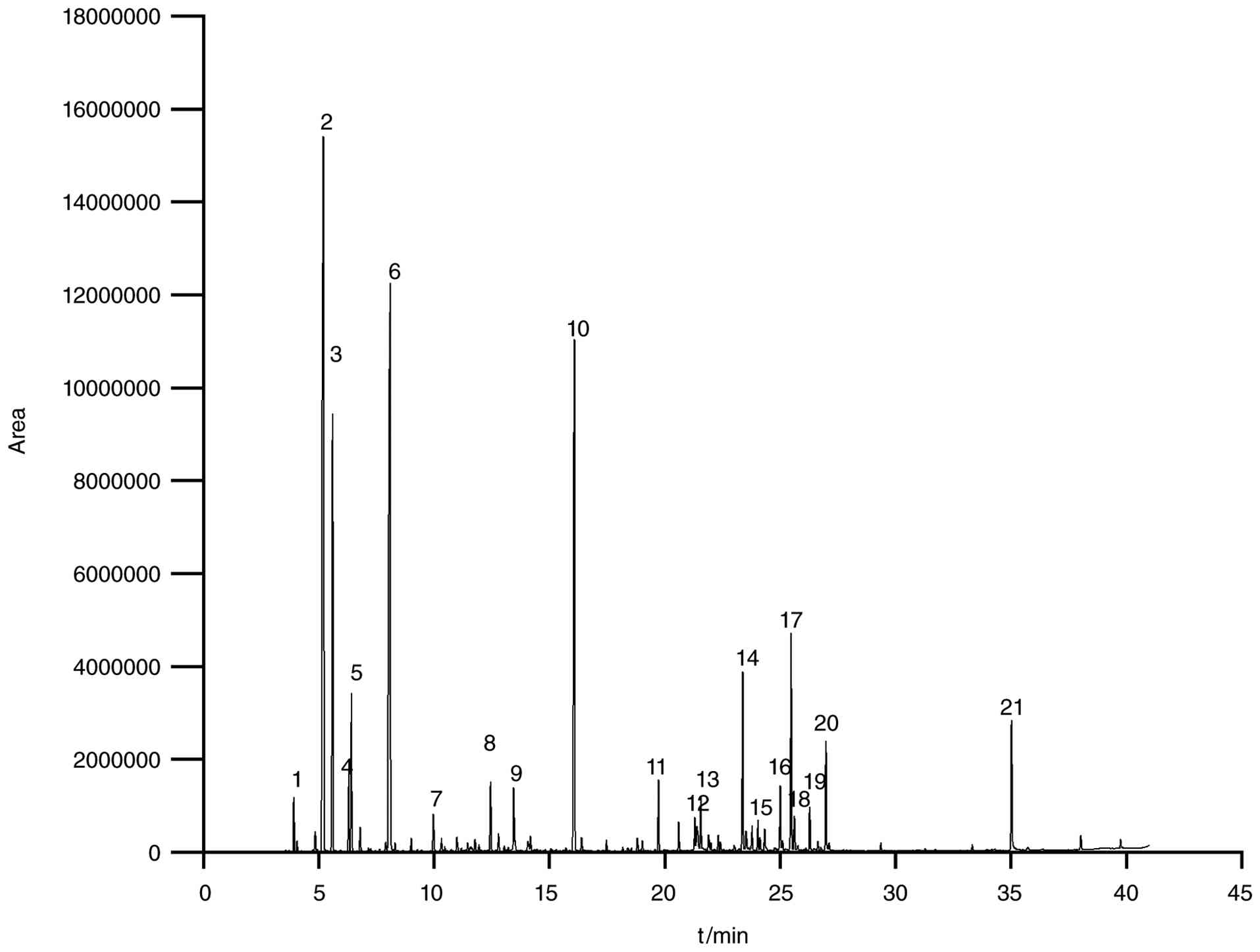
GC-MS analysis
The active components of Cnidium monnieri
essential oil were analyzed using GC-MS. The total ion chromatogram
is presented in Fig. 1. By
comparing with the NIST database and retention indices, 21 active
compounds were identified as listed in Table IX, with their chemical structures
depicted in Fig. 2.
![Compound structure. (A) Butanoic
acid, 2-methyl-, 1-methylethyl ester; (B)
(1R)-2,6,6-Trimethylbicyclo[3.1.1]hept-2-ene; (C) Camphene; (D)
Bicyclo[3.1.0]hexane, 4-methylene-1-(1-methylethyl)-; (E)
Bicyclo[3.1.1]heptane, 6,6-dimethyl-2-methylene-, (1S)-; (F)
d-limonene; (G) Cyclohexene, 1-methyl-4-(1-methylethylidene)-; (H)
endo-borneol; (I) Estragole; (J) Bicyclo[2.2.1]heptan-2-ol,
1,7,7-trimethyl-, acetate, (1S-endo)-; (K) Caryophyllene; (L)
(1R,2S,6S,7S,8S)-8-Isopropyl-1-methyl-3-methylenetricyclo[4.4.0.02,7]decane-rel-;
(M) trans-α-Bergamotene; (N) Perillyl isobutyrate; (O) Hexadecane;
(P) (6E)-8-Methyl-6-nonenoic acid; (Q) Bicyclopentyl-1,1'-diene;
(R) Chrysantenyl 2-methuylbutanoate; (S) cis-Chrysanthenyl
propionate; (T) 2-Cyclohexen-1-ol, 2-methyl-5-(1-methylethenyl)-,
propanoate; (U) Osthole)](/article_images/br/25/2/br-25-02-02164-g01.jpg) | Figure 2Compound structure. (A) Butanoic
acid, 2-methyl-, 1-methylethyl ester; (B)
(1R)-2,6,6-Trimethylbicyclo[3.1.1]hept-2-ene; (C) Camphene; (D)
Bicyclo[3.1.0]hexane, 4-methylene-1-(1-methylethyl)-; (E)
Bicyclo[3.1.1]heptane, 6,6-dimethyl-2-methylene-, (1S)-; (F)
d-limonene; (G) Cyclohexene, 1-methyl-4-(1-methylethylidene)-; (H)
endo-borneol; (I) Estragole; (J) Bicyclo[2.2.1]heptan-2-ol,
1,7,7-trimethyl-, acetate, (1S-endo)-; (K) Caryophyllene; (L)
(1R,2S,6S,7S,8S)-8-Isopropyl-1-methyl-3-methylenetricyclo[4.4.0.02,7]decane-rel-;
(M) trans-α-Bergamotene; (N) Perillyl isobutyrate; (O) Hexadecane;
(P) (6E)-8-Methyl-6-nonenoic acid; (Q) Bicyclopentyl-1,1'-diene;
(R) Chrysantenyl 2-methuylbutanoate; (S) cis-Chrysanthenyl
propionate; (T) 2-Cyclohexen-1-ol, 2-methyl-5-(1-methylethenyl)-,
propanoate; (U) Osthole) |
| Table IXActive components of Cnidium
monnieri volatile oil. |
Table IX
Active components of Cnidium
monnieri volatile oil.
| No. | Compound | CAS | Total, % |
|---|
| 1 | Butanoic acid,
2-methyl-, 1-methylethyl ester | 066576-71-4 | 0.836 |
| 2 |
(1R)-2,6,6-Trimethylbicyclo[3.1.1]hept-2-ene | 007785-70-8 | 25.585 |
| 3 | Camphene | 000079-92-5 | 9.976 |
| 4 |
Bicyclo[3.1.0]hexane,
4-methylene-1-(1-methylethyl)- | 003387-41-5 | 1.363 |
| 5 |
Bicyclo[3.1.1]heptane,
6,6-dimethyl-2-methylene-, (1S)- | 018172-67-3 | 3.01 |
| 6 | D-Limonene | 005989-27-5 | 22.22 |
| 7 | Cyclohexene,
1-methyl-4-(1-methylethylidene)- | 000586-62-9 | 0.766 |
| 8 | endo-Borneol | 000507-70-0 | 1.366 |
| 9 | Estragole | 000140-67-0 | 1.469 |
| 10 |
Bicyclo[2.2.1]heptan-2-ol,
1,7,7-trimethyl-, acetate, (1S-endo)- | 005655-61-8 | 14.792 |
| 11 | Caryophyllene | 000087-44-5 | 1.387 |
| 12 |
(1R,2S,6S,7S,8S)-8-Isopropyl-1-methyl-3-methylenetricyclo
[4.4.0.02,7]decane-rel- | 018252-44-3 | 0.6 |
| 13 |
trans-α-bergamotene | 013474-59-4 | 0.885 |
| 14 | Perillyl
isobutyrate | 1000429-32-1 | 3.265 |
| 15 | Hexadecane | 000544-76-3 | 0.525 |
| 16 |
(6E)-8-Methyl-6-nonenoic acid | 059320-77-3 | 1.16 |
| 17 |
Bicyclopentyl-1,1'-diene | 000934-02-1 | 4.222 |
| 18 | Chrysantenyl
2-methuylbutanoate | 053820-13-6 | 0.628 |
| 19 | cis-Chrysanthenyl
propionate | 070470-69-8 | 0.832 |
| 20 | 2-Cyclohexen-1-ol,
2-methyl-5-(1-methylethenyl)-, propanoate | 000097-45-0 | 2.185 |
| 21 | Osthole | 000484-12-8 | 2.929 |
Preparation of cream. Single-factor
analysis experiment
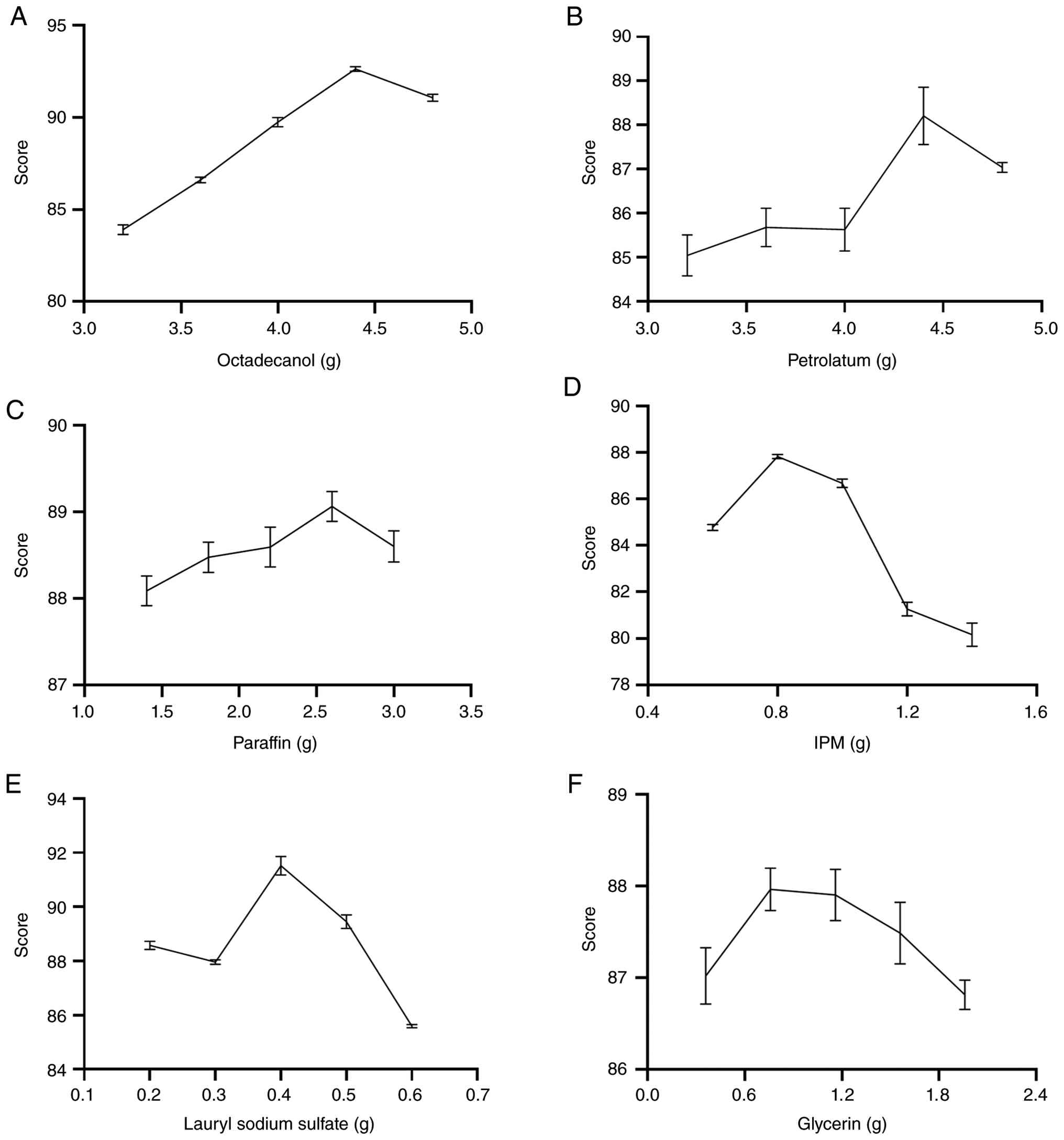
Fig. 3 illustrates
the effect of varying base component levels on the final evaluation
scores. Octyldodecanol, IPM and sodium lauryl sulfate markedly
influenced the scores. Among these, octyldodecanol (4.40 g)
provided optimal cream stability; lower levels led to oil-water
separation, while higher levels resulted in hard,
difficult-to-spread creams. IPM (0.80 g) exhibited optimal skin
affinity; lower concentrations caused excessive greasiness, while
higher levels disrupted the cream structure, leading to a granular
texture. Sodium lauryl sulfate (0.40 g) achieved maximum
emulsifying efficacy; lower concentrations prevented cream
formation, while higher levels increased viscosity. By contrast,
petrolatum, liquid paraffin and glycerin exhibited markedly lower
variability, with no significant changes observed across their five
respective concentration levels. As lubricants and moisturizers,
their moderate dosages exerted no discernible effect on the cream
formulation. The experimental variables comprised (A) octadecanol,
(B) petrolatum, (C) liquid paraffin, (D) IPM, (E) lauryl sodium
sulfate and (F) glycerin, with detailed experimental procedures and
results outlined in Tables X and
XI.
| Table XSingle-factor experiment score
distribution (n=3). |
Table X
Single-factor experiment score
distribution (n=3).
| | External
properties | Stability | Particle size | |
|---|
| Group | Sense | Glossiness | Granularity | Skin feeling | Coating
property | Centrifugal
stability | Heatproof
level |
Cold-resistance | Size | Distribution | Moisture
retention | Score |
|---|
| A1 | 5.82 | 5.90 | 5.77 | 5.83 | 5.78 | 9.80 | 7.47 | 4.13 | 8.30 | 7.60 | 17.51 | 83.91 |
| A2 | 5.69 | 5.95 | 5.78 | 5.70 | 5.61 | 9.80 | 8.88 | 4.95 | 8.20 | 8.00 | 18.05 | 86.60 |
| A3 | 5.91 | 5.93 | 5.83 | 5.86 | 5.82 | 9.80 | 8.43 | 7.55 | 8.65 | 8.51 | 17.44 | 89.74 |
| A4 | 5.84 | 5.94 | 5.89 | 5.83 | 5.92 | 9.80 | 9.61 | 8.87 | 8.84 | 8.67 | 17.41 | 92.63 |
| A5 | 5.69 | 5.83 | 5.85 | 5.70 | 5.63 | 9.80 | 9.59 | 8.42 | 8.41 | 8.30 | 17.86 | 91.07 |
| B1 | 5.74 | 5.91 | 5.91 | 5.82 | 5.65 | 9.80 | 8.33 | 4.53 | 7.85 | 8.29 | 17.21 | 85.04 |
| B2 | 5.85 | 5.95 | 5.92 | 5.91 | 5.89 | 9.80 | 8.50 | 4.50 | 8.30 | 7.56 | 17.49 | 85.68 |
| B3 | 5.80 | 5.95 | 5.92 | 5.88 | 5.81 | 9.80 | 8.50 | 4.53 | 7.53 | 8.27 | 17.63 | 85.63 |
| B4 | 5.83 | 5.95 | 5.93 | 5.88 | 5.87 | 9.80 | 8.98 | 6.07 | 8.00 | 7.93 | 17.97 | 88.21 |
| B5 | 5.78 | 5.95 | 5.92 | 5.84 | 5.81 | 9.80 | 7.42 | 6.60 | 7.80 | 7.81 | 18.31 | 87.04 |
| C1 | 5.83 | 5.95 | 5.93 | 5.79 | 5.82 | 9.80 | 8.76 | 5.52 | 8.01 | 7.96 | 18.72 | 88.09 |
| C2 | 5.84 | 5.95 | 5.93 | 5.84 | 5.84 | 9.80 | 9.37 | 5.87 | 7.50 | 8.30 | 18.24 | 88.48 |
| C3 | 5.85 | 5.95 | 5.94 | 5.82 | 5.73 | 9.80 | 8.73 | 5.47 | 8.00 | 8.41 | 18.89 | 88.59 |
| C4 | 5.85 | 5.95 | 5.95 | 5.87 | 5.88 | 9.80 | 8.57 | 5.77 | 8.60 | 8.45 | 18.38 | 89.06 |
| C5 | 5.76 | 5.94 | 5.95 | 5.70 | 5.80 | 9.80 | 7.38 | 6.89 | 8.30 | 8.20 | 18.89 | 88.60 |
| D1 | 5.76 | 5.94 | 5.96 | 5.76 | 5.81 | 9.80 | 7.32 | 8.45 | 6.91 | 5.12 | 17.95 | 84.78 |
| D2 | 5.78 | 5.93 | 5.82 | 5.95 | 5.83 | 9.80 | 9.11 | 7.39 | 7.50 | 6.81 | 17.91 | 87.82 |
| D3 | 5.78 | 5.80 | 5.82 | 5.86 | 5.83 | 9.80 | 8.24 | 7.89 | 7.50 | 6.58 | 17.56 | 86.67 |
| D4 | 5.76 | 5.93 | 5.78 | 5.85 | 5.85 | 9.80 | 8.99 | 9.22 | 3.00 | 3.00 | 18.09 | 81.27 |
| D5 | 5.79 | 5.93 | 5.91 | 5.75 | 5.69 | 9.80 | 8.66 | 8.23 | 3.20 | 3.50 | 17.70 | 80.17 |
| E1 | 5.77 | 5.80 | 5.81 | 5.91 | 5.84 | 9.80 | 9.21 | 9.35 | 6.89 | 6.67 | 17.53 | 88.58 |
| E2 | 5.70 | 5.93 | 5.95 | 5.70 | 5.65 | 9.80 | 8.62 | 5.03 | 8.52 | 8.34 | 18.71 | 87.96 |
| E3 | 5.90 | 5.86 | 5.88 | 5.75 | 5.79 | 9.80 | 9.30 | 8.10 | 8.12 | 8.30 | 18.72 | 91.52 |
| E4 | 5.82 | 5.93 | 5.92 | 5.79 | 5.81 | 9.80 | 9.13 | 6.13 | 8.39 | 7.70 | 19.03 | 89.45 |
| E5 | 5.78 | 5.80 | 5.82 | 5.84 | 5.86 | 9.80 | 8.12 | 7.35 | 7.01 | 6.57 | 17.66 | 85.60 |
| F1 | 5.63 | 5.93 | 5.87 | 5.57 | 5.63 | 9.80 | 8.97 | 6.23 | 7.64 | 7.43 | 18.31 | 87.02 |
| F2 | 5.69 | 5.93 | 5.90 | 5.62 | 5.64 | 9.80 | 9.13 | 6.59 | 7.82 | 7.43 | 18.40 | 87.96 |
| F3 | 5.76 | 5.93 | 5.92 | 5.72 | 5.72 | 9.80 | 9.23 | 7.53 | 6.80 | 7.21 | 18.28 | 87.90 |
| F4 | 5.72 | 5.93 | 5.86 | 5.68 | 5.68 | 9.80 | 9.45 | 6.28 | 7.56 | 7.10 | 18.44 | 87.49 |
| F5 | 5.63 | 5.93 | 5.89 | 5.60 | 5.58 | 9.80 | 8.92 | 6.64 | 7.55 | 6.95 | 18.33 | 86.81 |
| Table XISingle-factor experiment results
(n=3). |
Table XI
Single-factor experiment results
(n=3).
| Level | A | B | C | D | E | F |
|---|
| 1 | 83.91 | 85.04 | 88.09 | 84.78 | 88.58 | 87.02 |
| 2 | 86.60 | 85.68 | 88.48 | 87.82 | 87.96 | 87.96 |
| 3 | 89.74 | 85.63 | 88.59 | 86.67 | 91.52 | 87.90 |
| 4 | 92.63 | 88.21 | 89.06 | 81.27 | 89.45 | 87.49 |
| 5 | 91.07 | 87.04 | 88.60 | 80.17 | 85.60 | 86.81 |
Experimental results of response
surface optimization. Response surface experiment results
A Box-Behnken response surface design was conducted
using Design-Expert 13 software. The experimental details and
results are shown in Table
XII.
| Table XIIResponse surface experiment
results. |
Table XII
Response surface experiment
results.
| Standard | Run | A | B | C | Score |
|---|
| 7 | 1 | 4.00 | 0.80 | 0.50 | 85.55 |
| 14 | 2 | 4.40 | 0.80 | 0.40 | 91.63 |
| 9 | 3 | 4.40 | 0.60 | 0.30 | 86.59 |
| 15 | 4 | 4.80 | 0.80 | 0.30 | 91.58 |
| 3 | 5 | 4.00 | 1.00 | 0.40 | 85.72 |
| 17 | 6 | 4.80 | 0.80 | 0.50 | 92.15 |
| 11 | 7 | 4.40 | 0.60 | 0.50 | 86.18 |
| 4 | 8 | 4.80 | 1.00 | 0.40 | 87.07 |
| 12 | 9 | 4.40 | 1.00 | 0.50 | 86.42 |
| 13 | 10 | 4.40 | 0.80 | 0.40 | 92.48 |
| 16 | 11 | 4.40 | 0.80 | 0.40 | 92.26 |
| 6 | 12 | 4.80 | 0.80 | 0.30 | 87.61 |
| 5 | 13 | 4.00 | 0.80 | 0.30 | 85.98 |
| 10 | 14 | 4.40 | 1.00 | 0.30 | 87.52 |
| 8 | 15 | 4.80 | 0.80 | 0.50 | 86.95 |
| 1 | 16 | 4.00 | 0.60 | 0.40 | 84.14 |
| 2 | 17 | 4.80 | 0.60 | 0.40 | 86.35 |
Model fitting and analysis of
variance
Based on the experimental scores, regression
analysis yielded the following multiple quadratic regression
equation: Y=92.02 + 0.8238*A +
0.4338*B-0.3250*C-0.2150*
AB-0.0575*AC-0.1725*BC-3.18*A²-3.02*B²-2.32*C².
The results of the analysis of variance are
presented in Table XIII. The
model was significant (P<0.0001), with a non-significant
lack-of-fit term (P=0.7267>0.05), indicating that the model
fitted well with minimal interference from other factors.
Additionally, R²=0.9932 and adjusted R²=0.9844 further demonstrated
the model's high accuracy. The F-value revealed the following
influence order: octadecanol (A) > IPM (B) > sodium lauryl
sulfate (C).
| Table XIIIResponse surface regression model
analysis of variance. |
Table XIII
Response surface regression model
analysis of variance.
| Source | Sum of Squares | Degrees of
freedom | Mean Square | F-value | P-value | |
|---|
| Model | 123.63 | 9 | 13.74 | 113.32 | <0.0001 | Significant |
| A-octadecanol | 5.43 | 1 | 5.43 | 44.78 | 0.0003 | |
| B-IPM | 1.51 | 1 | 1.51 | 12.42 | 0.0097 | |
| C-lauryl sodium
sulfate | 0.8450 | 1 | 0.8450 | 6.97 | 0.0334 | |
| AB | 0.1849 | 1 | 0.1849 | 1.53 | 0.2567 | |
| AC | 0.0132 | 1 | 0.0132 | 0.1091 | 0.7508 | |
| BC | 0.1190 | 1 | 0.1190 | 0.9819 | 0.3547 | |
| A² | 42.51 | 1 | 42.51 | 350.68 | <0.0001 | |
| B² | 38.47 | 1 | 38.47 | 317.30 | <0.0001 | |
| C² | 22.66 | 1 | 22.66 | 186.95 | <0.0001 | |
| Residual | 0.8486 | 7 | 0.1212 | | | |
| Lack of Fit | 0.2168 | 3 | 0.0723 | 0.4575 | 0.7267 | Not
significant |
| Pure Errot | 0.6318 | 4 | 0.1580 | | | |
| Cor Total | 124.48 | 16 | | | | |
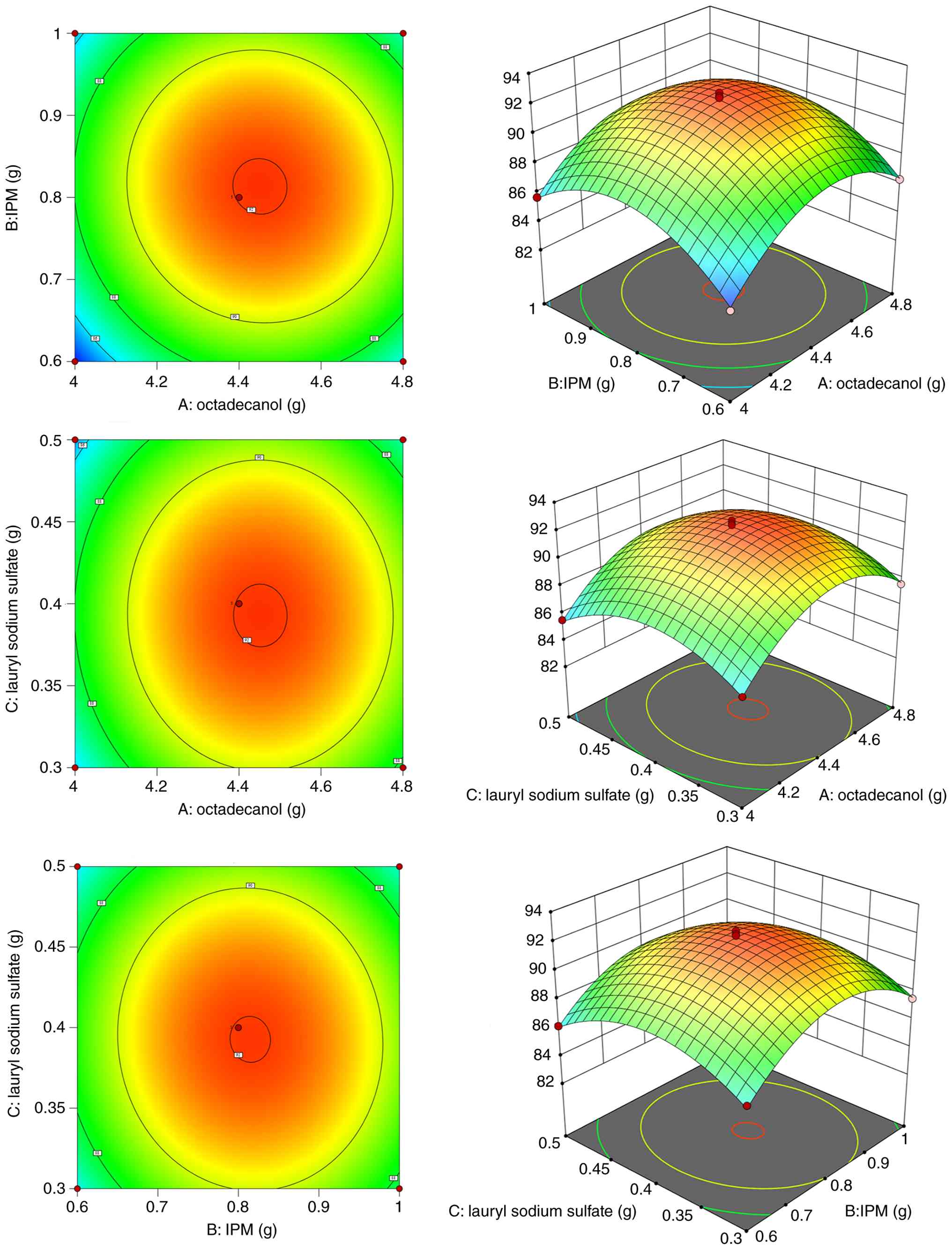
Model optimization and prediction
Based on the multiple quadratic regression equation,
Design-Expert 13 was used to generate contour plots and response
surfaces, which visually depicted the effect of factor interactions
on cream scores, as illustrated in Fig.
4. These interactions further elucidated the intricate
relationships within the formulation matrix. In the AB interaction,
cream scores initially rose and then declined with increasing
octadecyl alcohol content. Similarly, when IPM content rose,
octadecanol scores followed an up-and-down pattern, suggesting a
non-significant interaction between octadecyl alcohol and IPM. The
highest score was attained at an octadecanol content of 4.00 g and
an IPM content of 0.60 g. For the AC interaction, the score
initially increased and subsequently decreased with rising sodium
lauryl sulfate content. When sodium lauryl sulfate content
increased, the trend of octadecyl alcohol scores remained
unchanged, similarly indicating a non-significant interaction
between the two. The highest score was recorded at an octadecanol
content of 4.40 g and a sodium lauryl sulfate content of 0.40 g. In
the BC interaction, both IPM and sodium lauryl sulfate content
exhibited scores that initially increased and then decreased,
pointing to a non-significant interaction between them. The highest
score was achieved at an IPM content of 0.80 g and a sodium lauryl
sulfate content of 0.40 g. Response surface prediction pinpointed
the optimal formulation for preparing CMVO ointment: octadecanol at
4.45 g, IPM at 0.81 g and sodium lauryl sulfate at 0.39 g, yielding
a comprehensive score of 92.10 points.
Verification of the optimal
formulation
The total assessment scores are detailed in Table XIV. The average scores were
92.46±0.26, 92.48±0.2 and 91.48±0.27, closely aligning with the
model-predicted value of 92.10. This demonstrated a strong
concordance between the experimental results and the predicted
values.
| Table XIVResults of validation experiments
(n=3). |
Table XIV
Results of validation experiments
(n=3).
| | External
properties | Stability | Particle size | |
|---|
| Group | Sense | Glossiness | Granularity | Skin feeling | Coating
property | Centrifugal
stability | Heatproof
level |
Cold-resistance | Size | Distribution | Moisture
retention | Score |
|---|
| 1-1 | 5.58 | 5.91 | 5.52 | 5.63 | 5.74 | 9.80 | 8.91 | 9.54 | 8.92 | 8.53 | 18.13 | 92.21 |
| 1-2 | 5.51 | 5.91 | 5.43 | 5.65 | 5.78 | 9.80 | 8.79 | 9.51 | 8.99 | 8.79 | 18.27 | 92.43 |
| 1-3 | 5.59 | 5.91 | 5.61 | 5.69 | 5.75 | 9.80 | 8.95 | 9.46 | 9.01 | 8.85 | 18.11 | 92.73 |
| 2-1 | 5.65 | 5.91 | 5.47 | 5.51 | 5.67 | 9.80 | 8.71 | 9.15 | 9.35 | 8.58 | 18.48 | 92.28 |
| 2-2 | 5.72 | 5.91 | 5.51 | 5.69 | 5.83 | 9.80 | 8.96 | 9.07 | 9.21 | 8.63 | 18.34 | 92.67 |
| 2-3 | 5.64 | 5.91 | 5.36 | 5.67 | 5.82 | 9.80 | 8.85 | 9.22 | 9.27 | 8.69 | 18.26 | 92.49 |
| 3-1 | 5.21 | 5.91 | 5.52 | 5.50 | 5.69 | 9.80 | 9.59 | 9.36 | 8.47 | 8.26 | 18.42 | 91.73 |
| 3-2 | 5.51 | 5.91 | 5.17 | 5.49 | 5.64 | 9.80 | 9.29 | 9.18 | 8.53 | 8.31 | 18.37 | 91.20 |
| 3-3 | 5.41 | 5.91 | 5.23 | 5.47 | 5.68 | 9.80 | 9.34 | 9.27 | 8.91 | 8.25 | 18.25 | 91.52 |
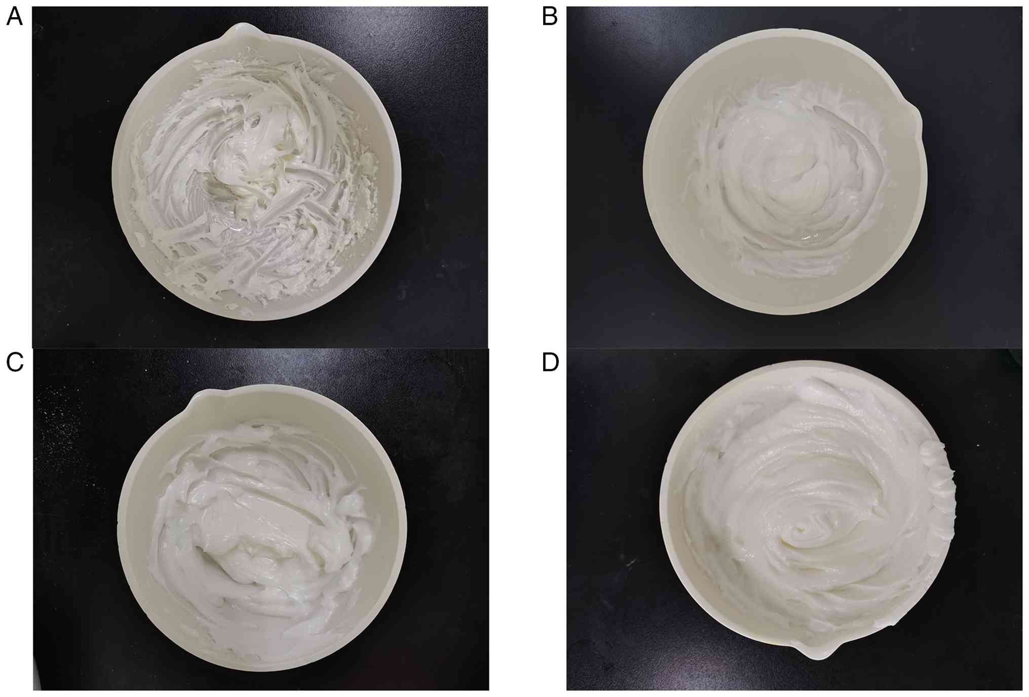
Quality evaluation. Appearance
characteristics
As presented in Fig.
5, these are the appearances of four cream formulations. In the
selection of a suitable cream, priority should be given to those
with uniform texture, appropriate consistency and the absence of
noticeable particles or separation.
Physical stability. Centrifugal
stability
After centrifugation at 735 x g for 30 min (25±2˚C),
none of the formulations exhibited oil-water separation, with no
significant differences observed among groups. These findings
demonstrate that the formulation exhibited good centrifugal
stability.
Heat stability
Following storage at 60˚C for 6 h and subsequent
cooling to room temperature, the cream maintained a uniform texture
and color. It remained smooth, refreshing and easy to apply,
without any signs of thinning, clumping, or oil-water separation,
demonstrating the cream's good heat resistance.
Cold stability
After refrigeration at 4˚C for 2 h and freezing at
-20˚C for 24 h, the cream exhibited no significant changes in color
or texture upon returning to room temperature. It remained light,
smooth and easy to apply, with no signs of thinning, clumping, or
oil-water separation, confirming the cream's strong cold
resistance.
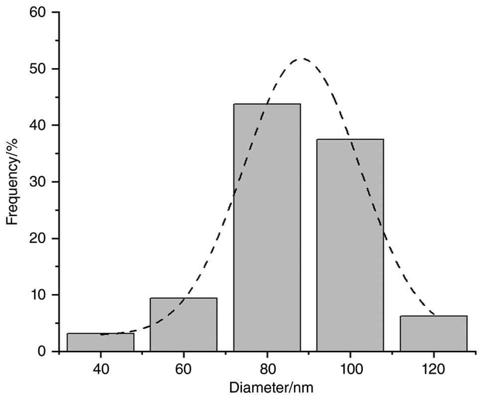
Droplet diameter
Particle size measurements of the drug-loaded
ointment revealed that 3 to 5 particle sizes were marked in each
group across low, medium and high dosage formulations (n=3). The
average particle size was ~85.50±15.33 nm, meeting the preparation
standard (ointment particle size <180 nm). The particle size
distribution is detailed in Fig. 6
and Table XV.
| Table XVQuality evaluation results of
different doses of Cnidium monnieri volatile oil cream. |
Table XV
Quality evaluation results of
different doses of Cnidium monnieri volatile oil cream.
| Drug Content | Particle size
(nm) | pH | Viscosity
(mPa∙S) |
|---|
| Low-dose group | 94.50±16.88 | 6.92±0.00 | 50,553 |
| Medium-dose
group | 77.77±14.92 | 6.91±0.02 | 50,533 |
| High-dose
group | 84.80±6.87 | 6.91±0.01 | 49,955 |
pH
The pH of the cream was measured using a PHS-3C pH
meter at different drug loading doses, with results shown in
Table XV. After analysis, it can
be seen that the average pH values for the low-, medium- and
high-dose groups were 6.92±0.00, 6.91±0.02 and 6.91±0.01,
respectively. Overall, the cream was weakly acidic, aligning with
the pH range suitable for skin application.
Viscosity
Viscosity measurements conducted at room
temperature, with results shown in Table XV, revealed similar viscosity
values across different doses, with no significant difference
observed. Moreover, this viscosity falls within the range suitable
for external skin application, ensuring the cream's applicability
and stability while preventing phenomena such as
stratification.
Observation of mouse skin
condition
A scoring system for assessing the severity of skin
lesions in mice was established based on the EASI criteria, as
detailed in Table XVI. On Day 4,
mice exhibited mild eczema symptoms. From Days 7 to 10, symptoms
intensified but remained below severe levels due to the
administration of the medication. By Days 13 to 16, significant
improvement was observed, indicating the medication's effectiveness
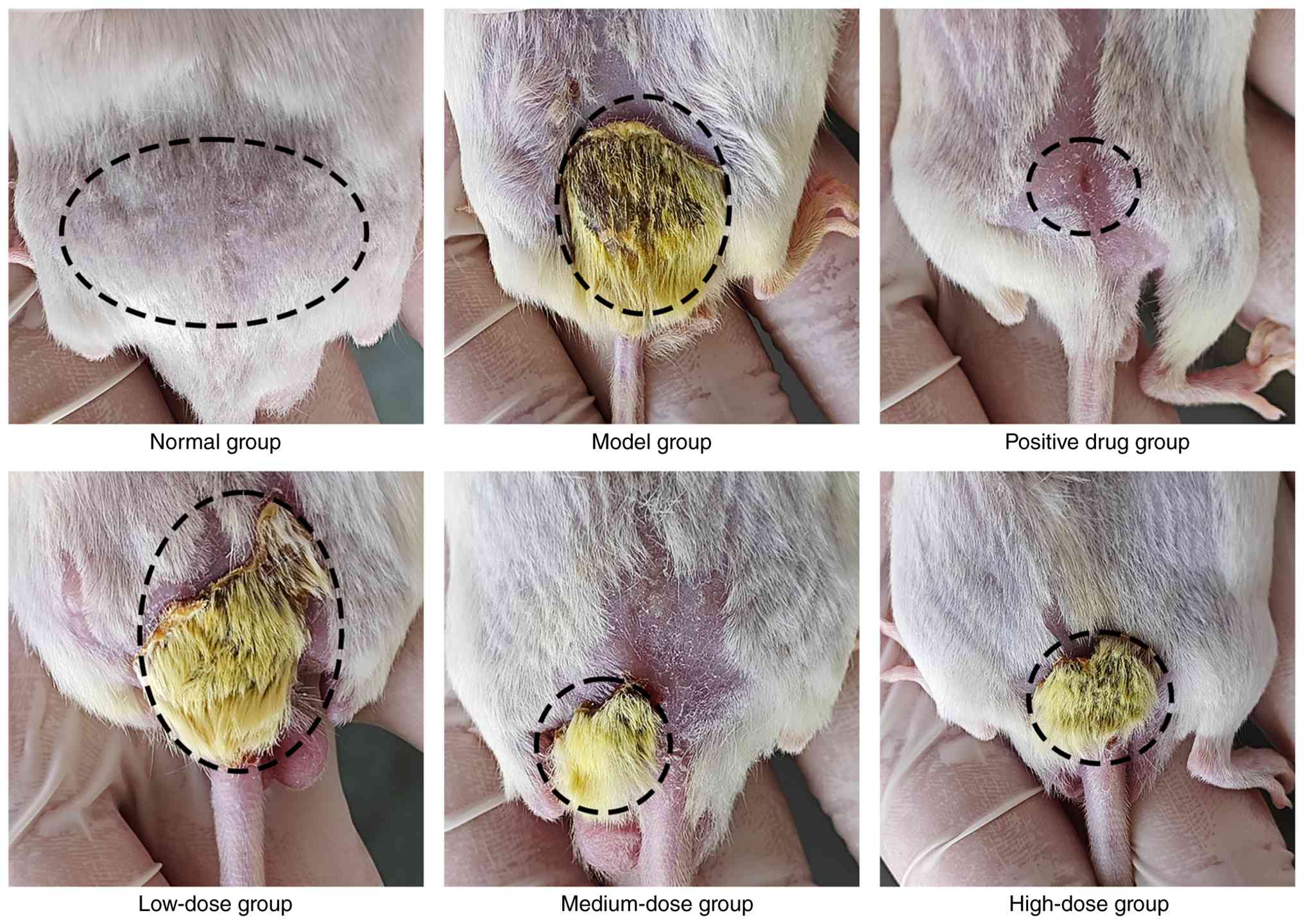
in alleviating eczema symptoms. As illustrated in Fig. 7, following DNCB induction, mice
displayed clear signs of eczema. However, treatment with CMVO
ointment mitigated skin tissue damage. Visual inspection revealed
that the control group's skin appeared pale pink without
abnormalities, while the model group exhibited pronounced skin
lesions and swelling, confirming successful DNCB-induced eczema
modeling. All treatment groups showed marked improvements in skin
condition. Although both the model and treated groups displayed
skin lesions, redness, swelling and scabbing, the model group
showed the most pronounced lesions and swelling. In the positive
control group, scabs had completely fallen off, revealing pale pink
skin without swelling or hyperemia. Mice in the low-dose cream
group still exhibited slight redness and swelling, with limited
desquamation of crusted areas. The medium-dose cream group
exhibited partial crust desquamation, exposing pale pink skin, as
did the high-dose cream group.
| Table XVIEASI score (n=10). |
Table XVI
EASI score (n=10).
| Group | Day 4 | Day 7 | Day 10 | Day 13 | Day 16 |
|---|
| Normal | 0.00±0.00 | 0.00±0.00 | 0.00±0.00 | 0.00±0.00 | 0.00±0.00 |
| Model | 2.94±0.13 | 4.98±0.68 | 6.08±0.28 | 4.88±0.32 | 3.44±0.34 |
| Positive | 2.82±0.20 | 5.12±0.55 | 3.80±0.92 | 2.20±0.88 | 1.69±0.51 |
| Low (2%) | 2.91±0.12 | 5.61±0.42 | 4.43±0.46 | 2.87±0.73 | 2.21±0.72 |
| Medium (4%) | 2.98±0.15 | 5.32±0.62 | 4.14±0.56 | 2.56±0.47 | 1.76±0.46 |
| High (8%) | 3.09±0.16 | 5.44±0.16 | 4.07±0.80 | 2.79±0.45 | 1.91±0.45 |
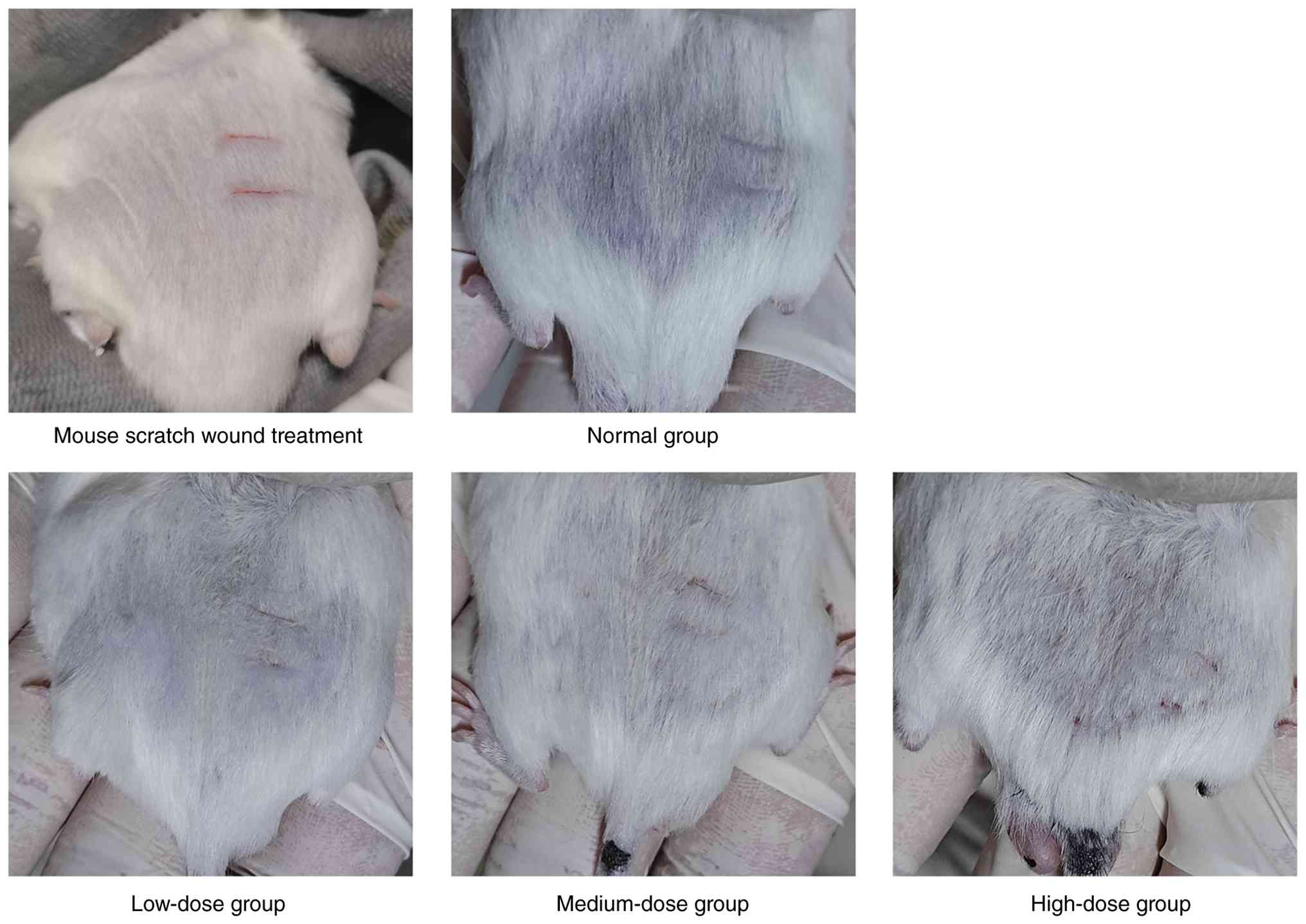
Analysis of security detection
As depicted in Fig.
8, mild erythema emerged in the high-dose group of the
experimental group after seven days of use. Calculations revealed
average scores per animal of 0.04 in the high-dose control group
and 0.26 in the skin-lesion group, both below the 0.5 threshold.
Based on these local irritation findings, the test was deemed
non-irritating according to the scoring criteria. The specific
scoring results are outlined in Table XVII.
| Table XVIIResults of the irritation test. |
Table XVII
Results of the irritation test.
| Group/Time | Day 1 | Day 2 | Day 3 | Day 4 | Day 5 | Day 6 | Day 7 | Day 8 | Day 9 | Day 10 | Day 11 | Day 12 | Day 13 | Day 14 | Average points | Overall rating |
|---|
| Normal control skin
group | 0 | 0 | 0 | 0 | 0 | 0 | 0 | 0 | 0 | 0 | 0 | 0 | 0 | 0 | 0 | Non-irritating |
| Normal control
damaged group | 0 | 0 | 0 | 0 | 0 | 0 | 0 | 0 | 0 | 0 | 0 | 0 | 0 | 0 | 0 | Non-irritating |
| Low-dose treatment
normal group | 0 | 0 | 0 | 0 | 0 | 0 | 0 | 0 | 0 | 0 | 0 | 0 | 0 | 0 | 0 | Non-irritating |
| Low-dose treatment
damaged group | 0 | 0 | 0 | 0 | 0 | 0 | 0 | 0 | 0 | 0 | 0 | 0 | 0 | 0 | 0 | Non-irritating |
| Medium-dose
treatment normal group | 0 | 0 | 0 | 0 | 0 | 0 | 0 | 0 | 0 | 0 | 0 | 0 | 0 | 0 | 0 | Non-irritating |
| Medium-dose
treatment damaged group | 0 | 0 | 0 | 0 | 0 | 0 | 0 | 0 | 0 | 0 | 0 | 0 | 0 | 0 | 0 | Non-irritating |
| High-dose treatment
normal group | 0 | 0 | 0 | 0 | 0 | 0 | 0 | 0 | 0 | 0 | 0 | 0 | 0 | 0 | 0.04 | Non-irritating |
| High-dose treatment
damaged group | 0 | 0 | 0 | 0 | 0 | 0 | 0 | 0 | 0 | 0 | 0 | 0 | 0 | 0 | 0.26 | Non-irritating |
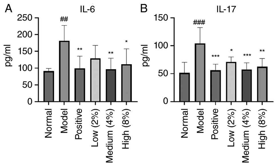
Determination of IL-6 and IL-17 by
ELISA
As shown in Fig. 9,
the model group exhibited markedly elevated levels of the
pro-inflammatory cytokines IL-6 and IL-17 compared with the normal
group. By contrast, the positive control group and the medium- and
high-dose groups of CMVO ointment showed marked reductions in these
proinflammatory factors compared with the model group.
Skin dyeing
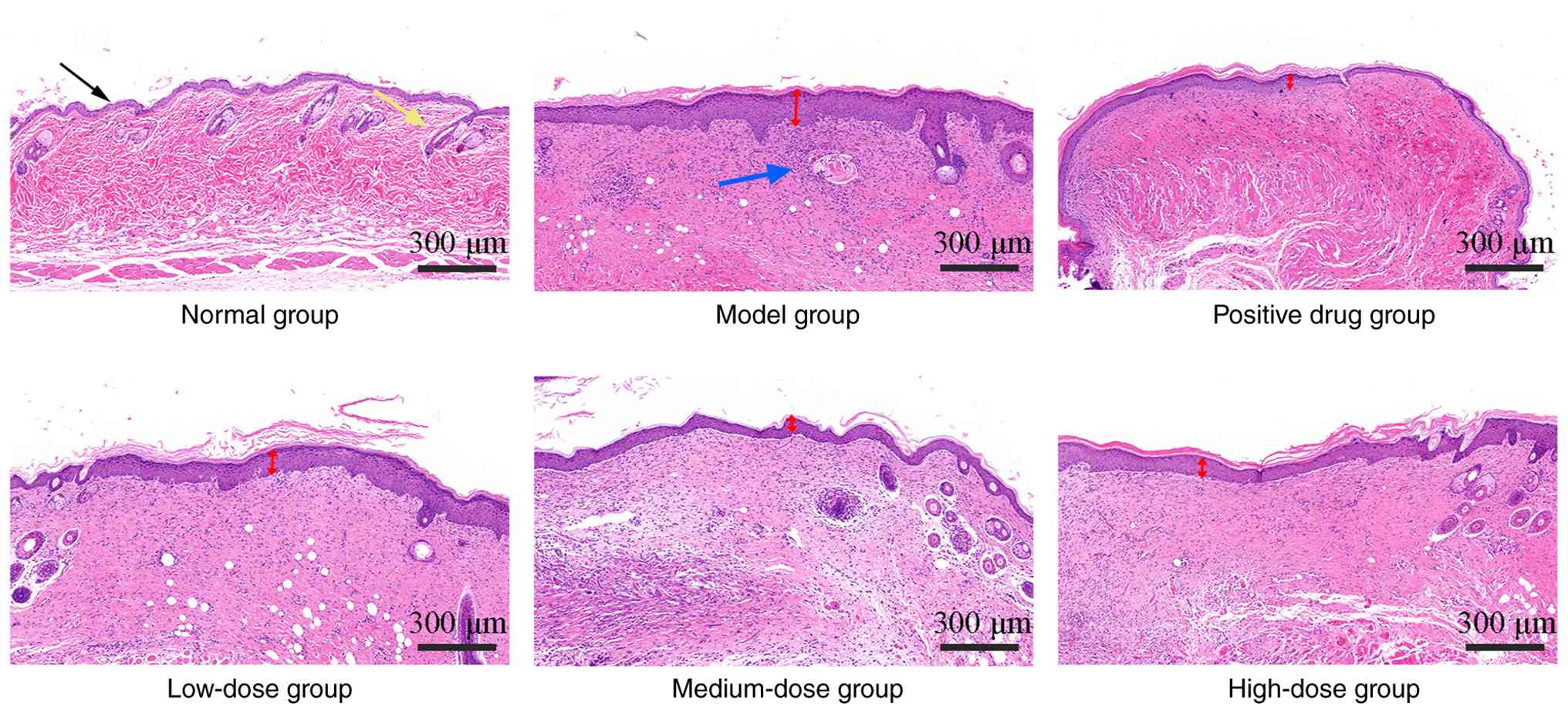
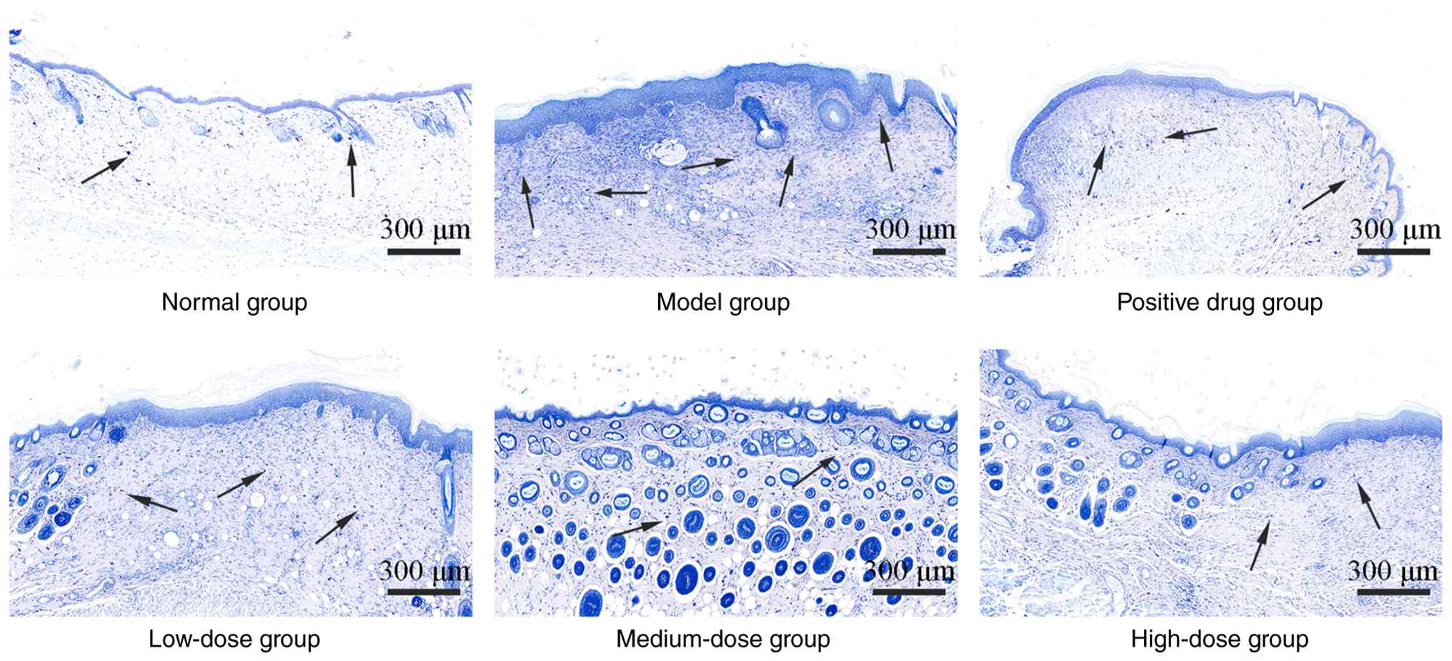
Figs. 10 and
11 depict the results of H&E
staining and toluidine blue staining of mouse skin tissue,
reflecting relevant pathological conditions. H&E staining
results revealed that the skin structure in the normal group was
intact, with no inflammatory cell infiltration in the dermis. By
contrast, the model group showed thickening of the granular and
spinous layers, intercellular edema and inflammatory cell
infiltration in the dermis. Treatment with different drugs led to
thinner granular and spinous layers and a significant reduction in
intercellular edema and inflammatory cell infiltration in the
dermis.
Toluidine blue staining highlighted clear
differences in mast cell distribution among the groups. The control
group had very few, sparsely distributed mast cells, while the
model group exhibited numerous densely clustered mast cells. After
drug administration, the positive group demonstrated a significant
reduction in mast cell numbers with a more scattered distribution.
Among the different dosage groups, mast cell counts decreased
progressively with increasing drug concentration.
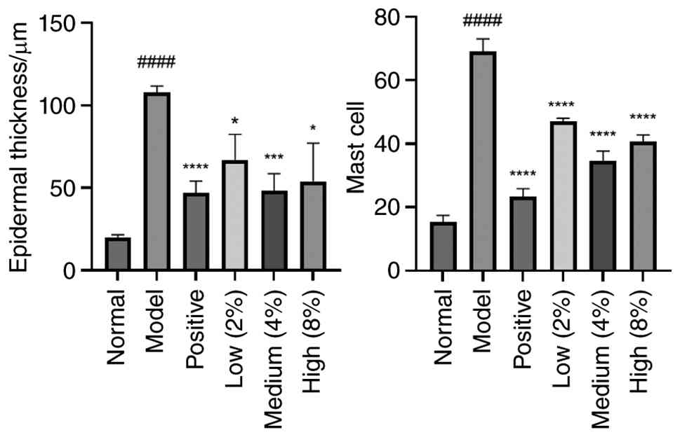
Quantitative analysis of epidermal thickness and
mast cell counts is presented in Fig.
12. Compared with the control group, the model group exhibited
increased epidermal thickness (P<0.0001) and mast cell counts
(P<0.0001). Compared with the model group, the treatment groups
exhibited reduced epidermal thickness and mast cell counts
(P<0.0001, P<0.001, P<0.05), reflecting the inflammatory
status in each group.
Immunohistochemistry
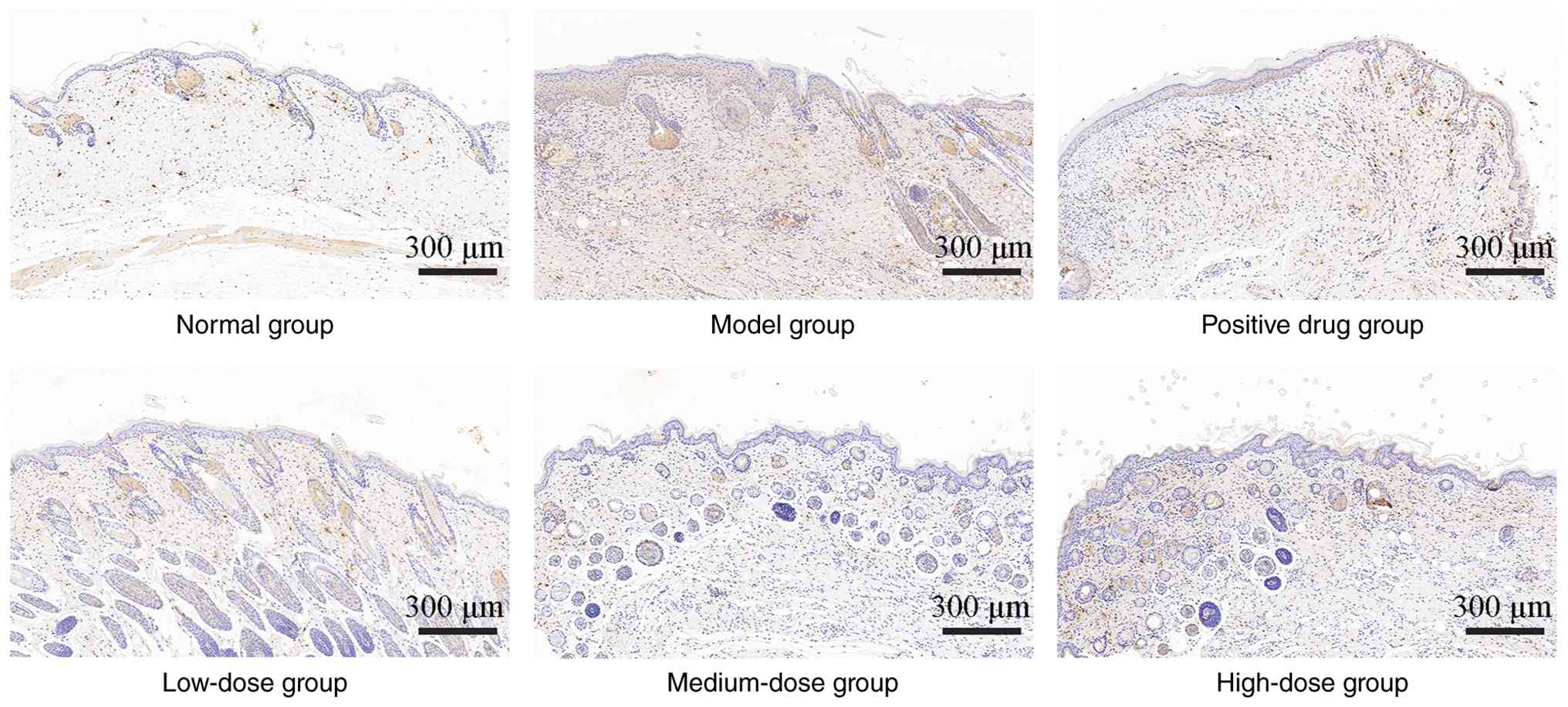
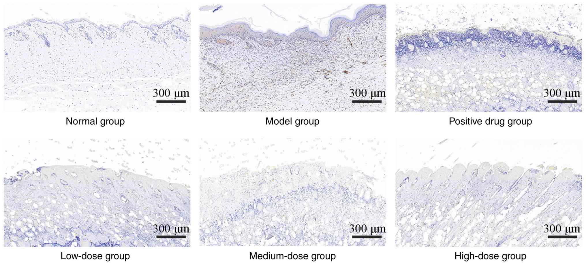
Immunohistochemical staining revealed JAK2
expression as a brownish-yellow color in the cytoplasm, while STAT3
was detected in both the cytoplasm and nucleus, also displaying a
brownish-yellow hue. In the normal group, JAK2 expression was weak,
confined to the basal layer of the epidermis and hair follicles,
while STAT3 remained inactive with minimal detectable staining.
Compared with the normal group, the model group exhibited thickened
stratum spinosum, with strong cytoplasmic JAK2 expression appearing
as brownish-yellow. Infiltrating inflammatory cells in the
superficial dermis exhibited brownish-gray staining. STAT3 showed
both cytoplasmic brownish-yellow positivity and brownish-gray
granular nuclear staining in inflammatory cells of the superficial
dermis. Overall, JAK2 and STAT3 expression levels were markedly
elevated in the model group. By contrast, all treatment groups
demonstrated markedly reduced positive expression levels, narrower
cytoplasmic distribution, lighter staining intensity and
disappearance of STAT3 nuclear positive signals, indicating
effective suppression of pathway activation following
treatment.
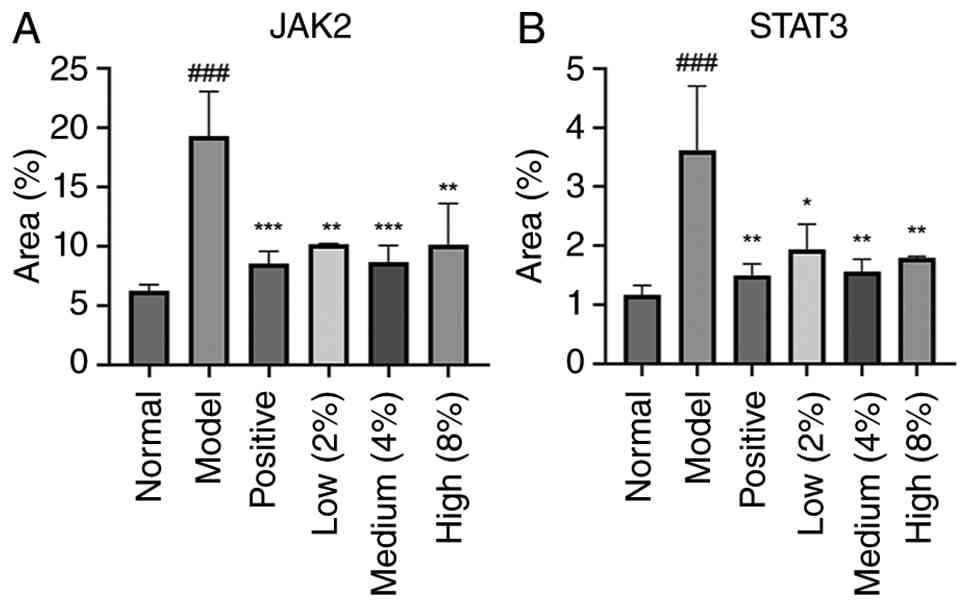
Analysis of results is shown in Fig. 13. Compared with the normal control
group, the model group exhibited markedly upregulated JAK2 and
STAT3 signaling (P<0.001), indicating altered molecular
expression of this pathway. Combined with H&E and toluidine
blue staining results, these findings suggested that the eczema
model successfully induced inflammation. Compared with the model
group, the positive drug group and different doses of CMVOC
administration groups showed significant differences in the
molecular expression of JAK2 and STAT3 (P<0.05, P<0.01,
P<0.001). These results suggested that the JAK2-STAT3 signaling
pathway may be one of the mechanisms by which CMVOC alleviates
eczema.
As illustrated in Fig.
14, JAK2 expression exhibited a brownish-yellow cytoplasmic
staining pattern. In the normal group, JAK2 expression was minimal
and distributed between the epidermis and dermis. Compared with the
normal group, the model group showed such as upregulated
JAK2-positive expression, with brownish-yellow cytoplasmic staining
in epidermal spinous layer cells and abundant inflammatory cell
infiltration in the dermis. Compared with the model group, the
positive group exhibited downregulated JAK2 expression, with fewer
brownish-yellow cytoplasmic cells and inflammatory cells in the
dermis. Similarly, all low-, medium- and high-dose groups treated
with CMVO ointment showed downregulated JAK2 signal expression,
with significant differences compared with the model group,
particularly in the medium- and high-dose groups.
As shown in Fig.
15, STAT3 expression appeared as a brownish-yellow cytoplasmic
staining. In the normal group, STAT3 signaling was minimal,
presenting as a pale yellow hue throughout the cytoplasm. Compared
with the normal group, the model group exhibited positive STAT3
expression, with deepened cytoplasmic staining in epidermal spinous
layer cells and dark brown nuclei in the dermis and epidermis.
Compared with the model group, the positive control group and the
low-, medium- and high-dose groups of CMVO ointment exhibited
lighter staining in the epidermal spinous layer, disappearance of
dark brown nuclei and weaker positive STAT3 signaling
expression.
Discussion
Eczema, a prevalent inflammatory skin disorder, is
characterized by multiple lesions, polymorphic manifestations and
intense itching (2). Environmental
stimuli trigger local and systemic immune responses via the skin's
neuro-immune-endocrine system, leading to various pathological
changes on the skin surface (6).
This condition often causes significant physical discomfort and
psychological distress. While traditional topical corticosteroids
are effective and widely used, their hormonal nature is associated
with notable adverse effects (12,13).
CMVO was selected for the present study for its anti-inflammatory
and antipruritic properties, aimed at alleviating eczema symptoms
(25). The first phase of this
research focused on analyzing the chemical composition of
Cnidium monnieri essential oil using GC-MS technology,
identifying 21 volatile components, including isopropyl
isobutyrate, (-)-β-pinene, caryophyllene, D-limonene, borneol and
isobornyl acetate. Among these constituents, (-)-β-pinene,
β-caryophyllene and cnidicin are known for their anti-inflammatory
and antibacterial activities and are likely key contributors to
CMVO's anti-inflammatory efficacy (18,19,21-23).
The second phase focused on screening and optimizing the
formulation of CMVO ointment by evaluating its appearance, physical
stability, moisturizing properties, particle size and other
characteristics. The selected cream base consists of cetyl alcohol,
petrolatum, liquid paraffin, IPM, sodium lauryl sulfate, glycerin,
ethyl nipagin and distilled water. Among these, cetyl alcohol acts
as a thickener to enhance cream stability; petrolatum and liquid
paraffin act as lubricants to regulate cream greasiness and improve
spreadability; IPM functions as a penetration enhancer to improve
skin affinity; sodium lauryl sulfate acts as a surfactant to ensure
emulsification between the oil and water phases; glycerin serves as
a humectant, maintaining skin hydration while preventing moisture
loss during storage; and propylene glycol ethyl ether functions as
a preservative to ensure shelf stability. By combining these base
characteristics, the fine texture exhibits excellent stability,
strong moisturizing properties and easy application. To further
optimize the formulation ratios, a single-factor experiment
identified cetyl alcohol, IPM and sodium lauryl sulfate as the most
influential factors. A response surface experiment was then
conducted to determine the optimal formulation. The composition
comprises: octadecanol (4.45 g), petrolatum (4 g), liquid paraffin
(2.20 g), IPM (0.81 g), CMVO (1.60 g), sodium lauryl sulfate (0.39
g), glycerin (1.16 g), ethyl nicotinate (0.04 g) and distilled
water (25.35 g).
The volatile oil extracted from C. monnieri
contains components such as β-caryophyllene and cnidicin, which
alleviate eczema-related itching and inflammation by inhibiting
inflammatory mediators (19,23).
Therefore, The present study further investigated the
pharmacodynamic effects of CMVO ointment using mouse models of
inflammatory responses and multiple analytical methods. The
DNCB-induced eczema model in mice revealed severe lesions and
erythema in the model group. Microscopic analysis showed epidermal
acanthosis, dermal intercellular edema and inflammatory cell
infiltration. By contrast, the positive control group treated with
compound dexamethasone acetate and the low-, medium- and high-dose
groups administered CMVOC showed reduced dorsal skin damage, with
slight improvements in skin condition correlating with increasing
CMVOC dosage. Microscopic examination further revealed a thinner
stratum spinosum and fewer inflammatory cells. ELISA assays
detected upregulated pro-inflammatory cytokines IL-6 and IL-17 in
the model group and downregulated levels in the treatment groups,
confirming CMVOC's effectiveness in suppressing eczema-related
inflammatory responses.
Recent research on inflammatory skin diseases such
as eczema has expanded such as due to their complex and diverse
underlying mechanisms. Natural products have been widely studied,
yielding notable progress. For instance, baicalin from
Scutellaria baicalensis root modulates anti-inflammatory and
immune responses by reducing the phosphorylation levels of JAK1,
STAT1, STAT2, STAT3, STAT5 and STAT6. Essential oils from citrus
plants inhibit inflammatory mediators such as IL-1β, IL-6, IL-8 and
TNF-α by reducing the phosphorylation levels of STAT1 and STAT3 in
the JAK-STAT pathway, as well as P-38, ERK and IKB-α in the MAPK
pathway (47). Extracts from
Ginkgo biloba leaves suppress JAK2 and STAT3
signaling-related neurotransmitters and inflammatory mediators to
control itching and inflammation (48). Tripterygin such as downregulates
inflammatory factors such as IL-4, TNF-α, IgE, IL-17 and IL-6,
upregulates SOCS1 expression, inhibits JAK-STAT3 pathway
activation, blocks T lymphocyte activation and improves
inflammatory responses (49). Due
to the well-established role of the JAK2-STAT3 signaling pathway in
inflammatory skin diseases (50,51),
this pathway was chosen for the mechanism study. In addition, key
inflammatory mediators evaluated in the present study are closely
associated with STAT3 signaling, supporting the relevance of this
pathway.
Based on the findings from the aforementioned
experiments and given that IL-17 induces increased IL-6 production,
which in turn activates the JAK-STAT pathway (42) and preliminary results from H&E
staining, toluidine blue staining and immunohistochemistry, CMVOC
may inhibit the expression levels of JAK2 and STAT3 signaling,
thereby suppressing the inflammatory response in eczema.
In the present study, a formulation of CMVO ointment
was developed and optimized and the formulation underwent
systematic response surface method optimization, demonstrating that
the CMVOC can such as alleviate the inflammatory response
associated with eczema. The volatile oil also appears to exhibit
analgesic and antipruritic effects, though these were not
thoroughly investigated in this experiment and warrant
comprehensive future research. However, in the pharmacodynamic
experiments, several serum samples in the control group were
affected by hemolysis due to technical factors during blood
collection. As a result, all serum-based analyses were conducted
using 36 mice (n=6 per group). The reduced sample size may limit
the statistical power of the present study and should be taken into
consideration when interpreting the results. Nevertheless,
consistent trends were observed across multiple pathological and
biochemical indicators and statistically significant differences
were detected between groups, which support the observed effects.
Future studies with larger sample sizes are warranted to further
validate and strengthen the conclusions of the present study.
Furthermore, the stability of the main anti-inflammatory active
substances of β-caryophyllene and cnidicin inferred from the GC-MS
results can be explored in greater depth using methods such as HPLC
and TLC in future research. In addition, a more comprehensive
quantitative analysis of major active components would further
improve the quality control of the formulation. At the same time,
long-term stability and microbial stability studies are currently
underway and will be reported in future work. To date, research on
CMVOC has focused on pharmacodynamic evaluation, with no
pharmacokinetic studies conducted yet.
However, the present study has several limitations.
First, the phosphorylation levels of JAK2 and STAT3, which are
critical indicators of pathway activation, were not evaluated. The
current analysis was limited to total protein expression assessed
by immunohistochemistry, which does not directly reflect the
activation state of the JAK2/STAT3 signaling pathway. Second,
pathway-specific intervention experiments (such as the use of JAK2
agonists/inhibitors or gene overexpression approaches) were not
performed. Therefore, a direct causal relationship between the
inhibition of JAK2-STAT3 signaling and the anti-inflammatory
effects of CMVOC cannot be definitively established. Last, other
inflammation-related signaling pathways, such as MAPK and NF-κB,
were not investigated and their potential contributions cannot be
excluded. Future investigations will focus on conducting pathway
intervention experiments through pharmacological regulation,
expanding the investigation to additional signaling pathways,
elucidating the underlying mechanisms of action, strengthen the
theoretical basis for the cream's efficacy in alleviating eczema
inflammation and validating the signaling mechanism through mRNA
expression analysis, western blotting analysis of phosphorylated
(p-)JAK2 and p-STAT3 proteins or exploration of other signaling
pathways or inflammatory factors. Clarifying relevant dose-response
and time-response relationships and optimizing the administration
regimen are also essential. Clinical trials are needed to confirm
the safety and efficacy of treating human eczema. While the
anti-inflammatory effects of CMVOC in eczema have been
preliminarily demonstrated, the ointment formulation technology
remains amenable to further refinement. Additionally, combination
studies with other medicinal herbs, such as Sophora
flavescens and Kochia scoparia, could enhance eczema
therapeutic efficacy and support the development of improved
treatments.
In conclusion, the present study demonstrated that
CMVOC is a safe and effective treatment for eczema, with consistent
quality and notable efficacy in alleviating eczema inflammatory
symptoms. These findings support further research and development,
positioning CMVOC as a promising novel therapeutic option. However,
clinical studies are required to validate its efficacy and optimize
its therapeutic use in humans.
Acknowledgements
Not applicable.
Funding
Funding: The present study was supported by Shaanxi Provincial
Department of Education Youth Innovation Team Scientific Research
Plan Project (grant no. 25JP047); Shaanxi Provincial Key Research
and Development Program Project (grant no. 2024CY-JJQ-36); Shaanxi
Provincial Traditional Chinese Medicine Science and Technology
Innovation Team (grant no. TZKN-CXTD-03); Shaanxi Provincial
Department of Science and Technology Project (grant nos.
2024ZC-YYDP-110 and 2025JC-YBMS-1056); Shaanxi Province Xianyang
City Science and Technology Project (grant no.
L2024-QCY-ZYYJJQ-X28); Shaanxi Provincial Administration of
Traditional Chinese Medicine (grant no. ZYJXG-Y23005); Key
Technological Innovation Team for Industrialization of Aromatic
Traditional Chinese Medicine; Shaanxi Provincial Engineering
Research Center for Traditional Chinese Medicine Aromatic Industry;
Key Discipline of High Level Traditional Chinese Medicine in
Shaanxi Province, Traditional Chinese Medicine Processing; Shaanxi
Provincial Department of Education Project (grant no. 24JP045);
Innovation and Entrepreneurship Training Program for College
Students (grant no. S202410716081). Youth Innovation Team of
Shaanxi Universities for Traditional Chinese Medicine Health-Care
Technologies in Elderly Chronic Disease Management; Qin Chuangyuan
Project for Innovation and Agglomeration of Traditional Chinese
Medicine Industry (grant no. L2024-QCY-ZYYJJQ-X69).
Availability of data and materials
The data generated in the present study are included
in the figures and/or tables of this article.
Authors' contributions
Experimental designer was TS. Experimental
procedures were performed by TS, LD, BZ, XD and XG. Result analysis
was by TS, JieW and JinW. TS drafted the initial manuscript. YW, XZ
and TS confirm the authenticity of all the raw data. HN, JS, YS,
DG, JZ and XS conducted data analysis and confirmed the results.
Manuscript revision and editing was by XZ, XS, YW, HN, JS, YS, DG
and JZ. YW proposed the research direction of the present study,
supervised the implementation of experiments, revised the
manuscript and addressed the comments raised by the reviewers. All
authors read and approved the final manuscript.
Ethics approval and consent to
participate
The animal studies were reviewed and approved by the
Laboratory Animal Welfare Ethics Committee, which was approved by
Shaanxi University of Chinese Medicine (approval no.
SUCMDL20250512001). ARRIVE guidelines were followed.
Patient consent for publication
Not applicable.
Competing interests
The authors declare that they have no competing
interests.
References
|
1
|
Chovatiya R: Atopic dermatitis (eczema).
JAMA. 329(268)2023.PubMed/NCBI View Article : Google Scholar
|
|
2
|
Tokura Y, Yunoki M, Kondo S and Otsuka M:
What is ‘eczema’? J Dermatol. 52:192–203. 2025.PubMed/NCBI View Article : Google Scholar
|
|
3
|
Schmitt J, Apfelbacher CJ and Flohr C:
Eczema. BMJ Clin Evid. 2011(1716)2011.PubMed/NCBI
|
|
4
|
Liu Y, Sun S, Zhang D, Li W, Duan Z and Lu
S: Effects of residential environment and lifestyle on atopic
eczema among preschool children in Shenzhen, China. Front Public
Health. 10(844832)2022.PubMed/NCBI View Article : Google Scholar
|
|
5
|
Long Q, Jin H, You X, Liu Y, Teng Z, Chen
Y, Zhu Y and Zeng Y: Eczema is a shared risk factor for anxiety and
depression: A meta-analysis and systematic review. PLoS One.
17(e0263334)2022.PubMed/NCBI View Article : Google Scholar
|
|
6
|
Slominski RM, Raman C, Jetten AM and
Slominski AT: Neuro-immuno-endocrinology of the skin: How
environment regulates body homeostasis. Nat Rev Endocrinol.
21:495–509. 2025.PubMed/NCBI View Article : Google Scholar
|
|
7
|
de Lusignan S, Alexander H, Broderick C,
Dennis J, McGovern A, Feeney C and Flohr C: The epidemiology of
eczema in children and adults in England: A population-based study
using primary care data. Clin Exp Allergy. 51:471–482.
2021.PubMed/NCBI View Article : Google Scholar
|
|
8
|
Zhang J, Zhu S, Yuan L, Yu X, Ling S,
Zhang J and Yang B: Assessing atopic dermatitis control in Chinese
patients: Validation of the chinese version of recap of atopic
eczema questionnaire (RECAP) and an investigation into its
interpretability. Acta Derm Venereol. 105(adv43458)2025.PubMed/NCBI View Article : Google Scholar
|
|
9
|
Ferreira MA, Vonk JM, Baurecht H,
Marenholz I, Tian C, Hoffman JD, Helmer Q, Tillander A, Ullemar V,
van Dongen J, et al: Shared genetic origin of asthma, hay fever and
eczema elucidates allergic disease biology. Nat Genet.
49:1752–1757. 2017.PubMed/NCBI View Article : Google Scholar
|
|
10
|
Marenholz I, Arnau-Soler A,
Rosillo-Salazar OD and Lee YA: New insights from genetic studies of
eczema. Med Genet. 35:33–45. 2023.PubMed/NCBI View Article : Google Scholar
|
|
11
|
Lax SJ, Harvey J, Axon E, Howells L,
Santer M, Ridd MJ, Lawton S, Langan S, Roberts A, Ahmed A, et al:
Strategies for using topical corticosteroids in children and adults
with eczema. Cochrane Database Syst Rev. 3(CD013356)2022.PubMed/NCBI View Article : Google Scholar
|
|
12
|
Ference JD and Last AR: Choosing topical
corticosteroids. Am Fam Physician. 79:135–140. 2009.PubMed/NCBI
|
|
13
|
Stacey SK and McEleney M: Topical
corticosteroids: Choice and application. Am Fam Physician.
103:337–343. 2021.PubMed/NCBI
|
|
14
|
Sun Y, Yang AWH and Lenon GB: .
Phytochemistry, Ethnopharmacology, Pharmacokinetics and Toxicology
of Cnidium monnieri (L.) Cusson. Int J Mol Sci.
21(1006)2020.PubMed/NCBI View Article : Google Scholar
|
|
15
|
Yuan S, Liu M, Duan X and Zheng Y:
Research progress on the processing history, chemical constituents
and pharmacological effects of cnidii fructus. Chin Wild Plant
Resour. 42:74–81. 2023.
|
|
16
|
Arizmendi N, Alam SB, Azyat K, Makeiff D,
Befus AD and Kulka M: The complexity of sesquiterpene chemistry
dictates its pleiotropic biologic effects on inflammation.
Molecules. 27(2450)2022.PubMed/NCBI View Article : Google Scholar
|
|
17
|
Paço A, Brás T, Santos JO, Sampaio P,
Gomes AC and Duarte MF: Anti-inflammatory and immunoregulatory
action of sesquiterpene lactones. Molecules.
27(1142)2022.PubMed/NCBI View Article : Google Scholar
|
|
18
|
Ahn SS, Yeo H, Jung E, Ou S, Lee YH, Lim Y
and Shin SY: β-Caryophyllene ameliorates
2,4-dinitrochlorobenzene-induced atopic dermatitis through the
downregulation of mitogen-activated protein kinase/EGR1/TSLP
signaling axis. Int J Mol Sci. 23(14861)2022.PubMed/NCBI View Article : Google Scholar
|
|
19
|
Scandiffio R, Geddo F, Cottone E, Querio
G, Antoniotti S, Gallo MP, Maffei ME and Bovolin P: Protective
effects of (E)-β-caryophyllene (BCP) in chronic inflammation.
Nutrients. 12(3273)2020.PubMed/NCBI View Article : Google Scholar
|
|
20
|
Kotan R, Kordali S and Cakir A: Screening
of antibacterial activities of twenty-one oxygenated monoterpenes.
Z Naturforsch C J Biosci. 62:507–513. 2007.PubMed/NCBI View Article : Google Scholar
|
|
21
|
Salehi B, Upadhyay S, Erdogan Orhan I,
Kumar Jugran A, L D Jayaweera S, A Dias D, Sharopov F, Taheri Y,
Martins N, Baghalpour N, et al: Therapeutic potential of α- and
β-pinene: A miracle gift of nature. Biomolecules.
9(738)2019.PubMed/NCBI View Article : Google Scholar
|
|
22
|
Sun M, Sun M and Zhang J: Osthole: An
overview of its sources, biological activities, and modification
development. Med Chem Res. 30:1767–1794. 2021.PubMed/NCBI View Article : Google Scholar
|
|
23
|
He S, Liang X, Chen W, Nima Y, Li Y, Gu Z,
Lai S, Zhong F, Qiu C, Mo Y, et al: Osthole ameliorates chronic
pruritus in 2,4-dichloronitrobenzene-induced atopic dermatitis by
inhibiting IL-31 production. Chin Herb Med. 17:368–379.
2024.PubMed/NCBI View Article : Google Scholar
|
|
24
|
Feng X: Research progress on the
applicability of essential oils in promoting permeation. Chin
Tradit Pat Med. 35:157–161, 164. 2013.(In Chinese). https://qikan.cqvip.com/Qikan/Article/Detail?id=44468738.
|
|
25
|
Wang XD, Bai JL, Ma ZJ, Fan J, Kong WB,
Wang JL, Zhang J and Liang JY: . Analysis of chemical composition,
antibacterial activity, antioxidant properties, and cytotoxicity of
essential oils from four plant fruits. Microb Pathog.
209(108133)2025.PubMed/NCBI View Article : Google Scholar
|
|
26
|
Sattayakhom A, Wichit S and Koomhin P: The
effects of essential oils on the nervous system: A scoping review.
Molecules. 28(3771)2023.PubMed/NCBI View Article : Google Scholar
|
|
27
|
Sharmeen JB, Mahomoodally FM, Zengin G and
Maggi F: Essential oils as natural sources of fragrance compounds
for cosmetics and cosmeceuticals. Molecules. 26(666)2021.PubMed/NCBI View Article : Google Scholar
|
|
28
|
Zhang X, Huang J, Xie Z, Chen F, Chen H,
Liao Y, Xu J and Yue P: Research progress on stabilization
techniques of volatile oils from traditional Chinese medicine and
their preparations application. Chin Herb Drugs. 56:5258–5266.
2025.
|
|
29
|
Sawant A, Kamath S, Kg H and Kulyadi GP:
Solid-in-oil-in-water emulsion: An innovative paradigm to improve
drug stability and biological activity. AAPS PharmSciTech.
22(199)2021.PubMed/NCBI View Article : Google Scholar
|
|
30
|
Li C: Study on preparation technology and
quality standard of Longzi Xiaozhong Cream (unpublished thesis).
Yunnan University of Chinese Medicine, 2023.
|
|
31
|
Ji X, Li X, Yang R, Zhao H, Zhai X, Ji L
and Yang L: Study on preparation of Aijiaohuang Cream and
improvement of psoriasis mice skin lesions. Shizhen Guoyi Guoyao.
35:625–628. 2024.(In Chinese). https://doi.org/10.3969/j.issn.1008-0805.2024.03.26.
|
|
32
|
He L, Li H, Li W, Li C and Zhang J:
Preparation technology of anti-inflammatory acne cream. J Shanxi
Med. 51:2171–2175. 2022.(In Chinese). https://qikan.cqvip.com/Qikan/Article/Detail?id=7108662780.
|
|
33
|
Yang J, Wang J, Wang L, Dong Y and Li X:
Study on insect-proof cream based on plant essential oil. Guangzhou
Chem Ind. 51:37–39. 2023.(In Chinese). https://qikan.cqvip.com/Qikan/Article/Detail?id=7110941216.
|
|
34
|
Liu H, Cai Y, Chu Y, Yu X, Song F, Wang H,
Zhang H and Sun X: Formulation of chrysomycin A cream for the
treatment of skin infections. Molecules. 27(4613)2022.PubMed/NCBI View Article : Google Scholar
|
|
35
|
Salehi N, Mortazavi SM and Moghimi H:
Investigating the changes in cream properties following topical
application and their influence on the product efficiency. Iran J
Pharm Res. 21(e123946)2022.PubMed/NCBI View Article : Google Scholar
|
|
36
|
Liao J, Lei Y, Chen J, Wei D, Zhou B,
Liang J, Su R and Chen G: Optimization of forming process of
Zhuangfang Xiaoji Yeti Cream by Box-Behnken response surface
methodology. J China Prescr Drug. 23:16–20. 2025.(In Chinese).
https://qikan.cqvip.com/Qikan/Article/Detail?id=7201293506.
|
|
37
|
Duan J, Li J, Wang Y, Zhoua P, Wanga X,
Xiaa N, Wanga J, Lia J, Wang W, Wang X, et al: Therapeutic effects
and mechanism of action of lavender essential oil on atopic
dermatitis by modulating the STAT3/RORγt pathway. Arab J Chem.
17(105525)2023.
|
|
38
|
Matsuki F, Suzuki S, Hirose T, Suzuki T,
Yoshida T, Onishi Y and Yamamoto R: Prescription and application
adequacy of topical corticosteroids based on the finger-tip unit
method in adult patients with atopic dermatitis: A cross-sectional
study. J Gen Fam Med. 26:547–554. 2025.PubMed/NCBI View Article : Google Scholar
|
|
39
|
Bakó E, Fehérvári P, Garami A, Dembrovszky
F, Gunther EE, Hegyi P, Csupor D and Böszörményi A: Efficacy of
topical essential oils in musculoskeletal disorders: Systematic
review and meta-analysis of randomized controlled trials.
Pharmaceuticals (Basel). 16(144)2023.PubMed/NCBI View Article : Google Scholar
|
|
40
|
Yang W: Study on the efficiency and safety
of Co-glycyrrhetinic acid cream (unpublished thesis). Hebei Medical
University, 2015.
|
|
41
|
Hanifin JM, Baghoomian W, Grinich E,
Leshem YA, Jacobson M and Simpson EL: The eczema area and severity
index-A practical guide. Dermatitis. 33:187–192. 2022.PubMed/NCBI View Article : Google Scholar
|
|
42
|
Cai Y, Zhao S, Yan W, Lin X, Gou W, Ye M,
Wu Y and Zhang W: Preliminary research on the effect of volatile
oil from Eugenia caryophyllata Thunb. on the skin of mice.
Chinese Ethnic and Folk Medicine. 32:23–27. 2023.(In Chinese).
https://qikan.cqvip.com/Qikan/Article/Detail?id=7110472857.
|
|
43
|
Fischer AH, Jacobson KA, Rose J and Zeller
R: Hematoxylin and eosin staining of tissue and cell sections. In:
Spector DL, Goldman RD (eds.). Basic Methods in Microscopy. Cold
Spring Harbor Laboratory Press, Cold Spring Harbor, NY, 2006, Ch.
4.
|
|
44
|
Raja N, Naikodi S, Govindarajan A and
Palanisamy K: Toluidine blue staining of murine mast cells and
quantitation by a novel, automated image analysis method using
whole slide skin images. J Histotechnol. 44:190–195.
2021.PubMed/NCBI View Article : Google Scholar
|
|
45
|
Hong S, Lee B, Kim JH, Kim EY, Kim M, Kwon
B, Cho HR, Sohn Y and Jung HS: Solanum nigrum Linne improves
DNCB-induced atopic dermatitis-like skin disease in BALB/c mice.
Mol Med Rep. 22:2878–2886. 2020.PubMed/NCBI View Article : Google Scholar
|
|
46
|
Oumarou Hama H, Aboudharam G, Barbieri R,
Lepidi H and Drancourt M: Immunohistochemical diagnosis of human
infectious diseases: A review. Diagn Pathol. 17(17)2022.PubMed/NCBI View Article : Google Scholar
|
|
47
|
Fan X, Liu Z, Yang W, Zhang H, Zhong H,
Pang Y, Ye X, Wu C and Li L: Advances in atopic dermatitis
treatment: From pathogenesis to natural product-based therapies.
Phytother Res. 39:4444–4473. 2025.PubMed/NCBI View Article : Google Scholar
|
|
48
|
Chummun Phul I, Gómez-Llonín A and
Bhaw-Luximon A: From traditional medicine to nanomedicine:
Potential of Ginkgo biloba extracts in treating inflammatory
skin diseases. RSC Med Chem. 15:2643–2656. 2024.PubMed/NCBI View Article : Google Scholar
|
|
49
|
Duan Y, Zhu L and Dong Y: Protective
effect and mechanism of tripterine on atopic dermatitis in mice
based on SOCS1/JAK-STAT3pathway. J Guangxi Med Univ. 39:290–297.
2022.
|
|
50
|
Zeng H, Zhao B, Zhang D, Rui X, Hou X,
Chen X, Zhang B, Yuan Y, Deng H and Ge G: Viola yedoensis Makino
formula alleviates DNCB-induced atopic dermatitis by activating
JAK2/STAT3 signaling pathway and promoting M2 macrophages
polarization. Phytomedicine. 103(154228)2022.PubMed/NCBI View Article : Google Scholar
|
|
51
|
Wen C, Yang X, Han Y, Fu Y, Chen W, Zhang
H, Zhang X, Xiao Z, Cheng G and Zhang J: Integrating network
pharmacology and experimental validation to elucidate the mechanism
of Xiao-bi decoction in psoriasis treatment: Inhibition of
JAK2/STAT3 signaling and rebalancing Th17/Treg responses. J
Ethnopharmacol. 363(121477)2026.PubMed/NCBI View Article : Google Scholar
|