Introduction
Globally, liver cancer ranks as the fifth most
commonly diagnosed cancer and the third leading cause of
cancer-related mortality. In 2021, there were ~906,000 new cases
and 830,000 cases of mortality worldwide (1). In China, liver cancer had the second
highest mortality rate among all malignant tumors, with an
estimated 316500 deaths in 2022, accounting for ~22.42% of all
cancer-associated mortality (2-4).
Although treatments, such as surgical resection, interventional
therapy [including transarterial chemoembolization (TACE), hepatic
artery infusion chemotherapy and ablation] and systemic therapy,
prolong patient survival, the 5-year survival rate remains between
25 and 39% (5). In addition, the
recurrence rate is up to 80% (6).
Medical ozone, which consists of a reactive mixture
of oxygen and ozone, has been documented to exhibit diverse
therapeutic properties and is widely used for the treatment of
conditions such as mucositis, psoriasis, acute pain, neurovascular
disease and cancer (7-9).
The potential of ozone in oncology was highlighted in 1980: Ozone
selectively inhibits the proliferation of lung, breast and uterine
tumor cells without affecting normal cells (10). Accumulating evidence has further
substantiated its role in cancer therapy (11-13).
Zänker and Kroczek (12)
demonstrated that ozone enhanced the efficacy of 5-fluorouracil by
reversing chemoresistance in breast cancer cells. In addition,
Cannizzaro et al (13)
showed that ozone induces apoptosis in the neuroblastoma cell line
SK-N-DZ via caspase-3 and poly-ADP ribose polymerase pathways,
whilst impeding cell cycle progression in SK-N-SH cells by
modulating cyclin B1/cyclin-dependent kinase 1 activity. In the
context of liver cancer, Li et al (14) demonstrated that medical ozone can
inhibit proliferation, migration and epithelial-mesenchymal
transition in hepatocellular carcinoma (HCC) cells via reactive
oxygen species (ROS) accumulation and suppression of the
PI3K/AKT/NF-κB pathway (14).
Similarly, Tang et al (15)
found that ozonated water suppresses HCC cell proliferation,
invasion and metastasis by regulating the high mobility group box 1
protein (HMGB1)/NF-κB/STAT3 pathway. Additionally, ozone induces
apoptosis in BEL7402 cells through the mitochondrial pathway
(16). Despite its diverse
advantages, such as anti-inflammatory effects, pain relief and
improving immune function, ozone gas has limited clinical
applications due to respiratory toxicity and instability. By
contrast, ozonated water is safer and easier to handle. Ma et
al (17) reported that
intratumoral injection of ozonated saline promotes necrosis and
suppresses tumor growth, potentially through the IL-6 and TNF-α
pathways. Kuroda et al (18)
demonstrated that local ozone injection dose-dependently induced
tumor apoptosis or necrosis without damaging normal tissue.
Consistent findings were also reported by Kızıltan et al
(19), Peirone et al
(20), and Yıldırım et al
(21), supporting the translational
potential of ozone therapy.
Chronic inflammation serves a key role in tumor
progression. Within the tumor microenvironment, key inflammatory
mediators, such as NF-κB and STAT3, can activate genes involved in
cell proliferation and angiogenesis, thereby promoting tumor growth
and metastasis (22). Notably,
NF-κB perpetuates inflammatory signaling by sustaining the
expression of proinflammatory cytokines, including IL-6 and
IL-8(23). This process not only
amplifies local inflammation, but also fosters genetic instability
and aberrant cell proliferation, establishing a microenvironment
that supports tumorigenesis. As a pleiotropic cytokine, IL-6 is
highly expressed in numerous types of malignancy, including HCC
(24) and colorectal (25) and breast cancer (26). It functions by binding to its
specific receptor, IL-6 receptor α, leading to the activation of
glycoprotein 130 and subsequent initiation of the JAK/STAT3
signaling pathway (27). Upon
activation, phosphorylated (p-)STAT3 translocates into the nucleus
and upregulates the expression of target genes, such as Bcl-2, VEGF
and MMP2 (28,29). These genes collectively contribute
to tumor progression by inhibiting apoptosis whilst promoting
proliferation, metastasis and angiogenesis.
Ozone reacts with biological components to induce
controlled oxidative stress, leading to the formation of reactive
species (30). This process
activates the antioxidant defense system. Moderate oxidative stress
stimulates the transcription of the antioxidant response element
(ARE), promoting the production of several antioxidant enzymes,
including glutathione S-transferase, catalase, heme oxygenase-1 and
NADPH quinone oxidoreductase 1(31). These enzymes protect cells from
inflammatory damage (32).
Concurrently, moderate levels of oxidative stress suppress the
NF-κB signaling pathway, attenuating inflammatory responses and
decreasing the levels of proinflammatory factors (such as IL-6) in
the microenvironment (33).
Based on the aforementioned findings, it was
hypothesized that ozone induces apoptosis in liver cancer cells by
decreasing IL-6 levels within the tumor microenvironment, thereby
inhibiting the IL-6/STAT3 signaling pathway and suppressing
anti-apoptotic factor release. To evaluate this hypothesis, HepG2
and Huh-7 cells were cultured in medical ozone-enriched medium to
assess its effects on cell proliferation, migration and invasion.
Apoptosis rates were measured using flow cytometry, whilst western
blot analysis was performed to determine whether ozone-mediated
apoptosis occurs through suppression of the IL-6/STAT3 pathway.
Materials and methods
Cell culture
Human HCC cell lines HepG2 and Huh-7 and the normal
hepatic cell line THLE-2 were obtained from Procell Life Science
& Technology Co., Ltd. Cell identity was authenticated via
short tandem repeat (STR) profiling (performed by the Cell Resource
Center of JENNlO Biological Technology and Zhejiang Baidi
Biotechnology Co., Ltd.). STR loci matched the American Type
Culture Collection reference profile with >98% similarity,
confirming no cross-contamination. Mycoplasma contamination was
tested monthly using the MycoAlert™ Mycoplasma Detection
kit (Lonza Walkersville, Inc) and all cultures were negative. HepG2
cells were cultured in MEM supplemented with 10% FBS (both Gibco;
Thermo Fisher Scientific, Inc.) and 1% penicillin-streptomycin.
Huh-7 cells were maintained in DMEM (Gibco; Thermo Fisher
Scientific, Inc.) containing 10% FBS and 1%
penicillin-streptomycin. THLE-2 cells were cultured in THLE-2 Cell
Complete Medium (Wuhan Pusainuo Life Science Co., Ltd.). All three
cell lines were cultured at 37˚C with 5% CO2. Cells were
divided into experimental and control groups. The experimental
group was treated with ozonated medium, while the control group
received ozone-free complete medium. The THLE-2 cell line was used
to assess the safety of ozonated water on normal hepatocytes.
Preparation of ozonated medium
Complete medium was aliquoted into a sterile 250 ml
saline bottle. After removing air from the bottle, it was stored at
4˚C. The ozonated medium was freshly prepared 1 h before
application using a Medozon compact ozone generator (Humares GmbH).
The ozone generator was calibrated monthly using a standard ozone
solution. Specifically, an oxygen (95%)/ozone (5%) gas mixture was
infused into the pre-chilled medium at a flow rate of 3 l/min for
20 min at room temperature. The final ozonated medium was stored at
4˚C. Ozone concentration was determined using an ozone colorimeter
(Huankai Microbial).
Determination of half-maximal
inhibitory concentration (IC50)
Cells in the logarithmic growth phase were seeded
into 96-well plates at 5x10³ cells/well. After 24 h incubation,
cells were treated at 37˚C with ozonated medium at concentrations
of 0, 5, 10, 20, 40 and 60 µg/ml, each with three replicates.
Following 0, 24 and 48 h treatment, cell viability was assessed
using Cell Counting Kit-8 (CCK-8; Nanjing KeyGen Biotech Co.,
Ltd.). Absorbance was measured at 450 nm using a microplate reader
(Thermo Fisher Scientific, Inc.) to calculate the IC50.
Cell proliferation assay
Cell proliferation was evaluated using the CCK-8
assay. HepG2, Huh-7 and THLE-2 cells were seeded into 96-well
plates at 5x10³ cells/well and treated at 37˚C with ozonated or
control medium for 0, 24, 48 and 72 h. Supernatant was removed and
replaced with 100 µl medium containing 10% CCK-8 reagent. Following
1.5 h incubation in the dark, absorbance was measured at 450 nm.
Each experimental condition included three replicate wells and a
blank control. The experiment was repeated three times.
Wound healing assay
HepG2 and Huh-7 cells were seeded at 37˚C into
6-well plates at 4x10⁵ cells/well (three
replicates/group). After 24 h, when cells reached 90% confluency,
three straight scratches were made in each well using a 20-µl
pipette tip. The cells were washed three times with PBS and
incubated in serum-free MEM and DMEM. Images were captured at 0 and
72 h using an inverted light microscope (Nikon Corporation), before
scratch areas were measured by Image J software (version 1.53k,
National Institutes of Health). The assay was performed in
triplicate.
Transwell invasion assay
Transwell chambers were pre-coated with 50 µl
Matrigel (1 µg/µl) at 37˚C for 2 h. HepG2 and Huh-7 cells were
serum-starved for 8 h, detached with EDTA-trypsin, washed and
resuspended in serum-free MEM and DMEM. In total, 200 µl cell
suspension (1x10⁵ cells) was added to the upper chamber
and 600 µl complete MEM or DMEM supplemented with 10% FBS), Gibco)
was added to the lower chamber. Following 24 h incubation at 37 ˚C
with 5% CO2, non-invading cells were removed with a
cotton swab. Invaded cells were fixed with 20% methanol at room
temperature for 15 min, stained with 0.1% crystal violet at room
temperature for 20 min and imaged under an inverted light
microscope. Each group had three replicates and the experiment was
repeated three times.
Apoptosis analysis by flow
cytometry
HepG2 cells were seeded into 6-well plates at
5x10⁵ cells/well (three replicates/group) and treated at
37˚C with ozonated or control medium for 24 h. Supernatant was
collected, cells were washed with PBS, detached using EDTA-free
trypsin and centrifuged at 300 x g for 5 min at 4˚C. The cell
pellet was resuspended in 500 µl loading buffer and stained with
Annexin V-FITC and PI (Nanjing KeyGen Biotech Co., Ltd.; cat. no.
KGA1012) for 15 min at 37˚C in the dark. Samples were analyzed
using a FACSCalibur flow cytometer and the data was analyzed by
Flow Jo software (version 10.8.1; both BD Biosciences).
Single-stained Annexin V-FITC and PI controls confirmed no signal
overlap, and gating excluded debris to ensure accurate apoptotic
cell counting. Apoptotic cells were defined as the sum of cells in
both the early and late (Annexin V-FITC-positive, PI-positive)
apoptotic quadrants. The experiment was repeated three times.
Western blot analysis
HepG2 cells were lysed with RIPA buffer (Shanghai
Epizyme Biomedical Technology Co., Ltd.) containing PMSF and
phosphatase inhibitors (100:1:1). Following centrifugation at 1,800
x g for 15 min at 4˚C, protein concentration was determined using a
BCA kit (Beyotime Institute of Biotechnology) with a Nanodrop 2000
spectrophotometer (Thermo Fisher Scientific, Inc.). Proteins (20
µg/lane) were separated by 10% SDS-PAGE and transferred to a PVDF
membrane. The membrane was blocked with 5% non-fat milk for 1 h at
room temperature and incubated overnight at 4˚C with the following
primary antibodies: Caspase-3 (1:2,000; cat. no. ET1602-39;
Hangzhou HuaAn Biotechnology Co., Ltd.), Bcl-2 (1:2,000; cat. no.
ab182858; ABcam), Bax (1:20,000; cat. no. ET1603-34; Hangzhou HuaAn
Biotechnology Co., Ltd.), cleaved caspase-3 (1:2,000; cat. no.
ET1602-47; Hangzhou HuaAn Biotechnology Co., Ltd.), JAK2 (1:5,000,
cat. no. ab108596; Abcam), p-JAK2 (1:2,000; cat. no. ab32101;
Abcam), STAT3 (1:2,000, cat. no. ab68153; Abcam), p-STAT3 (1:2,000,
cat. no. 05-485; Hangzhou HuaAn Biotechnology Co., Ltd.), IL-6
(1:1,000; cat. no. FNab04282) and GAPDH (1:2,000, cat. no.
FNab03342; both Wuhan Fine Biotech, Co., Ltd.). After washing four
times with PBST buffer, the membrane was incubated with
HRP-conjugated goat anti-rabbit secondary antibody (1:10,000, cat.
no. FNSA-0004; Wuhan Fine Biotech, Co., Ltd.) for 1 h at 37˚C.
Signals were detected using an enhanced chemiluminescence reagent
(MilliporeSigma; cat. no. WBKLS0100) and quantified with a
chemiluminescence imaging system (ChemiDoc Go; Bio-Rad
Laboratories, Inc.). Images were analyzed with Image Lab Touch
Software 2.4 (Bio-Rad Laboratories, Inc.). All signals were
normalized to GAPDH (loading control) to ensure equal protein
loading. A total of three independent experiments was
performed.
Statistical analysis
Data from three independent experiments are
presented as the mean ± SD. Statistical analyses were conducted
using SPSS 20.0 (IBM Corp.). Comparisons between two groups were
performed using an unpaired independent samples t-test. The
normality of data distribution was assessed using the Shapiro-Wilk
test with SPSS software. Non-normally distributed data were
analyzed by Mann-Whitney U test. P<0.05 was considered to
indicate a statistically significant difference. Graphs were
generated using GraphPad Prism 8.0 (Dotmatics).
Results
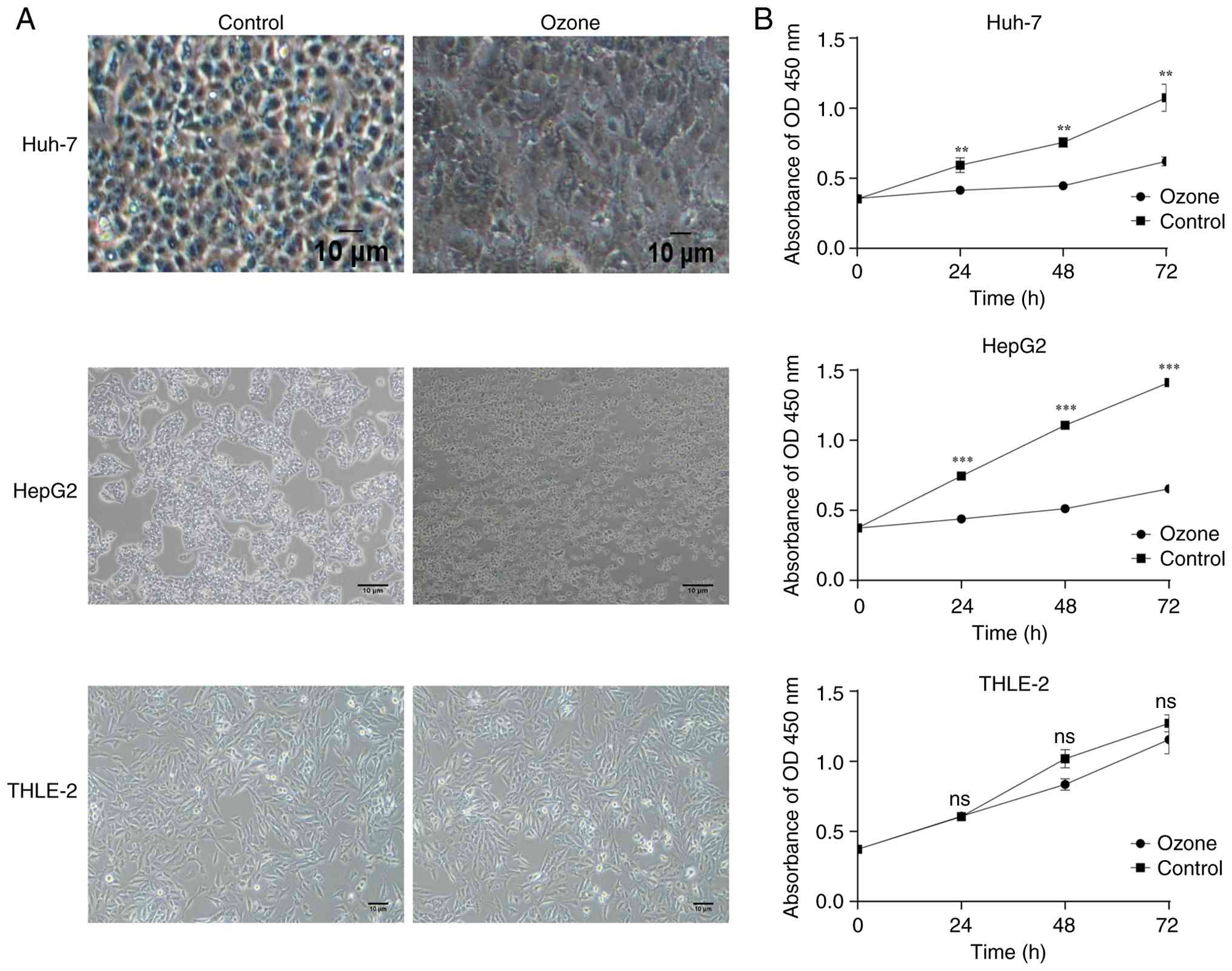
Ozone selectively inhibits
proliferation of liver cancer cells
Cell proliferation was evaluated using the CCK-8
assay. HepG2, Huh-7 and THLE-2 cells were treated with ozone at 0,
5, 10, 20, 40 and 60 µg/ml for 24, 48 and 72 h. Absorbance was
measured at each time point, where the IC50 was determined. Based
on comparative. Treatment with 20 µg/ml ozone for 0, 24, 48 and 72
h significantly suppressed the proliferation of HepG2 and Huh-7
cells in a time-dependent manner, while no significant effect was
observed on THLE-2 normal liver cells (Fig. 1). Therefore, 20 µg/ml was selected
for subsequent experiments.
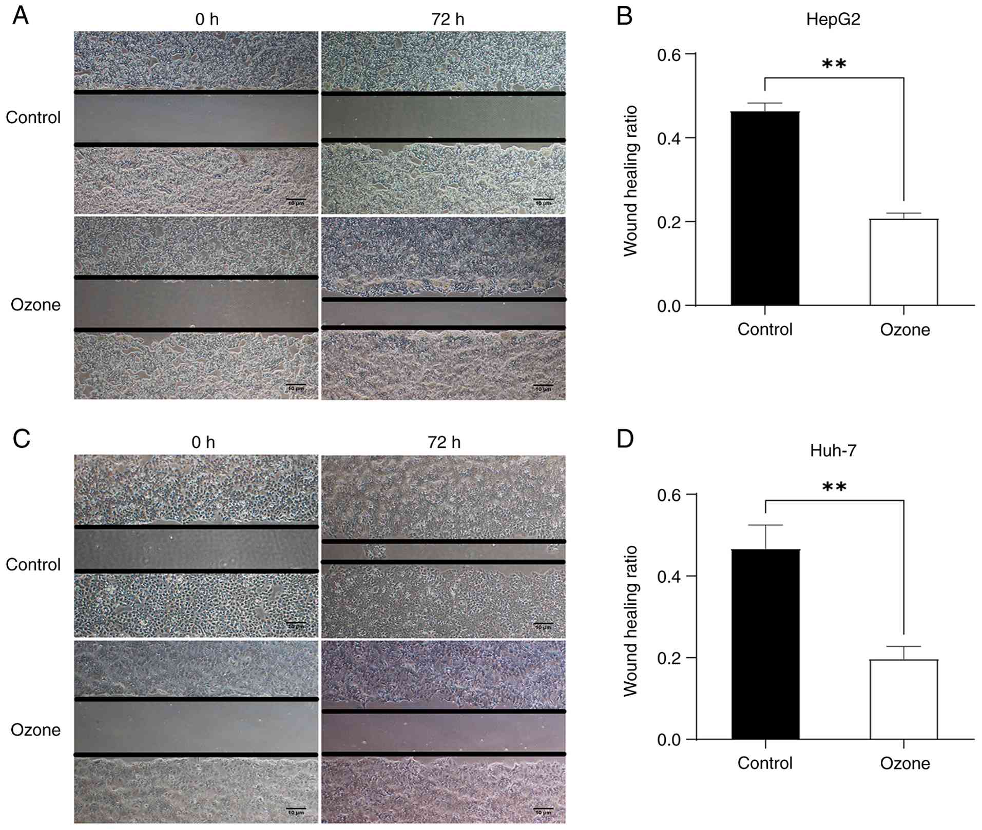
Ozone suppresses migration of liver
cancer cells
The effect of ozone on cell migration was assessed
using a wound healing assay. Ozone-treated HepG2 and Huh-7 cells
exhibited significantly decreased migration, with larger remaining
scratch areas compared with those in the control group (Fig. 2).
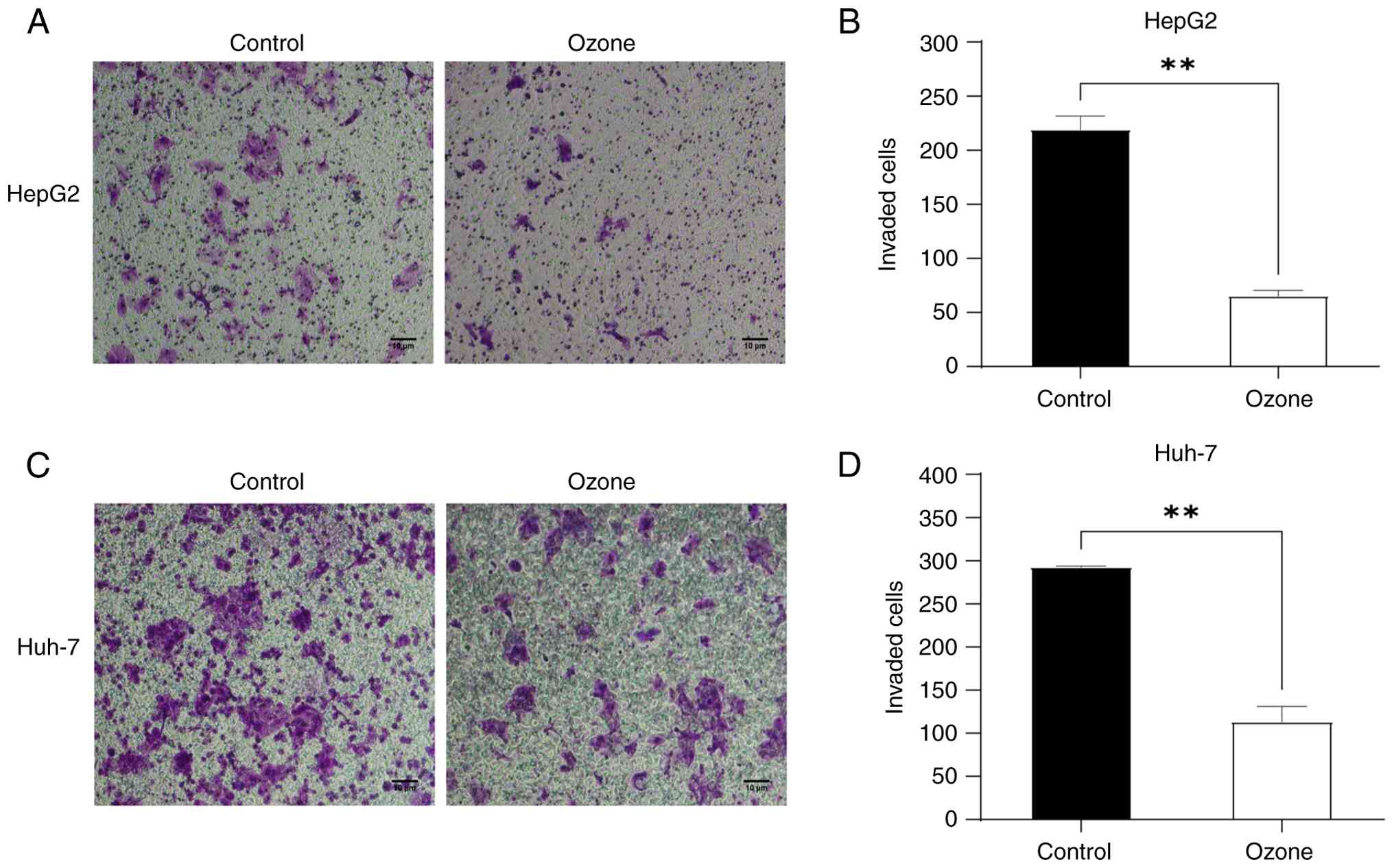
Ozone inhibits invasion of liver
cancer cells
A Transwell invasion assay was conducted to evaluate
the effect of ozone on the invasive capability of HepG2 and Huh-7
cells. Following 24 h of ozone treatment, the number of invading
cells was significantly lower compared with the control group,
indicating that ozone effectively inhibited the invasiveness of
liver cancer cells (Fig. 3).
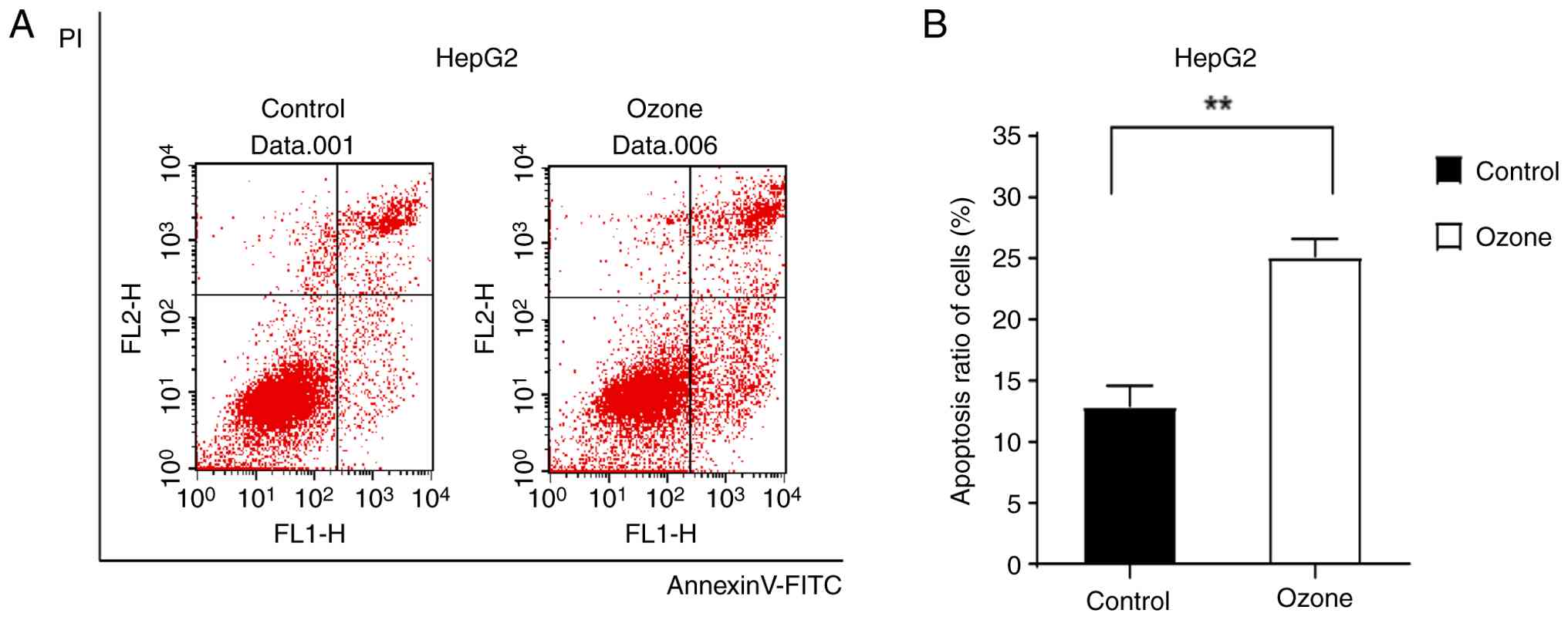
Ozone induces apoptosis in HepG2
cells
Flow cytometry was used to analyze apoptosis in
HepG2 cells following ozone treatment, which demonstrated a notable
increase in apoptotic cells in the treated group (Fig. 4A). The apoptosis rate was
significantly higher in the ozone group compared with that in the
control, demonstrating an induction of apoptosis (Fig. 4B).
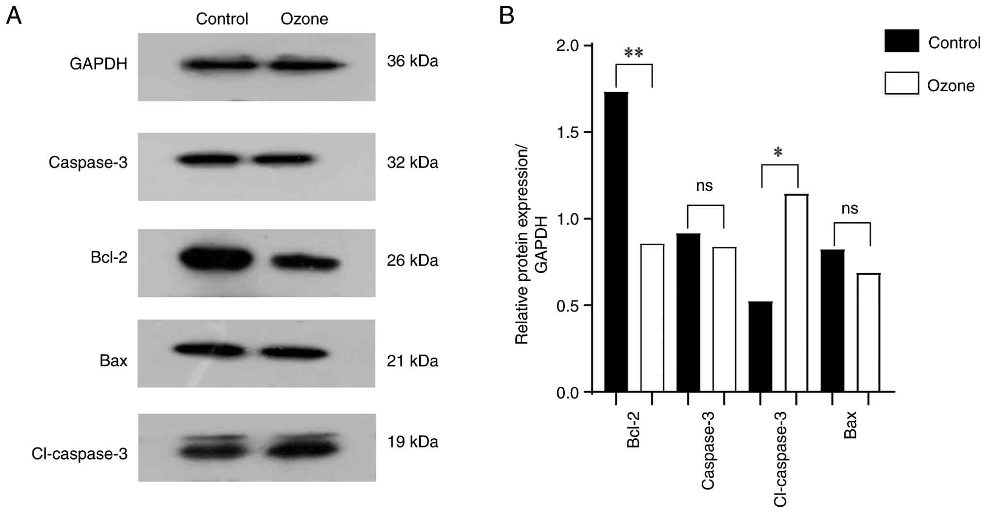
Western blot analysis revealed that ozone treatment
significantly upregulated cleaved caspase-3 and downregulated
Bcl-2, with a non-significant decrease in Bax expression (Fig. 5). Bcl-2/Bax ratio was decreased in
the ozone-treated compared to the control group. These results
indicated that ozone promoted apoptosis in HepG2 cells primarily
through the intrinsic mitochondrial pathway (Fig. 5).
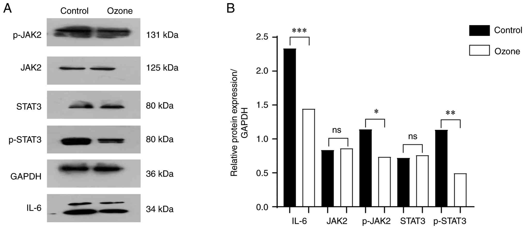
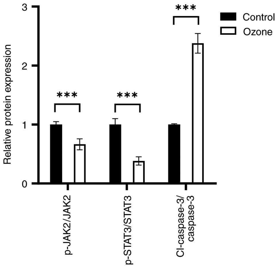
Ozone suppresses the IL-6/STAT3
signaling pathway
To investigate the molecular mechanism underlying
ozone-induced apoptosis, the IL-6/STAT3 pathway was examined using
western blotting. Ozone significantly reduced IL-6 expression and
decreased the phosphorylation levels of JAK2 and STAT3, while total
JAK2 and STAT3 expression levels remained unchanged. These findings
suggested that ozone inhibited IL-6/STAT3 signaling, contributing
to apoptosis induction in HepG2 cells (Figs. 6 and 7).
Discussion
The present study demonstrated that ozone inhibited
the proliferation of HepG2 and Huh-7 cells, while exhibiting no
significant cytotoxic effects on the normal hepatocyte line THLE-2.
This selective anti-tumor activity aligns with previous reports
(10-14)
that also indicate that ozone can effectively eliminate cancer
cells while sparing normal cells. Simonetti et al (34) reported time- and
concentration-dependent cytotoxic effects of ozone on HT-29 colon
cancer cells. A study by Schulz et al (35) found that intraperitoneal
insufflation of an ozone/oxygen mixture at an advanced disease
stage significantly improved survival, induced complete tumor
regression, and appeared to activate an immune-mediated antitumor
response.
In the present study, medical ozone water directly
inhibited the IL-6/STAT3 pathway, resulting in dose-dependent
decreases in p-STAT3 and Bcl-2, and subsequent apoptosis. Western
blot analysis confirmed downregulation of Bcl-2, upregulation of
cleaved caspase-3 and a reduced Bcl-2/Bax ratio, indicating
mitochondrial apoptotic pathway activation, consistent the
pro-apoptotic effects of ozone (36). Additionally, ozone water
significantly suppressed migration and invasion in HepG2 and Huh-7
cells, which may decrease metastasis. While Li et al
(14) and Tang et al
(15) described ozone-induced
suppression via ROS/PI3K/AKT/NF-κB and HMGB1/NF-κB/STAT3 pathways,
respectively, the present study provides novel insights into
IL-6/STAT3 pathway involvement. By contrast with Costanzo et
al (37), which found no effect
in HeLa cells, the present results emphasize cell type-dependent
responses. The present study demonstrated that ozone water had a
minimal impact on normal liver cell (THLE-2) morphology and
proliferation, highlighting its translational relevance.
Chronic inflammation serves a role in tumorigenesis
by fostering a microenvironment conducive to proliferation,
angiogenesis and immune evasion (38). Inflammatory mediators, such as ROS
and nitric oxide synthase, induce DNA damage and dysregulate
oncogenic signaling (39). On one
hand, these substances cause double-strand DNA breaks and induce
gene mutations; on the other hand, they promote the activation of
proto-oncogenes and inactivation of tumor suppressor genes
(40). NF-κB is a central
inflammatory transcription factor that can promote sustained IL-6
expression, which inhibits apoptosis and supports tumor survival
(41). Ozone exerts
anti-inflammatory effects through moderate oxidative stress. This
mechanism is initiated when ozone-derived ROS and lipid oxidation
products, such as 4-hydroxynonenal, act as signaling molecules
(30). These molecules modify
critical cysteine residues on the Keap1 protein, disrupting its
association with Nrf2. This leads to Nrf2 stabilization and
translocation into the nucleus. Upon binding to the ARE, Nrf2
drives the transcription of cytoprotective enzymes, including heme
oxygenase-1, NAD(P)H quinone dehydrogenase 1 and glutamate-cysteine
ligase (42,43). The upregulation of this antioxidant
repertoire effectively restores cellular redox homeostasis.
Consequently, this Nrf2-mediated adaptive response suppresses the
NF-κB signaling pathway, a master regulator of IL-6 transcription,
thereby inhibiting its activation. The net outcome is a notable
downregulation of IL-6 gene expression and protein synthesis
(44,45).
IL-6 is a key cytokine linking inflammation and
cancer. It activates the JAK/STAT3 pathway, leading to the
transcription of genes involved in proliferation (cyclin D1),
invasion (MMP2/3), apoptosis resistance (Bcl-2) and angiogenesis
(VEGF) (46,47). Aberrant activation of IL-6/STAT3
signaling is frequently observed in tumors and associated with poor
prognosis (48,49). The present results showed that ozone
downregulated IL-6 expression and decreased the phosphorylation of
JAK2 and STAT3, thereby inhibiting this oncogenic pathway (50,51).
This is consistent with studies across various types of cancer,
such as colorectal cancer, myeloma and gallbladder cancer; where
STAT3 inhibition suppresses tumor growth and induces apoptosis
(48,51).
Ozone is administered in various forms clinically,
including as gas (52,53), ozonated water, oil and by
autohemotherapy (54). Its
potential use in liver cancer management may include sensitizing
tumor cells to chemo- or radiotherapy, decreasing drug resistance
and enhancing the efficacy of TACE by increasing drug concentration
and decreasing tumor blood supply. In addition, ozone exhibits
immunomodulatory properties. Rossmann et al (55) observed that ozone treatment
increased tumor-infiltrating lymphocytes and confers transferable
anti-tumor immunity through peripheral blood mononuclear cells in a
VX2 rabbit model, suggesting that ozone may activate systemic
anti-tumor immune responses.
In summary, the present study demonstrated that
ozone inhibited migration, invasion and proliferation, while
promoting apoptosis in liver cancer cells in vitro. Ozone
suppresses the IL-6/STAT3 signaling pathway, likely through its
anti-inflammatory effects. However, limitations should be
acknowledged. The present study was conducted only on two liver
cancer cell lines (HepG2 and Huh-7), therefore generalizability to
other liver cancer subtypes remains unclear. In addition, the
impact of ozone on the cell cycle was not investigated. The role of
IL-6/STAT3 signaling was also not validated through genetic
approaches, such as silencing or overexpression. All experiments
were performed in vitro, precluding the determination of the
efficacy of ozone in in vivo settings. Another limitation of
the present study is the absence of transcriptome analysis based on
next-generation sequencing (NGS). The present study focused on the
role of the IL6/STAT3 signaling pathway in ozone water-induced
apoptosis of liver cancer cells, but apoptosis regulation typically
involves complex interactions among multiple signaling networks
(such as MAPK, PI3K/Akt or NF-κB pathways). The lack of NGS data
restricts understanding of the mechanism of action, preventing
systematic identification of differentially expressed genes,
verification of whether the effects are specifically mediated by
IL6/STAT3 or involve broader signaling networks, and discovery of
potential synergistic targets. Therefore, future studies should
integrate transcriptomics to expand mechanistic insights and animal
models are necessary to evaluate the therapeutic potential of ozone
in vivo. Further research is warranted to explore the
effects of ozone on a broader panel of liver cancer models, its
influence on cell cycle progression and its efficacy in combination
with existing therapy.
Acknowledgements
Not applicable.
Funding
Funding: The present study was supported by the school-level
youth research project of Chuanbei Medical College (grant no.
CBY23-QNA17).
Availability of data and materials
The data generated in the present study may be
requested from the corresponding author.
Authors' contributions
LZ conceived and designed the study and analyzed and
interpreted data. SQ, CC, ZZ, NY, DD and JH performed experiments
and analyzed data. SQ and CC confirm the authenticity of all the
raw data. All authors have read and approved the final
manuscript.
Ethics approval and consent to
participate
Not applicable.
Patient consent for publication
Not applicable.
Competing interests
The authors declare that they have no competing
interests.
References
|
1
|
Sung H, Ferlay J, Siegel RL, Laversanne M,
Soerjomataram I, Jemal A and Bray F: Global cancer statistics 2020:
GLOBOCAN estimates of incidence and mortality worldwide for 36
cancers in 185 countries. CA Cancer J Clin. 71:209–249.
2021.PubMed/NCBI View Article : Google Scholar
|
|
2
|
Yang Y, Sun J, Cai J, Chen M, Dai C, Wen
T, Xia J, Ying M, Zhang Z and Zhang X: , et al; Chinese
Association of Liver Cancer and Chinese Medical Doctor Association:
Chinese expert consensus on the whole-course management of
hepatocellular carcinoma (2023 edition). Liver Cancer. 14:311–333.
2024.PubMed/NCBI View Article : Google Scholar
|
|
3
|
Chen W, Zheng R, Baade PD, Zhang S, Zeng
H, Bray F, Jemal A, Yu XQ and He J: Cancer statistics in China,
2015. CA Cancer J Clin. 66:115–132. 2016.PubMed/NCBI View Article : Google Scholar
|
|
4
|
Han B, Zheng R, Zeng H, Wang S, Sun K,
Chen R, Li L, Wei W and He J: Cancer incidence and mortality in
China, 2022. J Natl Cancer Cent. 4:47–53. 2024.PubMed/NCBI View Article : Google Scholar
|
|
5
|
Nishida N: Long-term prognosis and
management of hepatocellular carcinoma after curative treatment.
Clin Mol Hepatol. 26:480–483. 2020.PubMed/NCBI View Article : Google Scholar
|
|
6
|
Reig M, Forner A, Rimola J, Ferrer-Fàbrega
J, Burrel M, Garcia-Criado Á, Kelley RK, Galle PR, Mazzaferro V,
Salem R, et al: BCLC strategy for prognosis prediction and
treatment recommendation: The 2022 update. J Hepatol. 76:681–693.
2022.PubMed/NCBI View Article : Google Scholar
|
|
7
|
Smith NL, Wilson AL, Gandhi J, Vatsia S
and Khan SA: Ozone therapy: An overview of pharmacodynamics,
current research, and clinical utility. Med Gas Res. 7:212–219.
2017.PubMed/NCBI View Article : Google Scholar
|
|
8
|
Bocci V: The Clinical Application of Ozone
Therapy. Springer Netherlands, pp97-232, 2010.
|
|
9
|
Bocci V, Zanardi I and Travagli V:
Oxygen/ozone as a medical gas mixture. A critical evaluation of the
various methods clarifies positive and negative aspects. Med Gas
Res. 1(6)2011.PubMed/NCBI View Article : Google Scholar
|
|
10
|
Sweet F, Kao MS, Lee SC, Hagar WL and
Sweet WE: Ozone selectively inhibits growth of human cancer cells.
Science. 209:931–933. 1980.PubMed/NCBI View Article : Google Scholar
|
|
11
|
Clavo B, Pérez JL, López L, Suárez G,
Lloret M, Rodríguez V, Macías D, Santana M, Hernández MA,
Martín-Oliva R and Robaina F: Ozone therapy for tumor oxygenation:
A pilot study. Evid Based Complement Alternat Med. 1:93–98.
2004.PubMed/NCBI View Article : Google Scholar
|
|
12
|
Zänker KS and Kroczek R: In vitro
synergistic activity of 5-fluorouracil with low-dose ozone against
a chemoresistant tumor cell line and fresh human tumor cells.
Chemotherapy. 36:147–154. 1990.PubMed/NCBI View Article : Google Scholar
|
|
13
|
Cannizzaro A, Verga Falzacappa CV,
Martinelli M, Misiti S, Brunetti E and Bucci B: O(2/3) exposure
inhibits cell progression affecting cyclin B1/cdk1 activity in
SK-N-SH while induces apoptosis in SK-N-DZ neuroblastoma cells. J
Cell Physiol. 213:115–125. 2007.PubMed/NCBI View Article : Google Scholar
|
|
14
|
Li J, Zeng T, Tang S, Zhong M, Huang Q, Li
X and He X: Medical ozone induces proliferation and migration
inhibition through ROS accumulation and PI3K/AKT/NF-κB suppression
in human liver cancer cells in vitro. Clin Transl Oncol.
23:1847–1856. 2021.PubMed/NCBI View Article : Google Scholar
|
|
15
|
Tang S, Xu B, Pang H, Xiao L, Mei Q and He
X: Ozonated water inhibits hepatocellular carcinoma invasion and
metastasis by regulating the HMGB1/NF-κB/STAT3 signaling pathway. J
Hepatocell Carcinoma. 10:203–215. 2023.PubMed/NCBI View Article : Google Scholar
|
|
16
|
Tang S, Xu B, Li J, Zhong M, Hong Z, Zhao
W, Zeng T and He X: Ozone induces BEL7402 cell apoptosis by
increasing reactive oxygen species production and activating JNK.
Ann Transl Med. 9(1257)2021.PubMed/NCBI View Article : Google Scholar
|
|
17
|
Ma Q, Yang C, Jiang X, Liu J, Shi Y, Li H,
Liu H and Yang J: Effectiveness of ozonated saline in the treatment
of VX2 tumors in rabbits. J Interv Med. 1:143–149. 2019.PubMed/NCBI View Article : Google Scholar
|
|
18
|
Kuroda K, Azuma K, Mori T, Kawamoto K,
Murahata Y, Tsuka T, Osaki T, Ito N, Imagawa T, Itoh F and Okamoto
Y: The safety and anti-tumor effects of ozonated water in vivo. Int
J Mol Sci. 16:25108–25120. 2015.PubMed/NCBI View Article : Google Scholar
|
|
19
|
Kızıltan HŞ, Bayir AG, Yucesan G, Eris AH,
İdin K, Karatoprak C, Aydin T, Akcakaya A and Mayadagli A: Medical
ozone and radiotherapy in a peritoneal, Erlich-ascites, tumor-cell
model. Altern Ther Health Med. 21:24–29. 2015.PubMed/NCBI
|
|
20
|
Peirone C, Mestre VF, Medeiros-Fonseca B,
Colaço B, Pires MJ, Martins T, Gil da Costa RM, Neuparth MJ,
Medeiros R, Bastos MMSM, et al: Ozone therapy prevents the onset of
dysplasia in HPV16-transgenic mice-A pre-clinical efficacy and
safety analysis. Biomed Pharmacother. 104:275–279. 2018.PubMed/NCBI View Article : Google Scholar
|
|
21
|
Yıldırım M, Erkişi S, Yılmaz H, Ünsal N,
İnaç E, Tanrıver Y and Koçak P: The apoptotic effect of ozone
therapy on mitochondrial activity of highly metastatic breast
cancer cell line MDA-MB-231 using in vitro approaches. J Interv
Med. 5:64–71. 2022.PubMed/NCBI View Article : Google Scholar
|
|
22
|
Zhou Y, Xia L, Liu Q, Wang H, Lin J, Oyang
L, Chen X, Luo X, Tan S, Tian Y, et al: Induction of
pro-inflammatory response via activated macrophage-mediated NF-κB
and STAT3 pathways in gastric cancer cells. Cell Physiol Biochem.
47:1399–1410. 2018.PubMed/NCBI View Article : Google Scholar
|
|
23
|
Slattery ML, Mullany LE, Sakoda L,
Samowitz WS, Wolff RK, Stevens JR and Herrick JS: The NF-κB
signalling pathway in colorectal cancer: Associations between
dysregulated gene and miRNA expression. J Cancer Res Clin Oncol.
144:269–283. 2018.PubMed/NCBI View Article : Google Scholar
|
|
24
|
Zhuang PY, Zhang KW, Wang JD, Zhou XP, Liu
YB, Quan ZW and Shen J: Effect of TALEN-mediated IL-6 knockout on
cell proliferation, apoptosis, invasion and anti-cancer therapy in
hepatocellular carcinoma (HCC-LM3) cells. Oncotarget.
8:77915–77927. 2017.PubMed/NCBI View Article : Google Scholar
|
|
25
|
Świerczyński M, Szymaszkiewicz A, Fichna J
and Zielińska M: New insights into molecular pathways in colorectal
cancer: Adiponectin, interleukin-6 and opioid signaling. Biochim
Biophys Acta Rev Cancer. 1875(188460)2021.PubMed/NCBI View Article : Google Scholar
|
|
26
|
He JY, Wei XH, Li SJ, Liu Y, Hu HL, Li ZZ,
Kuang XH, Wang L, Shi X, Yuan ST and Sun L: Adipocyte-derived IL-6
and leptin promote breast cancer metastasis via upregulation of
Lysyl Hydroxylase-2 expression. Cell Commun Signal.
16(100)2018.PubMed/NCBI View Article : Google Scholar
|
|
27
|
Mihara M, Hashizume M, Yoshida H, Suzuki M
and Shiina M: IL-6/IL-6 receptor system and its role in
physiological and pathological conditions. Clin Sci (Lond).
122:143–159. 2012.PubMed/NCBI View Article : Google Scholar
|
|
28
|
Fu XL, Duan W, Su CY, Mao FY, Lv YP, Teng
YS, Yu PW, Zhuang Y and Zhao YL: Interleukin 6 induces M2
macrophage differentiation by STAT3 activation that correlates with
gastric cancer progression. Cancer Immunol Immunother.
66:1597–1608. 2017.PubMed/NCBI View Article : Google Scholar
|
|
29
|
Yu H, Lee H, Herrmann A, Buettner R and
Jove R: Revisiting STAT3 signalling in cancer: New and unexpected
biological functions. Nat Rev Cancer. 14:736–746. 2014.PubMed/NCBI View
Article : Google Scholar
|
|
30
|
Bocci V, Borrelli E, Travagli V and
Zanardi I: The ozone paradox: ozone is a strong oxidant as well as
a medical drug. Med Res Rev. 29:646–682. 2009.PubMed/NCBI View Article : Google Scholar
|
|
31
|
Inal M, Dokumacioglu A, Özcelik E and Ucar
O: The effects of ozone therapy and coenzyme Q10
combination on oxidative stress markers in healthy subjects. Ir J
Med Sci. 180:703–707. 2011.PubMed/NCBI View Article : Google Scholar
|
|
32
|
Narayanan KB: Enzyme-based
anti-inflammatory therapeutics for inflammatory diseases.
Pharmaceutics. 17(606)2025.PubMed/NCBI View Article : Google Scholar
|
|
33
|
Gong G, Xiang L, Yuan L, Hu L, Wu W, Cai
L, Yin L and Dong H: Protective effect of glycyrrhizin, a direct
HMGB1 inhibitor, on focal cerebral ischemia/reperfusion-induced
inflammation, oxidative stress, and apoptosis in rats. PLoS One.
9(e89450)2014.PubMed/NCBI View Article : Google Scholar
|
|
34
|
Simonetti V, Quagliariello V, Giustetto P,
Franzini M and Iaffaioli RV: Association of ozone with
5-fluorouracil and cisplatin in regulation of human colon cancer
cell viability: In vitro anti-inflammatory properties of ozone in
colon cancer cells exposed to lipopolysaccharides. Evid Based
Complement Alternat Med. 2017(7414083)2017.PubMed/NCBI View Article : Google Scholar
|
|
35
|
Schulz S, Häussler U, Mandic R, Heverhagen
JT, Neubauer A, Dünne AA, Werner JA, Weihe E and Bette M: Treatment
with ozone/oxygen-pneumoperitoneum results in complete remission of
rabbit squamous cell carcinomas. Int J Cancer. 122:2360–2367.
2008.PubMed/NCBI View Article : Google Scholar
|
|
36
|
Luongo M, Marinelli O, Zeppa L, Aguzzi C,
Morelli MB, Amantini C, Frassineti A, di Costanzo M, Fanelli A,
Santoni G and Nabissi M: Cannabidiol and oxygen-ozone combination
induce cytotoxicity in human pancreatic ductal adenocarcinoma cell
lines. Cancers (Basel). 12(2774)2020.PubMed/NCBI View Article : Google Scholar
|
|
37
|
Costanzo M, Romeo A, Cisterna B, Calderan
L, Bernardi P, Covi V, Tabaracci G and Malatesta M: Ozone at low
concentrations does not affect motility and proliferation of cancer
cells in vitro. Eur J Histochem. 64(3119)2020.PubMed/NCBI View Article : Google Scholar
|
|
38
|
Sohrab SS, Raj R, Nagar A, Hawthorne S,
Paiva-Santos AC, Kamal MA, El-Daly MM, Azhar EI and Sharma A:
Chronic inflammation's transformation to cancer: A nanotherapeutic
paradigm. Molecules. 28(4413)2023.PubMed/NCBI View Article : Google Scholar
|
|
39
|
Singh N, Baby D, Rajguru JP, Patil PB,
Thakkannavar SS and Pujari VB: Inflammation and cancer. Ann Afr
Med. 18:121–126. 2019.PubMed/NCBI View Article : Google Scholar
|
|
40
|
Meira LB, Bugni JM, Green SL, Lee CW, Pang
B, Borenshtein D, Rickman BH, Rogers AB, Moroski-Erkul CA, McFaline
JL, et al: DNA damage induced by chronic inflammation contributes
to colon carcinogenesis in mice. J Clin Invest. 118:2516–2525.
2008.PubMed/NCBI View Article : Google Scholar
|
|
41
|
Taniguchi K and Karin M: IL-6 and related
cytokines as the critical lynchpins between inflammation and
cancer. Semin Immunol. 26:54–74. 2014.PubMed/NCBI View Article : Google Scholar
|
|
42
|
Ahmed SM, Luo L, Namani A, Wang XJ and
Tang X: Nrf2 signaling pathway: Pivotal roles in inflammation.
Biochim Biophys Acta Mol Basis Dis. 1863:585–597. 2017.PubMed/NCBI View Article : Google Scholar
|
|
43
|
Wardyn JD, Ponsford AH and Sanderson CM:
Dissecting molecular cross-talk between Nrf2 and NF-κB response
pathways. Biochem Soc Trans. 43:621–626. 2015.PubMed/NCBI View Article : Google Scholar
|
|
44
|
Zeng J, Lei L, Zeng Q, Yao Y, Wu Y, Li Q,
Gao L, Du H, Xie Y, Huang J, et al: Ozone therapy attenuates
NF-κB-mediated local inflammatory response and activation of Th17
cells in treatment for psoriasis. Int J Biol Sci. 16:1833–1845.
2020.PubMed/NCBI View Article : Google Scholar
|
|
45
|
Sagai M and Bocci V: Mechanisms of action
involved in ozone therapy: Is healing induced via a mild oxidative
stress? Med Gas Res. 1(29)2011.PubMed/NCBI View Article : Google Scholar
|
|
46
|
Rokavec M, Öner MG, Li H, Jackstadt R,
Jiang L, Lodygin D, Kaller M, Horst D, Ziegler PK, Schwitalla S, et
al: IL-6R/STAT3/miR-34a feedback loop promotes EMT-mediated
colorectal cancer invasion and metastasis. J Clin Invest.
124:1853–1867. 2014.PubMed/NCBI View Article : Google Scholar
|
|
47
|
Marcucci F, Stassi G and De Maria R:
Epithelial-mesenchymal transition: A new target in anticancer drug
discovery. Nat Rev Drug Discov. 15:311–325. 2016.PubMed/NCBI View Article : Google Scholar
|
|
48
|
Zhang H, Li G, Ni L, Wang R, Peng L, Zang
L, Lu C, Wang Z and Liu J: Berberine hydrochloride inhibits the
proliferation and metastasis of p53 mutant gallbladder cancer cells
by regulating the IL6/STAT3 pathway. Sci Rep.
15(26808)2025.PubMed/NCBI View Article : Google Scholar
|
|
49
|
Baker K: Organoids provide an important
window on inflammation in cancer. Cancers (Basel).
10(151)2018.PubMed/NCBI View Article : Google Scholar
|
|
50
|
Yuan J, Feng Z, Wang Q, Han L, Guan S, Liu
L, Ye H, Xu L and Han X: 3'UTR of SARS-CoV-2 spike gene hijack host
miR-296 or miR-520h to disturb cell proliferation and cytokine
signaling. Front Immunol. 13(924667)2022.PubMed/NCBI View Article : Google Scholar
|
|
51
|
Shain KH, Yarde DN, Meads MB, Huang M,
Jove R, Hazlehurst LA and Dalton WS: Beta1 integrin adhesion
enhances IL-6-mediated STAT3 signaling in myeloma cells:
Implications for microenvironment influence on tumor survival and
proliferation. Cancer Res. 69:1009–1015. 2009.PubMed/NCBI View Article : Google Scholar
|
|
52
|
Yu H and Jove R: The STATs of cancer-new
molecular targets come of age. Nat Rev Cancer. 4:97–105.
2004.PubMed/NCBI View Article : Google Scholar
|
|
53
|
Clavo B, Ceballos D, Gutierrez D, Rovira
G, Suarez G, Lopez L, Pinar B, Cabezon A, Morales V, Oliva E, et
al: Long-term control of refractory hemorrhagic radiation proctitis
with ozone therapy. J Pain Symptom Manage. 46:106–112.
2013.PubMed/NCBI View Article : Google Scholar
|
|
54
|
Viebahn-Hänsler R, Viebahn-Hänsler OS and
Fahmy Z: Ozone in medicine: The low-dose ozone concept-guidelines
and treatment strategies. Ozone Sci Eng. 34:408–424. 2012.
|
|
55
|
Rossmann A, Mandic R, Heinis J, Höffken H,
Küssner O, Kinscherf R, Weihe E and Bette M: Intraperitoneal
oxidative stress in rabbits with papillomavirus-associated head and
neck cancer induces tumoricidal immune response that is adoptively
transferable. Clin Cancer Res. 20:4289–4301. 2014.PubMed/NCBI View Article : Google Scholar
|