Introduction
The emerging field of bionanotechnology-where
nanotechnology meets biotechnology-offers exciting new
possibilities for various applications (1). Recent advances have driven major
breakthroughs across different technological domains. In
particular, nanoparticles (NPs)-tiny engineered materials between
1-100 nanometers in size-have captured researchers' attention due
to their unique properties (2-4).
Metal NPs especially stand out because they are highly stable, easy
to modify chemically and effective at enhancing permeability
(5).
The demand for NPs is rapidly increasing across
various sectors due to their versatile applications (6). These applications span multiple
industries, including biomedical fields, energy sectors and
chemistry (7,8). Silver (Ag) and gold (Au) NPs are
particularly promising for biomedical uses. AuNPs, for example,
help detect and kill cancer cells. They can effectively heat and
destroy cancer cells after being absorbed, even triggering cell
death in lymphocytic leukemia (9,10).
Studies have also shown that AuNPs derived from Suaeda
monoica and Nerium oleander leaves extract have powerful
antioxidant effects (11,12). AgNPs are similarly versatile-they're
used in everything from biological labeling to antibacterial
treatments, sensors, electrodes and integrated circuits. They've
proven especially effective against various disease-causing
pathogens (13).
AgNPs and AuNPs can be synthesized using chemical,
physical and biological methods (14,15).
Successful NP synthesis relies on selecting eco-friendly solvents,
efficient reducing agents and non-toxic stabilizing materials.
Among the various synthesis techniques, physical, chemical and
biosynthetic methods are commonly used. While traditional chemical
approaches work, they often involve harsh chemicals and can be
costly (16). By contrast,
biosynthetic methods using plants and microorganisms offer a
gentler approach that's especially valuable for medical
applications (17-19).
Plants are particularly useful in this process because they contain
natural compounds such as flavonoids and saponins that can both
reduce metal salts and stabilize the resulting NPs (20). This green approach is gaining
popularity because it's cost-effective, environmentally friendly
and produces stable, high-quality NPs without harmful contamination
(13,21).
The Annona squamosa (A. squamosa)
plant, commonly known as custard apple, has proven particularly
valuable in this field. Several studies have utilized different
parts of the A. squamosa plant, including leaves (22-24),
seeds (25-27),
fruits (23,28) and peel extract (29) for the biosynthesis of AgNPs and
AuNPs, and have examined their antibacterial, anticancer,
antioxidant, anti-arthritic and larvicidal activities (28-30).
Of note, while numerous parts of the plant have been studied, to
the best of our knowledge, no one has yet explored using the seeds
to make AuNPs or tested seed-derived AgNPs against cancer and
non-cancerous cells. The present study fills this gap by creating
both silver and gold nanoparticles from A. squamosa seeds
and testing their effectiveness against bacteria, breast cancer
cells (MCF-7) and normal/non-carcinogenic cells (293 cells).
Materials and methods
Synthesis of AgNPs
A. squamosa seed extract was procured from
Anhui Minmetals Development Ltd. To prepare the A. squamosa
seed extract solution, 0.3 g of the powder was dissolved in 100 ml
of ddH2O. For synthesizing AgNPs, 33 ml of A.
squamosa seeds extract solution was added with 33 ml of a 0.1 M
of silver nitrate (AgNO3). The pH of the mixture was
adjusted to 7.0 and the mixture was heated at 60˚C for 5 min and
incubated under the exclusion of light for 24 h. The successful
formation of of AgNPs was marked by a color change from light to
dark brown.
Synthesis of AuNPs
AuNP synthesis begins with making a gold ion
solution by mixing 100 mg of HAuCl4 (Smart Lab,
Indonesia) in 100 ml of ddH2O. To produce AuNPs, 100 ml
of HAuCl4 solution was combined with 100 ml of A.
squamosa seed extract solution. The mixture was then incubated
under the exclusion of light for 24 h. The appearance of AuNPs was
marked by a color change from light brown to purple.
Characterization of AgNPs and
AuNPs
The synthesis of AgNPs and AuNPs was independently
repeated at least three times to assess reproducibility. The
consistency and formation of the NPs were confirmed by UV-vis
spectrophotometry and particle size analysis (PSA). The presence of
AgNPs and AuNPs was initially verified based on their
characteristic surface plasmon resonance absorption bands.
Functional groups associated with the biomolecules responsible for
the reduction and stabilization processes were analyzed using a
Spectrum 100 FT-IR spectrometer (PerkinElmer, Inc.) within the
wavenumber range of 4,000-400/cm. Furthermore, PSA was employed to
determine the hydrodynamic size distribution of the NPs in the
liquid phase. This method was commenced with an analysis of
refractive index and viscosity in the test samples and was crucial
for understanding NP properties such as chemical receptivity,
stability and biocompatibility. Subsequently, 2 ml of sample was
introduced into a PSA analysis cuvette, where a laser beam was
emitted into the sample and the intensity of the reflected light
was measured to ascertain particle size and distribution (30). Additionally, NP size distribution
was measured using a ViewSizer 3000 NP Size Analyzer (Horiba
Scientific). Measurements were performed using blue (B, 445 nm),
green (G, 520 nm) and red (R, 635 nm) lasers with pulse durations
of 15 msec for each channel (B/G/R: 15/15/15 msec). Transmission
electron microscopy (TEM) was used for morphology analysis of the
NPs (FEI Tecnai G2 F20 X-TWIN; Thermo Fisher Scientific, Inc.). A
small aliquot of NPs was deposited onto a copper grid, allowed to
dry at room temperature and subsequently analyzed by TEM. TEM
images were obtained from the electron beam transmitted through the
thin NP sample. The crystalline properties of the biosynthesized
NPs were characterized using a PANalytical Empyrean X-ray
Diffractometer (EA Almelo). Before X-ray diffraction (XRD)
analysis, the NPs were freeze-dried with a Lyovapor L-200 (Buchi)
at temperatures between -55 and -57˚C under a pressure of 2 mbar
for 24-48 h. The freeze-dried samples were then subjected to XRD
analysis using Cu Kα radiation, with diffraction data collected
over a 2θ range of 5˚-80˚.
Metal content evaluation of AgNPs
The Ag content of the NPs was quantified using an
Agilent 5100 Inductively Coupled Plasma-Optical Emission
Spectrometer (ICP-OES; Agilent Technologies, Inc.). Prior to
analysis, the AgNPs samples were digested in an acidic medium
prepared from ultrapure water containing 12% (v/v) hydrochloric
acid (37%) and 4% (v/v) nitric acid (65%). The digestion process
was carried out at 95±2˚C for 2 h, after which the solution volume
was brought to 50 ml using the same acid mixture. The concentration
of Ag in the NPs was determined according to the following equation
according to the 2018 study by Shahid et al (31):
where Cs represents the Ag concentration measured in
the digested solution by ICP-OES (Agilent 5100; Agilent
Technologies, Inc.), W is the final volume of the digestion
solution (ml) and V denotes the mass of the dried sample used for
analysis (0.025 g).
Sterility evaluation of NPs
Sterile blank discs injected with 20 µl of AgNPs and
AuNPs at a concentration of 100 mg/ml were placed onto sterile
Mueller-Hinton Agar (MHA) plates media (Sisco Research
Laboratories). Sterile blank discs injected with 20 µl of S.
aureus and E. coli culture (108 CFU/ml) were
used as a positive control, while sterile nuclease-free water
(Promega Corp.) served as a negative control. The plates were then
incubated for 24 h at 37˚C, after which bacterial growth was
observed.
Inoculum preparation of S. aureus and
E. coli
S. aureus [American Type Culture Collection
(ATCC) 25923] and E. coli (ATCC 25922) were obtained from
the Applied Microbiology Laboratory at the University-CoE Research
Center for Biomolecule Engineering, Universitas Airlangga. To
prepare an overnight culture, a single colony of each strain was
inoculated into Mueller-Hinton Broth (MHB) (Sisco Research
Laboratories) and incubated at 37˚C while shaking at 150 rpm for 24
h. Following this incubation, 60 µl of each overnight culture was
transferred into 3 ml of sterile MHB and cultured at 37˚C while
shaking at 150 rpm until the optical density (OD) at 600 nm
(OD600) reached 0.1. The OD was measured using a
NanoPhotometer NP80 (Implen GmbH) (32).
Antibacterial evaluation of AgNPs and
AuNPs
A modified method based on the Clinical and
Laboratory Standards Institute document M02-ed14(32) was used to assess the antibacterial
activity of the A. squamosa seeds extract, AuNPs and AgNPs.
Each assay was performed in triplicate. Using a cotton swab, 100 µl
of cultured test bacteria were spread onto MHA plates. A total of
20 µl of an antibiotic or NPs at different concentrations were
applied to sterile blank discs, which were then placed on the agar
media inoculated with the test microbes. Gentamicin (40 mg/ml) was
used as a positive control. A caliper was used to measure the clear
zone resulting from the inhibition of microbial growth.
The minimum inhibitory concentration (MIC) test was
conducted in triplicate by adding different concentrations of the
AgNPs from 50 to 0.098 mg/ml to MHB media inoculated with
105 colony-forming units/ml E. coli or S.
aureus in a 96-well microplate. The OD at 600 nm was measured
using a microplate reader before and after incubation at 37˚C for
24 h. The MIC or the lowest concentration of the AgNPs that
prevents bacterial growth is observed when there is no increase in
OD600 value after 24 h of incubation. Meanwhile, the
minimum bactericidal concentration (MBC) was determined by
spreading 100 µl of bacterial culture treated by AgNPs at the MIC
concentration and two concentrations above it onto MHA plates. The
MBC is the lowest concentration that inhibits any bacterial growth
on the MHA plates.
Cell proliferation analysis and
half-maximal inhibitory concentration (IC50)
determination
To evaluate the anticancer activity and selective
cytotoxicity of the NPs, an MTT assay was performed. Human breast
adenocarcinoma cells (MCF-7) [American Type Culture Collection
(ATCC)] and 293 cells (ATCC) were cultured in Dulbecco's modified
Eagle's medium (cat. no. D6429; Sigma-Aldrich; Merck KGaA)
supplemented with 10% fetal bovine serum (FBS) (cat. no. F2442;
Sigma-Aldrich; Merck KGaA) and 1% penicillin-streptomycin. 293
cells were used as a non-malignant control cell line because they
are of human origin, exhibit stable growth characteristics and are
widely employed in cytotoxicity studies to evaluate the selectivity
and safety of anticancer agents (33). Cells were maintained at 37˚C in a
humidified incubator containing 5% CO2. For the
cytotoxicity assay, cells were seeded into 96-well plates at a
density of 5x10³ cells per well in 100 µl of complete medium and
allowed to attach for 24 h. The culture medium was subsequently
replaced with fresh medium containing NFW (control), doxorubicin
(10 µg/ml; positive control), AgNO3, AgNPs or AuNPs at
concentrations of 12.5, 25, 50, 100 and 200 µg/ml. Cells were
incubated with the treatments for 24, 48 and/or 72 h. Following the
treatment period, 10 µl of MTT solution (5 mg/ml in PBS) was added
to each well and incubated for 4 h at 37˚C. The resulting formazan
crystals were dissolved by adding 100 µl of DMSO (Merck KGaA),
followed by incubation in the dark until complete dissolution of
the crystals. The absorbance was measured at 570 nm using a
microplate reader.
The IC50 values were determined by
plotting the percentage of cell viability against the logarithm of
the NP concentration and fitting the data using nonlinear
regression analysis (four-parameter logistic model) in GraphPad
Prism 10 software (Dotmatics). The IC50 value was
defined as the concentration required to reduce cell viability by
50% compared with the untreated control. All experiments were
performed in triplicate.
Cell cycle and DNA content
analysis
Cellular DNA content was assessed by staining with
4',6-diamidino-2-phenylindole (DAPI). In brief, cells were fixed in
4% paraformaldehyde and incubated with 1 µg/ml DAPI C001 (ABP
Biosciences) for 20 min at room temperature in the dark. After
staining, cells were washed three times with PBS and subjected to
cell cycle analysis. Images were acquired using a Cell-Insight CX7
Pro High Content Screening (HCS) System (version 393216; Thermo
Fisher Scientific, Inc.) with the assay parameters set to
‘CellCycle.V4’, an autofocus interval of 2, a PRIMEBSI 1.53 camera
and a x20 objective lens. Image processing and cell cycle
quantification were conducted using Thermo Scientific HCS Studio
5.0 Cell Analysis Software (Thermo Fisher Scientific, Inc.). Cell
cycle phases were determined based on DNA content, where cells
containing 2N DNA were classified as G1 phase, those with DNA
content between 2N and 4N as S phase, and those with 4N DNA as G2/M
phase.
RNA isolation and gene expression
analysis
MCF-7 cells were cultured in 6-well plates and
divided into three groups for a 48-h treatment period: A control
group, a doxorubicin-treated group (10 µg/ml) and an AgNPs-treated
group (100 µg/ml), with each group prepared in triplicate. The
concentrations of AgNPs were determined close to the
IC50 value. The RNA was extracted using the Total RNA
Mini Kit (cat. no. RTD050; Geneaid). The cDNA was synthesized with
ReverTra Ace qPCR RT Master Mix (cat. no. FSQ-201; Toyobo)
according to the manufacturer's protocol, with 50 ng RNA contained
in each sample. Quantitative PCR (qPCR) analysis was performed
using Thunderbird Next SYBR qPCR Mix (cat. no. QPX-201; Toyobo)
containing Tris (hydroxymethyl)aminomethane, magnesium sulfate,
deoxyadenosine triphosphate, deoxycytidine triphosphate,
deoxyguanosine triphosphate, deoxythymidine triphosphate,
SYBR® Green I and DNA polymerase. Each qPCR reaction
mixture consisted of 10 µl of THUNDERBIRD™ Next SYBR®
qPCR Mix, 6 pmol each of forward and reverse primers, 4 ng of cDNA
template and nuclease-free water added to a final volume of 20 µl.
The qPCR was performed with MyGo Pro Real-Time PCR System (IT-IS
Life Science Ltd.) under the following cycling conditions:
Predenaturation at 95˚C for 60 sec, followed by 40 cycles of
denaturation at 95˚C for 5 sec and annealing/extension at 60˚C for
30 sec. The threshold value was determined automatically using MyGo
Pro PCR Software version 3.5 (IT-IS Life Science Ltd.). Forward and
reverse primers for c-Myc (MYC), cyclin D1 (CCND1), human epidermal
growth factor receptor-2 (HER-2), cyclooxygenase-2 (COX-2), catenin
delta-1 (CTNND1), tumor protein P53 (TP53), BCL2 associated X
(BAX), BCL2 binding component 3 (BBC3/PUMA), BCL2, actin-b and
GAPDH were referenced from previous studies (34-38).
The primer sequences for the genes analyzed in this study are
provided in Table I. Gene
expression analysis was performed using the relative quantification
method and the reliability of the qPCR results was verified through
melting curve analysis. The fold change in mRNA expression was
calculated using the ΔΔCq formula:
2-ΔΔCq=2-[(Cqtarget-Cqreference)
sample-(Cqtarget-Cqreference) calibrator], where
Cqreference refers to the housekeeping gene and
calibrator indicates the control sample (39).
 | Table IPrimer sequences for quantitative PCR
analysis. |
Table I
Primer sequences for quantitative PCR
analysis.
| Genes | Primer
sequence |
|---|
| MYC | F,
5'-GGAGGAACAAGAAGATGAGG-3' |
| | R,
5'-GTAGTTGTGCTGATGTGTGG-3' |
| CCND1 | F,
5'-AACTACCTGGACCGCTTCCT-3' |
| | R,
5'-CCACTTGAGCTTGTTCACCA-3' |
| HER2 | F,
5'-CCGAGGGCCGGTATACATTC-3' |
| | R,
5'-GCTTGCTGCACTTCTCACAC-3' |
| PTGS2
(encodes COX2) | F,
5'-CCGGGTACAATCGCACTTAT-3' |
| | R,
5'-GGCGCTCAGCCATACAG-3' |
| CTNND1 | F,
5'-TCTGCCATAGCTGACCTCCT-3' |
| | R,
5'-GGAGTTCTGCTGTCCTCCTG-3' |
| TP53 | F, 5'-TGC GTG TTT
GTG CCT GTC CT-3' |
| | R, 5'-GTG CTC
GCTTAGTGCTCCCT-3' |
| BBC3
(encodes PUMA) | F
5'-GACCTCAACGCACAGTACGAG-3' |
| | R,
5'-AGGAGTCCCATGATGAGATTGT-3' |
| BAX | F,
5'-TCTGACGGCAACTTCAACTG-3' |
| | R,
5'-TTGAGGAGTCTCACCCAACC-3' |
| BCL2 | F,
5'-CAGCATGCGGCCTCTGTT-3' |
| | R,
5'-GGGCCAAACTGAGCAGAGTCT-3' |
| ACTB | F,
5'-CCACACTGTGCCCATCTACG-3' |
| | R,
5'-AGGATCTTCATGAGGTAGTCAGTCAG-3' |
| GAPDH | F,
5'-GTCAGTGGTGGACCTGACCT-3' |
| | R,
5'-AGGGGTCTACATGGCAACTG-3' |
Flow cytometric analysis of apoptosis
using Annexin V-FITC staining
Apoptosis analysis was performed on MCF-7 breast
cancer cells using the Annexin V-FITC staining method followed by
flow cytometric analysis. In brief, MCF-7 cells treated with AgNPs
(100 µg/ml) and untreated control cells were harvested after the
designated incubation period. Culture media containing floating
cells were first collected into centrifuge tubes to preserve
non-adherent apoptotic cells. The remaining adherent cells were
washed with PBS and detached using TrypLE™ Express (cat. no.
12604013; Gibco; Thermo Fisher Scientific, Inc.). The enzymatic
reaction was neutralized by adding complete culture medium
supplemented with FBS. The collected cell suspension was
centrifuged at 447 x g for 5 min at 4˚C and the resulting cell
pellet was washed twice with cold PBS. Subsequently, the cell
pellet was resuspended in 100 µl of 1X Annexin V Binding Buffer to
obtain a final cell concentration of
1x10⁵-1x10⁶ cells/ml. A total of 5 µl of
Annexin V-FITC reagent was added to each sample, followed by gentle
mixing and incubation for 15 min at room temperature (20-25˚C) in
the dark. After incubation, 400 µl of 1X Annexin V Binding Buffer
was added to each sample and the stained cells were transferred
into fluorescence-assisted cell sorting tubes for analysis. Samples
were analyzed immediately using a flow cytometer. The distribution
of viable, early apoptotic, late apoptotic and necrotic cells was
determined based on quadrant analysis of the resulting dot plots,
which were generated according to Annexin V fluorescence
intensity.
Statistical analysis
Statistical analyses were performed using GraphPad
Prism version 10 (Dotmatics). Data are presented as the mean ±
standard deviation (SD) from three independent experiments (n=3),
unless otherwise stated. Prior to hypothesis testing, data
normality was assessed using the Shapiro-Wilk test. For cell
viability assays, DNA content analysis, nuclear morphometric
measurements and relative gene expression analyses, statistical
differences among groups were determined using one-way ANOVA,
followed by Dunnett's multiple-comparisons test. Apoptosis levels
between the untreated control and AgNP-treated groups were compared
using an unpaired two-tailed Student's t-test. Gene expression data
associated with apoptosis-related genes were analyzed using two-way
ANOVA followed by Tukey's multiple-comparisons test. Differences
were considered statistically significant at P<0.05.
Results
Synthesis and characterization of
AgNPs and AuNPs
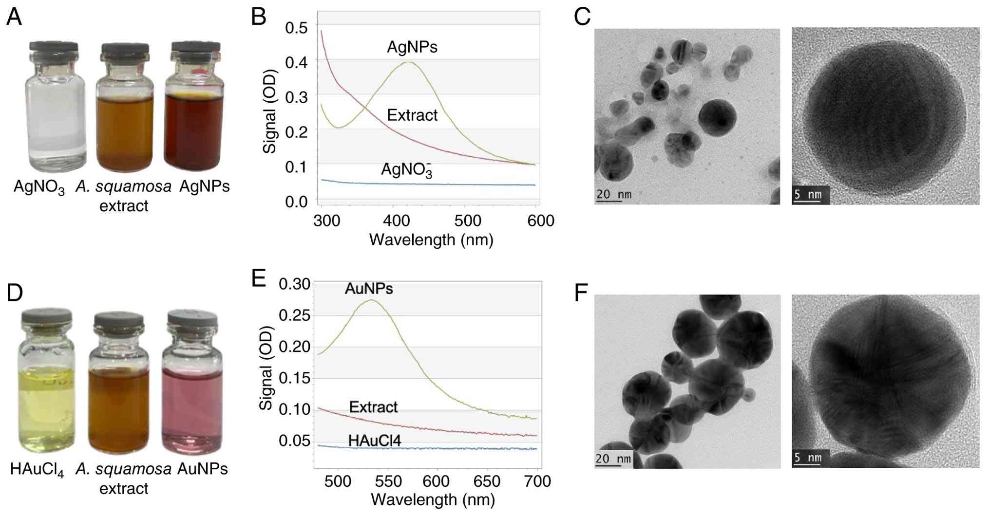
In the present study, a noticeable color transition
from light brown to dark brown was observed (Fig. 1A), and a prominent plasmon resonance
peak in the 410-420 nm range was detected, confirming the formation
of AgNPs (the A. squamosa seeds extract itself did not
exhibit any peaks in the 300-600 nm range) (Fig. 1B). This indicates that A.
squamosa seeds extract is a highly effective reducing agent for
converting silver ions to silver. For AuNPs, the successful
synthesis was visually confirmed by a distinct purple color
observed after A. squamosa seeds extract solution was mixed
with HAuCl4 solution (Fig.
1D). The plant extract efficiently reduced Au3+ ions to Au0.
The UV-Vis spectroscopy analysis of the resulting colloidal
solutions (Fig. 1E) revealed
absorption peaks in the 520-540 nm range, consistent with the
surface plasmon resonance of AuNPs, typically around 520 nm. This
resonance is influenced by the size of the NPs, affirming the
successful synthesis of AuNPs (30).
To further investigate the size, shape and crystal
structure of the NPs, TEM and PSA analyses were employed. TEM
images revealed distinct morphological characteristics of the AgNPs
and AuNPs. The AgNPs (Fig. 1C)
exhibited predominantly spherical morphology with moderate
polydispersity and occasional particle aggregation. High-resolution
(HR)TEM imaging showed well-defined lattice fringes, confirming the
crystalline nature of the NPs. By contrast, the AuNPs (Fig. 1F) appeared more uniform in shape and
size, displaying predominantly spherical to quasi-spherical
morphologies. The HRTEM image of AuNPs revealed pronounced lattice
fringes with a characteristic radial pattern, indicating high
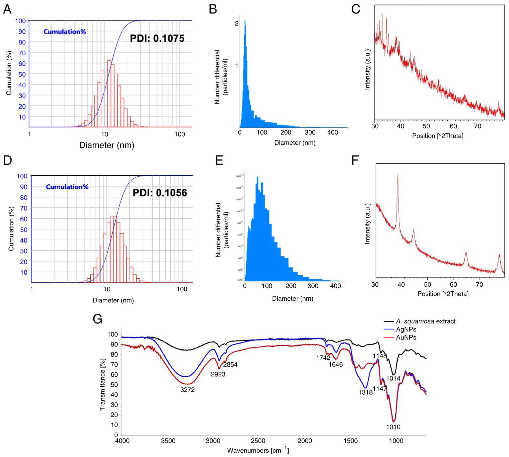
crystallinity and ordered atomic arrangements. According to the PSA
analysis, the average diameter of AgNPs was 14.05 nm, with sizes
ranging from 6 to 25 nm, while AuNPs had an average size of 14.92
nm, ranging from 3.67 to 44.89 nm (Fig.
2A and D). Meanwhile,
measurements performed using a ViewSizer 3000 NP Size Analyzer; the
mean hydrodynamic diameters of the AgNPs and AuNPs were 40.60±34
and 72.65±46 nm, respectively. For AgNPs, the D10, D50 and D90
values were 22.0, 29.5 and 70.76 nm, respectively, indicating that
10, 50 and 90% of the particles had diameters below these values.
However, the D10, D50 and D90 values of AuNPs were 26.7, 61.2 and
132.6 nm, respectively (Fig. 2B and
E). X-ray diffraction analysis of
the synthesized AgNPs showed broad peaks at 38.29, 45.41, 64.40 and
77.54˚, corresponding to the 111, 200, 220 and 311 planes,
indicating a face-centered cubic structure. Similarly, the XRD
patterns of the biologically synthesized AuNPs revealed sharp peaks
at 38.20, 43.73, 64.77 and 77.72˚, which align with the 111, 200,
220 and 311 planes, confirming their face-centered cubic
crystalline structure (Fig. 2C and
F).
As shown in Fig. 2G,
the synthesized AgNPs and AuNPs had similar peaks to A.
squamosa seed extract, indicating that A. squamosa seed
extract contributed to the reduction of Ag and Au ions. A broad
peak was present at ~3,272/cm that comes from OH stretching, and a
smaller peak at 2,923/cm from aliphatic CH. The peaks at 1,010/cm
and 1,147/cm confirm the presence of secondary OH groups. These
functional groups interact with the silver and gold to form a
protective layer around the NPs, which keeps them from clumping
together. Previous studies have shown that hydroxyl and phenolic
groups tend to bind strongly to noble metals through polar and
coordinating interactions (40).
The increase in peak intensity and slight shifts in wavelength
observed in the NPs spectrum, for example at 3,272, 1,147 and
1,010/cm, probably occur because the seed extract is coordinating
with the metals (41). Of note, the
AgNPs show a unique strong peak at 1,318/cm that comes from C-O
aromatic ester stretching and OH bending, which was not present in
A. squamosa seeds extract or AuNPs.
Metal content of AgNPs
ICP-OES analysis revealed that the synthesized AgNPs
contained 6.60 µg/g Ag, confirming the presence of Ag in the NP
preparation.
Sterility evaluation of NPs
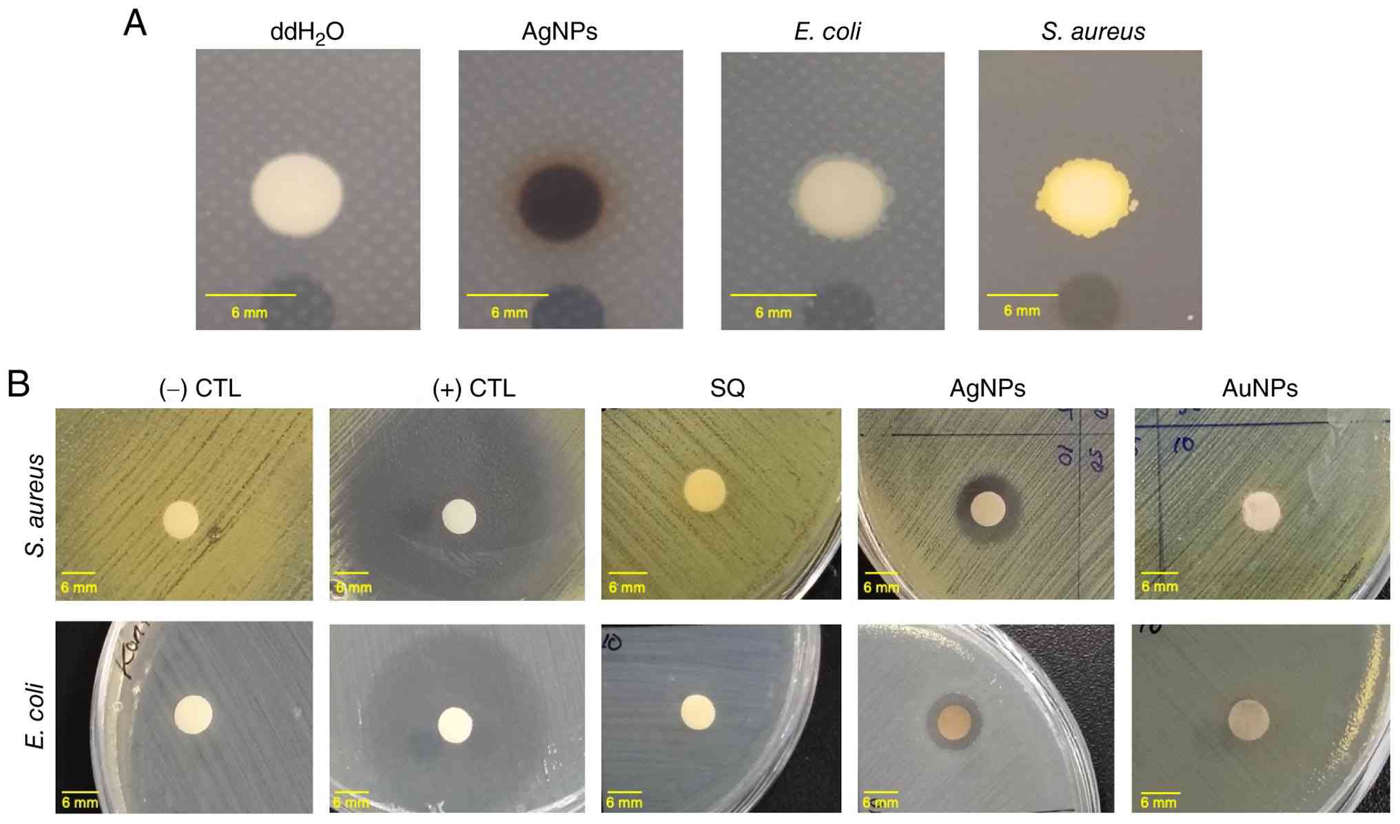
The sterility evaluation of AgNPs demonstrated that
AgNPs were sterile, as evidenced by the absence of bacterial growth
following inoculation onto MHA medium (Fig. 3A; Table
II).
| Table IISterility test of AgNPs. |
Table II
Sterility test of AgNPs.
| Sample | Bacterial
growth |
|---|
| AgNPs | - |
| S. aureus
culture | + |
| E. coli
culture | + |
| Sterile nuclease
free water | + |
Antibacterial potency of AgNPs and
AuNPs
To investigate the antibacterial activity of AgNPs
and AuNPs, disk diffusion assays were performed against E.
coli and S. aureus. Growth inhibition was observed on
plates with 100, 50, 25 and 10% concentrations of 100 mg/ml AgNPs,
but not with AuNPs or A. squamosa seeds extract (control)
after 24 h of incubation. Table
III and Fig. 3B reveal that
E. coli had a smaller zone of inhibition compared to S.
aureus. The bacterial growth inhibition around the wells was
attributed to the diffusion of AgNPs to the MHA plates.
| Table IIIResults of disc diffusion test of
AgNPs and AuNPs. |
Table III
Results of disc diffusion test of
AgNPs and AuNPs.
| | | Inhibition zone
diameter, mm |
|---|
| Organism | Concentration
(mg/ml) | AgNP | AuNP |
|---|
| Staphylococcus
aureus | 10 | 9.91±0.29 | 0.0 |
| | 25 | 11.57±0.68 | 0.0 |
| | 50 | 11.89±0.37 | 0.0 |
| | 100 | 13.63±2.86 | 0.0 |
| | + (Gentamycin) | 34.43±3.40 | 36.45±0.42 |
| | - (sterile
ddH2O) | 0.0 | 0.0 |
| | A. squamosa
seeds extract all concentrations | 0.0 | 0.0 |
| Escherichia
coli | 10 | 8.41±0.91 | 0.0 |
| | 25 | 7.89±0.37 | 0.0 |
| | 50 | 8.28±1.10 | 0.0 |
| | 100 | 8.53±1.03 | 0.0 |
| | + (Gentamycin) | 18.09±0.43 | 19.67±0.60 |
| | - (sterile
ddH2O) | 0.0 | 0.0 |
| | A. squamosa
seeds extract all concentrations | 0.0 | 0.0 |
Furthermore, the MIC and MBC values of AgNPs were
determined (Table IV). The MIC and
MBC are important metrics in antimicrobial evaluation, indicating
an agent's capacity to inhibit microbial proliferation and
eradicate pathogens, respectively. By definition, the MIC
represents the lowest concentration of an antimicrobial agent that
prevents visible microbial growth, whereas the MBC is the lowest
concentration required to kill 99.9% of the microbial population
(42). The correlation between
these values reveals the agent's mode of action: An MBC value close
to the MIC suggests bactericidal activity, whereas a substantially
higher MBC denotes a bacteriostatic effect (43). As presented in Table IV, the AgNPs exhibited
antibacterial activity against both tested microorganisms. The MIC
values were 0.781 mg/ml for Staphylococcus aureus and 3.125
mg/ml for Escherichia coli. The MBC values for both bacteria
were 3.125 mg/ml. The lower MIC value observed for S. aureus
indicates that AgNP was more effective against this Gram-positive
bacterium than against E. coli, as a lower concentration was
required to inhibit bacterial growth. Furthermore, the identical
MBC values suggest that a concentration of 3.125 mg/ml was required
to achieve bactericidal activity against both bacterial species.
The AgNPs exhibited bacteriostatic activity against S.
aureus and bactericidal activity against E. coli, as
evidenced by the relationship between their MIC and MBC values. In
S. aureus, the MBC was 4x higher than the MIC, whereas in
E. coli, the MIC and MBC were identical, indicating that the
inhibitory concentration was also sufficient to exert a
bactericidal effect.
| Table IVMIC and the MBC values of AgNPs. |
Table IV
MIC and the MBC values of AgNPs.
| Tested
bacteria | MIC value
(mg/ml) | MBC value
(mg/ml) |
|---|
| Staphylococcus
aureus | 0.781 | 3.125 |
| Escherichia
coli | 3.125 | 3.125 |
Effect of AgNPs, AuNPs and
AgNO3 on the viability of MCF-7 and 293 cells
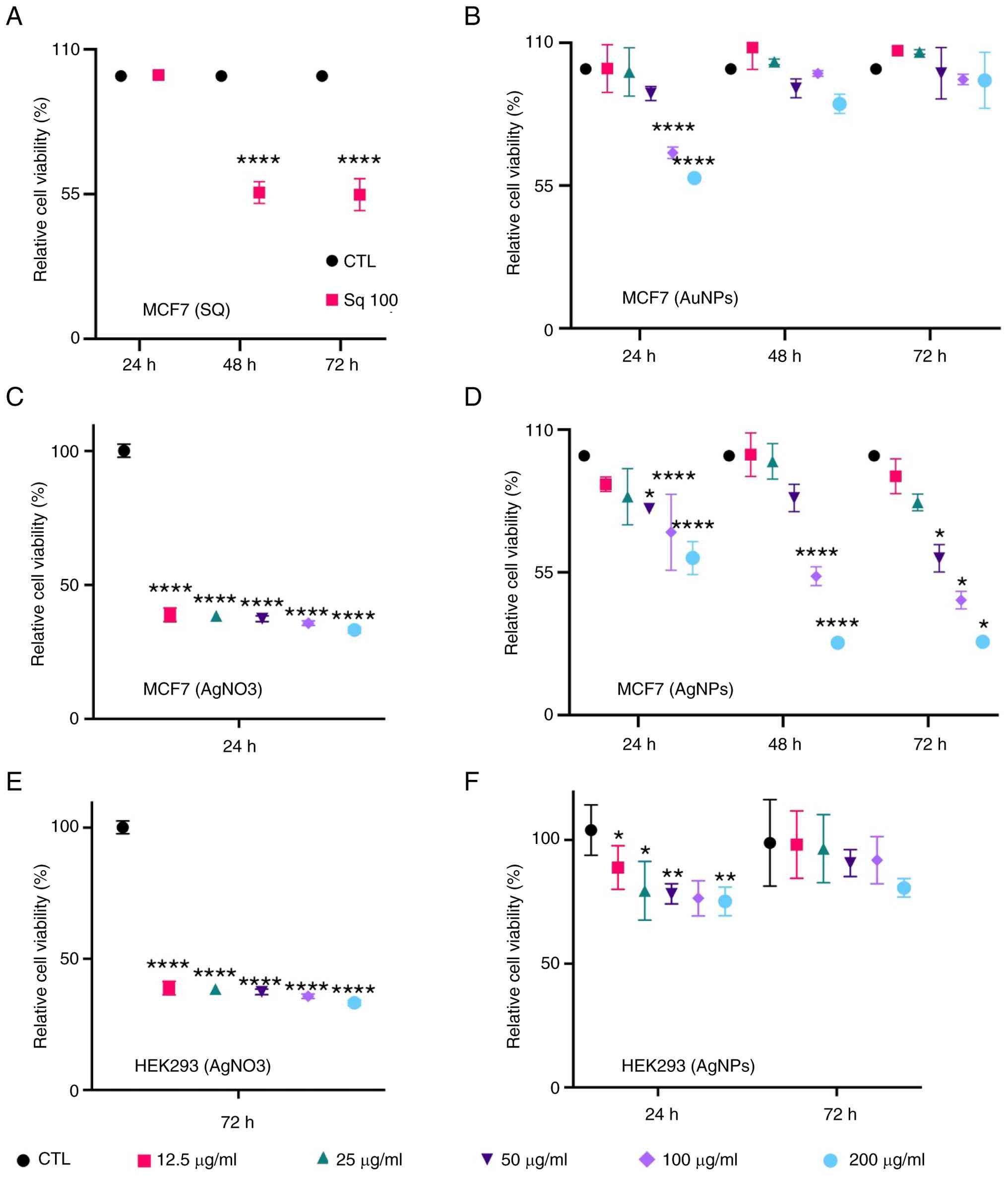
To evaluate the anticancer activity and selective
cytotoxicity of the biosynthesized NPs, an MTT assay was performed
on the breast cancer cell line MCF-7 and the non-carcinogenic 293
cells. Treatment with A. squamosa extract alone (100 µg/ml)
showed limited cytotoxic activity against MCF-7 cells at 24 h, with
the cell viability remaining comparable to the control group
(>90%). However, prolonged exposure significantly reduced the
cell viability to ~55% at both 48 and 72 h (P<0.0001),
indicating a time-dependent antiproliferative effect (Fig. 4A). By contrast, AuNPs only reduced
the cell viability after 24 h at 100 and 200 µg/ml. By 48 and 72 h,
there was no significant difference in cell viability between the
AuNP-treated cells and the control (Fig. 4B).
Treatment with AgNO3 produced strong
cytotoxic effects on MCF-7 cells, reducing cell viability to
~40-45% across all tested concentrations after 24 h of treatment
(P<0.0001) (Fig. 4C). Among all
tested treatments, AgNPs demonstrated the greatest anticancer
activity against MCF-7 cells. At 24 h, AgNPs reduced cell viability
in a concentration-dependent manner, with higher concentrations
decreasing viability to ~55-60% (P<0.0001). The cytotoxic effect
became more pronounced after 48 h, where the highest concentration
reduced viability to ~25-30% (P<0.0001). Similar effects were
maintained at 72 h, indicating sustained growth inhibition and
enhanced anticancer activity compared with SQ and AuNPs alone
(Fig. 4D).
To assess selectivity, the effects of
AgNO3 and AgNPs were also evaluated in 293 cells.
AgNO3 markedly reduced 293 cell viability, decreasing
the proportion of viable cells to ~30-40% at all tested
concentrations (Fig. 4E),
suggesting considerable toxicity toward normal cells. By contrast,
AgNPs displayed substantially lower toxicity toward 293 cells,
maintaining cell viability above 70-98% across most concentrations
and time-points (Fig. 4F). Although
a slight decline in viability was observed at higher
concentrations, the reduction was significantly less severe than
that observed in MCF-7 cells.
The MTT assay results showed that while the AuNPs
weren't toxic to the cells, the AgNPs had a clear effect, and
accordingly, the study further focused on measuring their
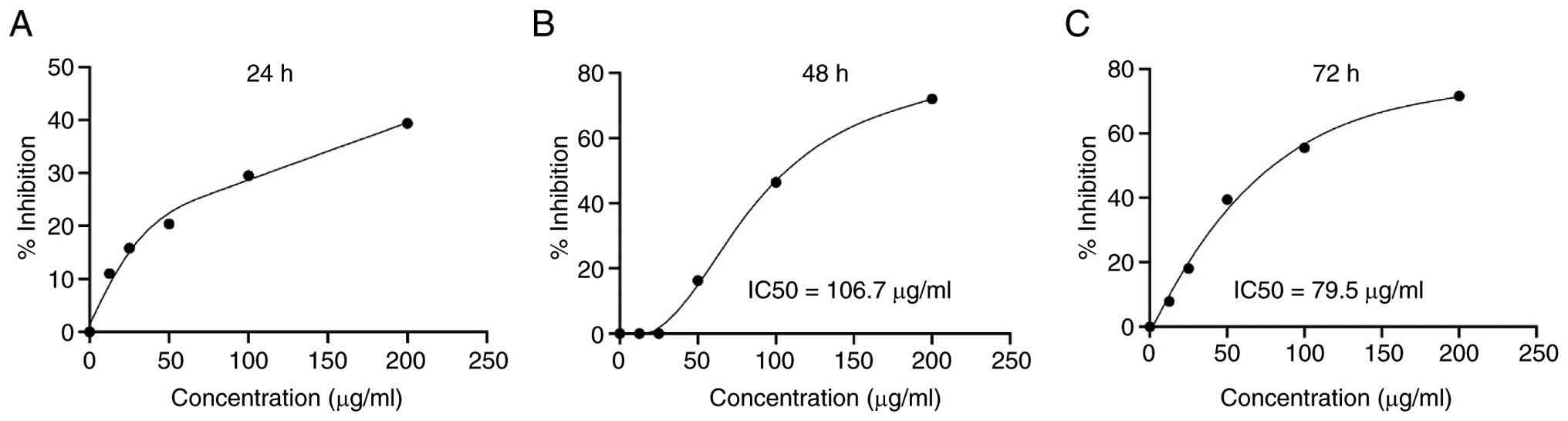
IC50 value. The IC50s of the AgNPs were
assessed through a detailed analysis involving five doses and three
incubation periods. As shown in Fig.
5, it was found that even high doses of AgNPs did not kill
enough cells to determine the IC50 after 24 h-it was
>200 µg/ml. However, at 48 and 72 h, the AgNPs were much more
effective, as evidenced by the significant reduction of cell
viability. The IC50 values were determined to be 106.7
µg/ml at 48 h and 79.5 µg/ml at 72 h. These results show that the
AgNPs have the potential to stop cancer cells from growing,
suggesting they could be useful in cancer treatments.
Effect of AgNPs on the cell cycle
phase and expression of several oncogenes
To investigate the possible molecular targets of
AgNPs in the MCF-7 cell line, cell cycle and RT-qPCR analysis of
several identified oncogenes in breast cancer (44-46)
was performed, including MYC, HER2, CCND1 and COX-2. The DNA
content analysis revealed that AgNPs treatment significantly
reduced the percentage of cells below 2N and increased the number
of cells with 4N DNA. The RT-qPCR analysis results indicated that
AgNPs derived from A. squamosa seed extract effectively
reduced the mRNA levels of MYC, CCND1, HER2, COX-2 and CTNND1
(Fig. 6). The Cq values are
presented in Table SI. These
findings underscore the potential of AgNPs to modulate the
expression of critical oncogenes and offer valuable insights into
their anti-proliferative mechanisms in cancer cells.
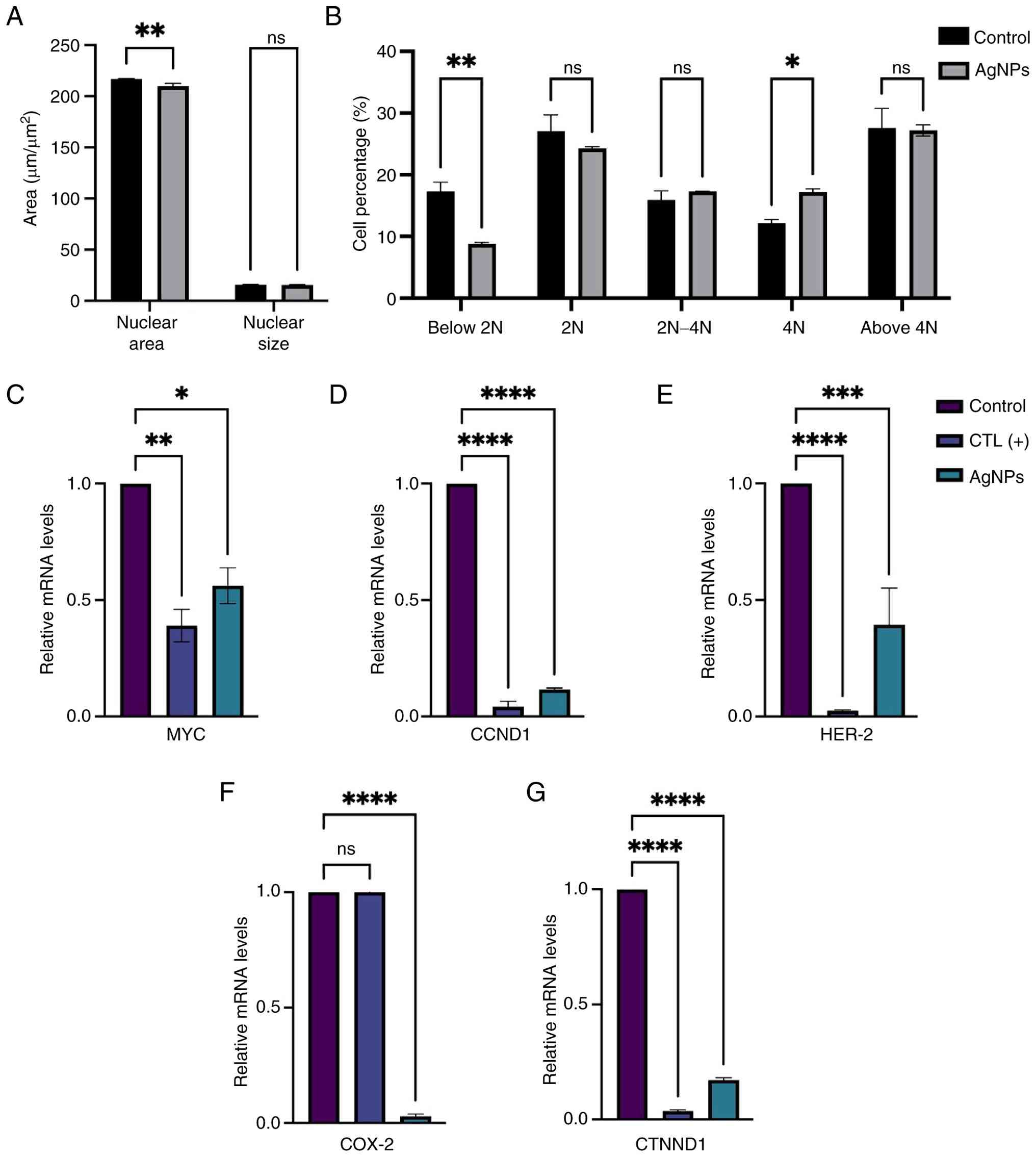 | Figure 6AgNPs treatment alters cell cycle
phase and several oncogene expressions in MCF-7 cells. (A) The
nuclear size and nuclear area of the MCF-7 cells following
treatment with 100 µg/ml of AgNPs for 24 h. (B) The DNA content
represents cell cycle phase of MCF-7 cells following treatment with
100 µg/ml of AgNPs for 24 h. (C) Relative mRNA levels of MYC, (D)
CCND1, (E) HER-2, (F) COX-2 and (G) CTNND1 in MCF-7 cells following
treatment with AgNPs (100 µg/ml) for 48 h. Data are presented as
mean ± standard deviation of three independent replicates (n=3).
Statistical analysis was performed using one-way ANOVA. Asterisks
indicate statistically significant differences compared with the
control group: *P<0.05, **P<0.01,
***P<0.001 and ****P<0.0001 vs. CTL.
ns, no significance; CTL, control; MYC, c-Myc; CCND1, cyclin D1;
HER2, human epidermal growth factor receptor-2; COX-2,
cyclooxygenase-2, CTNND1, catenin delta-1; AgNPs, silver
nanoparticles. |
AgNPs induce apoptosis in MCF-7
cells
To investigate whether the cytotoxic effect of AgNPs
was associated with apoptosis, MCF-7 cells were treated with AgNPs
(100 µg/ml) for 48 h and analyzed using Annexin V-FITC flow
cytometry. As shown in Fig. 7A and
B, AgNP treatment markedly
increased the population of Annexin V-positive cells compared with
the untreated control. Quantitative analysis revealed that the
percentage of apoptotic cells increased significantly from
21.25±0.07% in the control group to 27.3±0.32% following AgNP
treatment (P<0.001; Fig. 7B),
indicating that AgNPs effectively induced apoptosis in MCF-7
cells.
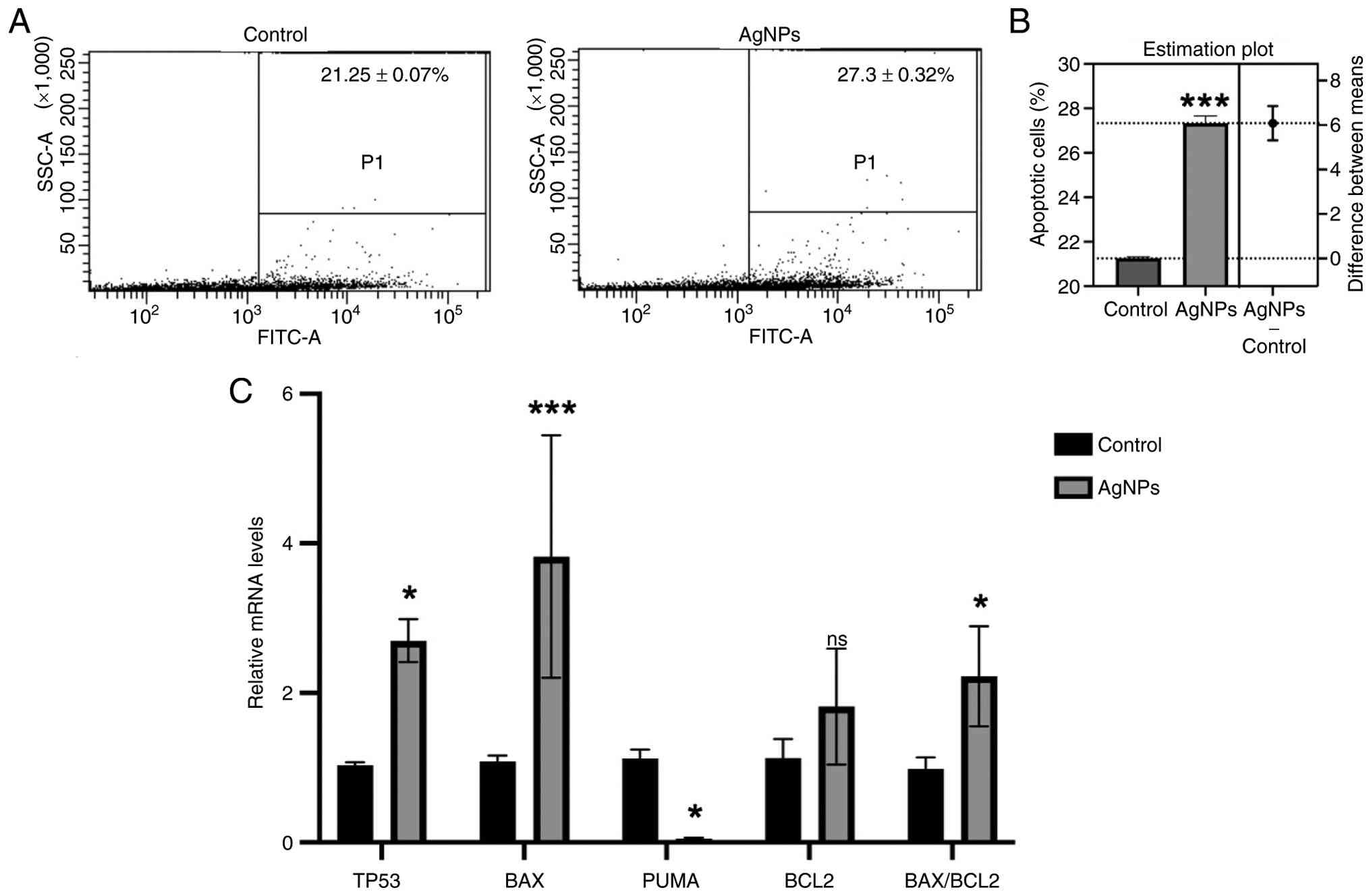 | Figure 7AgNP treatment induces apoptosis and
modulates the expression of apoptosis-related genes in MCF-7 cells.
(A) Representative flow cytometry dot plots of Annexin
V-FITC-stained MCF-7 cells showing apoptotic populations in the
untreated control and AgNP-treated (100 µg/ml, 48 h) groups. (B)
Quantification of apoptotic cells demonstrating a significant
increase in apoptosis following AgNP treatment compared with the
control group, as determined by Student's t-test. (C) Relative
expression levels of apoptosis-related genes following AgNP
exposure. AgNP treatment significantly upregulated p53, BAX and
BAX/BCL2 ratio, while significantly downregulating PUMA expression
compared with the control. Gene expression data were analyzed using
two-way ANOVA and are presented as the mean ± standard deviation
from three independent experiments (n=3). Statistical significance
is indicated as *P<0.05 and ***P<0.001
vs. control. ns, not significant; AgNPs, silver nanoparticles;
TP53, tumor protein P53; BAX, BCL2 associated X; BBC3/PUMA, BCL2
binding component 3; BCL2, BCL2 apoptosis regulator. |
To further elucidate the molecular mechanisms
underlying AgNP-induced apoptosis, the expression of several
apoptosis-related genes was evaluated by RT-qPCR (Fig. 7C), and the ΔΔCt values are presented
in Table SII. AgNP treatment
significantly upregulated the expression of p53 (2.7-fold), BAX
(3.8-fold), and the BAX/BCL2 ratio (2.2-fold) relative to the
control group (P<0.05-0.001). By contrast, the expression of
PUMA was markedly reduced to ~0.05-fold of the control level
(P<0.05). These findings suggest that AgNPs induce apoptosis in
MCF-7 cells through activation of the p53-mediated apoptotic
pathway.
Discussion
AgNPs are increasingly valued for their
antimicrobial properties in medical applications. Although the
precise mechanism behind the antibacterial activity of AgNPs is
still being studied, it is thought that both the size of the NPs
and the presence of phytochemical components contribute
synergistically to their effectiveness. The extensive surface area
and diverse phytochemical constituents of the seed extract
facilitate the reduction and stabilization of AgNPs, which are
typically <100 nm in size (47).
AgNPs can disrupt bacterial cells through various mechanisms,
including electrostatic interactions with the negatively charged
bacterial membranes, which leads to cell death (48). This interaction induces oxidative
stress through reactive oxygen species within the cell membrane
(49,50). The AgNPs don't stop there-they
actually break through the bacterial membrane, disrupt respiratory
chains and enter organelles where Ag+ ions interact with
DNA, ribosomes and enzymes. Of note, AgNPs prefer to attach to
nucleosides rather than phosphate groups, which leads to DNA damage
and ultimately cell death. AgNPs also accumulate in the membranes
of E. coli and S. aureus, increasing membrane
permeability and causing cell death.
Research utilizing Tectona grandis seed
extract for AgNP synthesis has revealed that NPs ranging from 10 to
30 nm in size can substantially inhibit S. aureus and E.
coli (51). In another study,
AgNPs produced using Trigonella foenum-graecum seed extract
demonstrated MIC values of 62.5 µg/ml for S. aureus and 125
µg/ml for E. coli (52).
While earlier studies have generally reported larger inhibition
zones for E. coli compared to S. aureus (52), the present findings indicate that
AgNPs exhibit greater effectiveness against S. aureus.
Meanwhile, a previous study on AgNPs synthesized using an aqueous
extract of Hypericum perforatum L. reported bactericidal
activity against S. aureus based on MIC and MBC evaluations
(53), which contrasts the present
findings, where a bacteriostatic effect was observed. This
discrepancy highlights the impact of the specific plant extract
used in AgNP synthesis on their antibacterial performance.
In addition to their antibacterial activity, the
present study demonstrated that AgNPs exert a selective
antiproliferative effect against cancer cells while exhibiting
minimal cytotoxicity toward normal 293 cells. This selective
cytotoxicity is highly desirable for anticancer agents, as it
minimizes damage to healthy tissues. Similar findings have been
reported previously, where AgNPs synthesized using walnut green
husk extract showed substantially greater cytotoxicity toward MCF-7
breast cancer cells than toward non-cancerous L-929 fibroblasts
(54). The preferential toxicity of
AgNPs toward cancer cells has been attributed to their enhanced
cellular uptake, which is facilitated by the altered metabolism,
rapid proliferation and increased endocytic activity characteristic
of malignant cells. These biological features render cancer cells
more susceptible to NP-induced oxidative stress and cellular damage
than normal cells (55).
Of note, the present results revealed a clear
difference between AgNO3 and AgNPs. While
AgNO3 exhibited cytotoxic effects on both MCF-7 and 293
cells, AgNPs selectively inhibited the growth of cancer cells with
limited toxicity toward normal cells. This selectivity may be
attributed to the controlled and sustained release of
Ag+ ions from the NP matrix, in contrast to the
immediate availability of free Ag+ ions from
AgNO3. Furthermore, the acidic tumor microenvironment
likely contributes to the enhanced anticancer activity of AgNPs
(56). Cancer cells generally
maintain a lower extracellular pH than normal cells, which promotes
increased dissolution of AgNPs and consequently a higher release of
Ag+ ions within tumor tissues. As a result, cancer cells
are exposed to greater intracellular silver ion concentrations,
leading to elevated oxidative stress, mitochondrial dysfunction and
apoptosis, while normal cells experience comparatively lower
toxicity (57). These mechanisms
collectively support the selective anticancer potential of AgNPs
observed in this study.
The anticancer activity of AgNPs may also be
associated with their ability to modulate the expression of genes
involved in cell proliferation and survival. In the present study,
dose-dependent cytotoxicity on MCF-7 cells treated with AgNPs was
observed, with IC50 values of 106.7 µg/ml at 48 h and
79.5 µg/ml at 72 h. These IC50 values are higher than
those found in a previous study, which reported IC50
values of 50 µg/ml at 24 h and 30 µg/ml at 48 h for AgNPs
synthesized using A. squamosa leaf extract against MCF-7
cells (24). The differences might
be attributed to the use of A. squamosa seed extract.
Another mechanism underlying the anticancer activity
of AgNPs is their ability to induce apoptosis in cancer cells. In
the present study, AgNP treatment significantly increased the
proportion of apoptotic MCF-7 cells compared with the control
group, indicating that apoptosis contributes to the observed
cytotoxic effect. This response is likely mediated through
activation of p53, a key regulator of programmed cell death.
Similar findings have been reported for green-synthesized AgNPs
from Allium ampeloprasum, which increased p53 and BAX
expression while decreasing BCL2 expression (58). Although BCL2 expression was also
elevated in the present study, the increase in BAX expression was
substantially greater, resulting in a higher BAX/BCL2 ratio. This
ratio is widely recognized as a key indicator of apoptosis,
reflecting the balance between pro-apoptotic and anti-apoptotic
signaling. Therefore, the elevated BAX/BCL2 ratio observed in
AgNP-treated cells supports the conclusion that AgNPs induce
apoptosis through activation of the intrinsic mitochondrial
apoptotic pathway (59).
When MCF-7 cells were exposed to AgNPs, significant
changes in several cancer-related genes were observed in the
present study. The treatment notably decreased the expression of
MYC, CCND1, COX-2, HER2 and
CTNND1. This is particularly interesting because c-Myc plays
a crucial role in driving breast cancer growth-it helps cancer
cells divide rapidly, and when it's overactive, patients tend to
have worse outcomes (60). c-Myc
promotes cell cycle progression by activating CCND1, which is
frequently overexpressed and amplified in breast cancer. CCND1
functions in the nucleus to regulate the transition from G1 to S
phase by interacting with CDK4 and CDK6(61). Meanwhile, COX-2 is often
overexpressed in breast cancer and its overexpression contributes
to tumor growth, invasion and metastasis (62). AgNPs effectively decreased
HER2 levels. HER2's role in breast cancer is particularly
concerning since it acts together with other EGFR family proteins
to trigger pathways that drive tumor growth and spread (63). A previous study reported lower
CTNND1 expression in breast cancer tissues from patients with
breast cancer compared to mammary gland cells from non-tumor
patients (64). This study is the
first to demonstrate that AgNPs synthesized with A. squamosa
seed extract can modulate several breast cancer-related genes.
Although AgNPs have good anticancer activity against MCF-7 cells,
it is still necessary to conduct further analyses to see the
effectiveness of AgNPs in animal models (in vivo
studies).
Despite the strong anticancer and antibacterial
effects of AgNPs, AuNPs synthesized using A. squamosa seed
extract did not exhibit comparable bioactivities. The differences
in the physicochemical properties of these nanomaterials may have
influenced their biological performance. Similar observations have
been reported in previous studies, where AuNPs showed minimal or no
cytotoxic effects in HeLa cells (65), A549 and Vero cells (66) and Caco-2 cells (67). The low cytotoxicity observed in the
present study suggests that A. squamosa-generated AuNPs may
have potential for biomedical applications where biocompatibility
is desirable. However, further studies are required to evaluate
their loading capacity, stability, targeting ability and delivery
efficiency before their suitability as drug-delivery vehicles can
be established. In general, AuNPs have attracted considerable
interest in drug-delivery research because of their tunable size
and shape, ease of surface functionalization and ability to
interact with a wide range of biomolecules. Previous studies have
explored the use of AuNPs for the delivery of drugs, genes,
proteins and vaccines (68-73).
Therapeutic agents, including antibiotics, can be associated with
AuNPs through ionic interactions, covalent conjugation or physical
adsorption.
Future studies should evaluate the anticancer
efficacy of the synthesized silver and gold NPs in additional
breast cancer subtypes, particularly triple-negative breast cancer
(MDA-MB-231), to determine whether their therapeutic effects extend
beyond hormone receptor-positive breast cancer. Preclinical in
vivo studies using appropriate animal models should then assess
antitumor efficacy, biodistribution, pharmacokinetics and long-term
safety, including the absorption, distribution, metabolism and
excretion profiles of the NPs. These studies will be essential for
establishing optimal dosing regimens, evaluating systemic toxicity
associated with prolonged exposure and supporting the clinical
translation of NP-based therapies. In addition, addressing public
perception and ethical considerations, together with collaboration
among researchers, clinicians and regulatory agencies, will
facilitate the safe and effective development of silver- and
gold-based nanomedicines.
In conclusion, A. squamosa seed extract
successfully mediated the green synthesis of AgNPs and AuNPs with
nanoscale size, spherical morphology and crystalline structures.
Among the synthesized NPs, the AgNPs exhibited bacteriostatic
activity against S. aureus and bactericidal activity against
E. coli, whereas the AuNPs showed no detectable
antibacterial effect. The AgNPs also demonstrated selective
anticancer activity by inhibiting the proliferation of MCF-7 breast
cancer cells while exerting minimal toxicity toward normal 293
cells. The cytotoxic effect was dose- and time-dependent, with
IC50 values of 106.704 and 79.519 µg/ml after 48 and 72
h of treatment, respectively. Mechanistically, AgNPs modulated the
expression of cancer-related genes, suppressed the expression of
several oncogenes and induced apoptosis through the activation of
p53-associated pathways, as evidenced by increased apoptotic cell
populations and an elevated BAX/BCL2 ratio. While these findings
underscore the therapeutic potential of A. squamosa seed
extract-biosynthesized NPs, further in vivo studies are
required to elucidate their biodistribution, pharmacokinetics,
mechanisms of action and long-term safety. Multidisciplinary
collaborations will be essential to advance their translation into
clinical applications.
Supplementary Material
ΔΔCq values of several oncogenes.
ΔΔCq values of several
apoptotic-related genes.
Acknowledgements
The authors are grateful to Mr. Bryan Khalfani,
Bachelor of Science (B.Sc) and Mr. Muhammad Sanda Amanullah, B.Sc
from the Biology Department, Faculty of Science and Technology,
Universitas Airlangga (Surabaya, Indonesia) for their technical
assistance with measuring inhibition zone diameters during
antibacterial analysis.
Funding
Funding: This research was supported by the International
Research Consortium (IRCON) 2024, Universitas Airlangga, Indonesia
(grant no. 178/UN3.LPPM/PT.01.03/2024) and Universiti Malaya
research grant (grant no. IF033-2024).
Availability of data and materials
The data generated in the present study are
included in the figures and/or tables of this article.
Authors' contributions
FRPD contributed to the conception of the study,
acquisition of data, analysis and interpretation of data,
confirmation of the authenticity of the raw data, manuscript
preparation and funding acquisition. AUR and AR contributed to
performing the experiments, and the acquisition and analysis of
data. AG confirmed the authenticity of the raw data and data
analysis. AKMJ contributed to funding acquisition, data acquisition
and manuscript preparation. TD contributed to the data analysis and
manuscript preparation. VRH and ACE contributed to performing the
experiments, data acquisition and analysis. All authors read and
approved the manuscript and agree to be accountable for all aspects
of the research, ensuring that any issues related to the accuracy
or integrity of any part of the work were appropriately
investigated and resolved.
Ethics approval and consent to
participate
Not applicable.
Patient consent for publication
Not applicable.
Competing interests
The authors declare that they have no competing
interests.
Use of artificial intelligence tools
During the preparation of this work, AI tools
(ChatGPT-5.5; https://chatgpt.com) were used to
improve the readability and language of the manuscript, and
subsequently, the authors revised and edited the content produced
by the AI tools as necessary, taking full responsibility for the
ultimate content of the present manuscript.
References
|
1
|
Antunes Filho S, Dos Santos MS, Dos Santos
OAL, Backx BP, Soran ML, Opriş O, Lung I, Stegarescu A and
Bououdina M: Biosynthesis of nanoparticles using plant extracts and
essential oils. Molecules. 28(3060)2023.PubMed/NCBI View Article : Google Scholar
|
|
2
|
Dewi FRP, Lim V, Rosyidah AL, Fatimah F,
Wahyuningsih SPA and Zubaidah U: Characterization of silver
nanoparticles (AgNPs) synthesized from Piper ornatum leaf extract
and its activity against food borne pathogen Staphylococcus
aureus. Biodivers J Biol Divers. 24:1742–1748. 2023.
|
|
3
|
Rashmi V and Sanjay KR: Green synthesis,
characterisation and bioactivity of plant-mediated silver
nanoparticles using Decalepis hamiltonii root extract. IET
Nanobiotechnol. 11:247–254. 2017.PubMed/NCBI View Article : Google Scholar
|
|
4
|
Al-Radadi NS, Al-Bishri WM, Salem NA and
ElShebiney SA: Plant-mediated green synthesis of gold nanoparticles
using an aqueous extract of Passiflora ligularis, optimization,
characterizations, and their neuroprotective effect on propionic
acid-induced autism in Wistar rats. Saudi Pharm J.
32(101921)2024.PubMed/NCBI View Article : Google Scholar
|
|
5
|
Beeram SR, Rodriguez E, Doddavenkatanna S,
Li Z, Pekarek A, Peev D, Goerl K, Trovato G, Hofmann T and Hage DS:
Nanomaterials as stationary phases and supports in liquid
chromatography. Electrophoresis. 38:2498–2512. 2017.PubMed/NCBI View Article : Google Scholar
|
|
6
|
Stark WJ, Stoessel PR, Wohlleben W and
Hafner A: Industrial applications of nanoparticles. Chem Soc Rev.
44:5793–5805. 2015.PubMed/NCBI View Article : Google Scholar
|
|
7
|
Subbiah R, Veerapandian M and Yun KS:
Nanoparticles: Functionalization and multifunctional applications
in biomedical sciences. Curr Med Chem. 17:4559–4577.
2010.PubMed/NCBI View Article : Google Scholar
|
|
8
|
Frey NA, Peng S, Cheng K and Sun S:
Magnetic nanoparticles: synthesis, functionalization, and
applications in bioimaging and magnetic energy storage. Chem Soc
Rev. 38:2532–2542. 2009.PubMed/NCBI View Article : Google Scholar
|
|
9
|
Bloise N, Strada S, Dacarro G and Visai L:
Gold nanoparticles contact with cancer cell: A brief update. Int J
Mol Sci. 23(7683)2022.PubMed/NCBI View Article : Google Scholar
|
|
10
|
Bishoyi AK, Nouri S, Hussen A, Bayani A,
Khaksari MN and Soleimani Samarkhazan H: Nanotechnology in leukemia
therapy: Revolutionizing targeted drug delivery and immune
modulation. Clin Exp Med. 25(166)2025.PubMed/NCBI View Article : Google Scholar
|
|
11
|
Arockiya Aarthi Rajathi F, Arumugam R,
Saravanan S and Anantharaman P: Phytofabrication of gold
nanoparticles assisted by leaves of Suaeda monoica and its
free radical scavenging property. J Photochem Photobiol B.
135:75–80. 2014.PubMed/NCBI View Article : Google Scholar
|
|
12
|
Tahir K, Nazir S, Li B, Khan AU, Khan ZUH,
Gong PY, Khan SU and Ahmad A: Nerium oleander leaves extract
mediated synthesis of gold nanoparticles and its antioxidant
activity. Mat Lett. 156:198–201. 2015.
|
|
13
|
Jadoun S, Arif R, Jangid NK and Meena RK:
Green synthesis of nanoparticles using plant extracts: A review.
Environ Chem Lett. 19:355–374. 2020.
|
|
14
|
Gahlawat G and Choudhury AR: A review on
the biosynthesis of metal and metal salt nanoparticles by microbes.
RSC Adv. 9:12944–12967. 2019.PubMed/NCBI View Article : Google Scholar
|
|
15
|
Velusamy P, Kumar GV, Jeyanthi V, Das J
and Pachaiappan R: Bio-inspired green nanoparticles: Synthesis,
mechanism, and antibacterial application. Toxicol Res. 32:95–102.
2016.PubMed/NCBI View Article : Google Scholar
|
|
16
|
Nath D and Banerjee P: Green
nanotechnology-a new hope for medical biology. Environ Toxicol
Pharmacol. 36:997–1014. 2013.PubMed/NCBI View Article : Google Scholar
|
|
17
|
Mohandoss S, Velu KS, Manoharadas S, Ahmad
N, Palanisamy S, You S, Akhtar MS and Lee YR: Synthesis,
characterization, and evaluation of silver nanoparticle-loaded
carboxymethyl chitosan with sulfobetaine methacrylate hydrogel
nanocomposites for biomedical applications. Polymers (Basel).
16(1513)2024.PubMed/NCBI View Article : Google Scholar
|
|
18
|
Thakur N, Manna P and Das J: Synthesis and
biomedical applications of nanoceria, a redox active nanoparticle.
J Nanobiotechnology. 17(84)2019.PubMed/NCBI View Article : Google Scholar
|
|
19
|
Wahab MA, Luming L, Matin MA, Karim MR,
Aijaz MO, Alharbi HF, Abdala A and Haque R: Silver
micro-nanoparticle-based nanoarchitectures: Synthesis routes,
biomedical applications, and mechanisms of action. Polymers
(Basel). 13(2870)2021.PubMed/NCBI View Article : Google Scholar
|
|
20
|
Khan SA, Shahid S and Lee CS: Green
synthesis of gold and silver nanoparticles using leaf extract of
clerodendrum inerme; characterization, antimicrobial, and
antioxidant activities. Biomolecules. 10(835)2020.PubMed/NCBI View Article : Google Scholar
|
|
21
|
Bharadwaj KK, Rabha B, Pati S, Sarkar T,
Choudhury BK, Barman A, Bhattacharjya D, Srivastava A, Baishya D,
Edinur HA, et al: Green synthesis of gold nanoparticles using plant
extracts as beneficial prospect for cancer theranostics. Molecules.
26(6389)2021.PubMed/NCBI View Article : Google Scholar
|
|
22
|
Jibrin F, Fanoro OT, Maluleke R, Lebepe
TC, Mgedle N, Mbaz GIM, Aladesuyi OA, Kalimuthu R, Odeku OA and
Oluwafemi OS: Biosynthesis, characterization, and antibacterial
activity of gold, silver, and bimetallic nanoparticles using
Annona squamosa L. leaves. Antibiotics (Basel).
13(1199)2024.PubMed/NCBI View Article : Google Scholar
|
|
23
|
Malik M, Iqbal MA, Malik M, Raza MA,
Shahid W, Choi JR and Pham PV: Biosynthesis and characterizations
of silver nanoparticles from Annona squamosa leaf and fruit
extracts for size-dependent biomedical applications. Nanomaterials
(Basel). 12(616)2022.PubMed/NCBI View Article : Google Scholar
|
|
24
|
Vivek R, Thangam R, Muthuchelian K,
Gunasekaran P, Kaveri K and Kannan S: Green biosynthesis of silver
nanoparticles from Annona squamosa leaf extract and its in
vitro cytotoxic effect on MCF-7 cells. Process Biochem.
47:2405–2410. 2012.
|
|
25
|
VA M, Ramesh S and Muthukrishnan L: Facile
fabrication of Annona squamosa L. seed extract mediated
silver nanoparticles challenged against biofilm forming oral
pathogens. Plant Nano Biol. 3(100023)2022.
|
|
26
|
Jose V, Raphel L, Aiswariya KS and Mathew
P: Green synthesis of silver nanoparticles using Annona
squamosa L. seed extract: Characterization, photocatalytic and
biological activity assay. Bioprocess Biosyst Eng. 44:1819–1829.
2021.PubMed/NCBI View Article : Google Scholar
|
|
27
|
Miao Y, Xu X, Yuan F, Shi Y, Chen Y, Chen
J and Li X: Four cytotoxic annonaceous acetogenins from the seeds
of Annona squamosa. Nat Prod Res. 30:1273–1279.
2016.PubMed/NCBI View Article : Google Scholar
|
|
28
|
Mokhtar FA, Selim NM, Elhawary SS, Abd El
Hadi SR, Hetta MH, Albalawi MA, Shati AA, Alfaifi MY, Elbehairi
SEI, Fahmy LI and Ibrahim RM: Green biosynthesis of silver
nanoparticles using annona glabra and Annona squamosa
extracts with antimicrobial, anticancer, apoptosis potentials,
assisted by in silico modeling, and metabolic profiling.
Pharmaceuticals (Basel). 15(1354)2022.PubMed/NCBI View Article : Google Scholar
|
|
29
|
Gangapuram BR, Bandi R, Alle M, Dadigala
R, Kotu GM and Guttena V: Microwave assisted rapid green synthesis
of gold nanoparticles using Annona squamosa L peel extract
for the efficient catalytic reduction of organic pollutants. J Mol
Struct. 1167:305–315. 2018.
|
|
30
|
Huang W, Wang L, Long D and Liu X:
Colorimetric determination and recycling of gold(III) ions using
label-free plasmonic H0.3MoO3 nanoparticles.
Microchimica Acta. 190(245)2023.PubMed/NCBI View Article : Google Scholar
|
|
31
|
Shahid M, Zhou Y, Cheng XW, Zar MS, Chen G
and Tang RC: Ferulic acid promoted in-situ generation of AgNPs@silk
as functional colorants. J Clean Prod. 176:736–744. 2018.
|
|
32
|
Lewis II JS: CLSI M100-Ed35 performance
standards for antimicrobial susceptibility testing standards. 35th
Edition. Clinical and Laboratory Standards Institute, 2025.
|
|
33
|
Twardowska M, Łyskowski A, Misiorek M,
Szymaszek Ż, Wołowiec S, Dąbrowska M and Uram Ł: Human embryonic
kidney HEK293 cells as a model to study SMVT-independent transport
of biotin and biotin-furnished nanoparticles in targeted therapy.
Int J Mol Sci. 26(1594)2025.PubMed/NCBI View Article : Google Scholar
|
|
34
|
Hua Z, Zhan Y, Zhang S, Dong Y, Jiang M,
Tan F, Liu Z, Thiele CJ and Li Z: P53/PUMA are potential targets
that mediate the protection of brain-derived neurotrophic factor
(BDNF)/TrkB from etoposide-induced cell death in neuroblastoma
(NB). Apoptosis. 23:408–419. 2018.PubMed/NCBI View Article : Google Scholar
|
|
35
|
Cao H and Sethumadhavan K: Identification
of Bcl2 as a stably expressed qPCR reference gene for human colon
cancer cells treated with cottonseed-derived gossypol and bioactive
extracts and bacteria-derived lipopolysaccharides. Molecules.
27(7560)2022.PubMed/NCBI View Article : Google Scholar
|
|
36
|
Zerbib J, Ippolito MR, Eliezer Y, De
Feudis G, Reuveni E, Savir Kadmon A, Martin S, Viganò S, Leor G,
Berstler J, et al: Human aneuploid cells depend on the RAF/MEK/ERK
pathway for overcoming increased DNA damage. Nat Commun.
15(7772)2024.PubMed/NCBI View Article : Google Scholar
|
|
37
|
Dewi FR, Rohmatika AU, Jamil AK, Demircan
T, Idris MF, Litazkiyyah L, Fahmi M, Rosyidah A, Hayati A and
Sugiharto S: Plant-based synthesis of gold and silver nanoparticles
using Artocarpus heterophyllus aqueous leaf extract and its
anticancer activities. Narra J. 5(e1770)2025.PubMed/NCBI View Article : Google Scholar
|
|
38
|
Dewi FRP, Geraldi A, Rohmatika AU, Satar
M, Hajar V, You YH, Elfentiana AC and Tan SC:
Phytochemical-mediated synthesis of silver and gold nanoparticles
from Piper ornatum: Characterization, antibacterial efficacy, and
anticancer mechanisms. J Nanotechnol. 2026(1225355)2026.
|
|
39
|
Livak KJ and Schmittgen TD: Analysis of
relative gene expression data using real-time quantitative PCR and
the 2(-Delta Delta C(T)) method. Methods. 25:402–408.
2001.PubMed/NCBI View Article : Google Scholar
|
|
40
|
Francis S, Joseph S, Koshy EP and Mathew
B: Microwave assisted green synthesis of silver nanoparticles using
leaf extract of elephantopus scaber and its environmental and
biological applications. Artif Cells Nanomed Biotechnol.
46:795–804. 2018.PubMed/NCBI View Article : Google Scholar
|
|
41
|
Baba IA, Awe OB, Mustapha S, Abubakar MA,
Abdulkareem AS, Tijani JO and Obayomi KS: Influence of
plant-derived extracts on the synthesis, physicochemical
properties, and applications of metal and metal oxide
nanoparticles. Hybrid Adv. 13(100666)2026.PubMed/NCBI View Article : Google Scholar
|
|
42
|
Geraldi A, Wardana AP, Aminah NS,
Kristanti AN, Sadila AY, Wijaya NH, Wijaya MRA, Diningrum NID,
Hajar VR and Manuhara YSW: Tropical medicinal plant extracts from
indonesia as antifungal agents against Candida Albicans. Front
Biosci (Landmark Ed). 27(274)2022.PubMed/NCBI View Article : Google Scholar
|
|
43
|
Rodríguez-Melcón C, Alonso-Calleja C,
García-Fernández C, Carballo J and Capita R: Minimum inhibitory
concentration (MIC) and minimum bactericidal concentration (MBC)
for twelve antimicrobials (biocides and antibiotics) in eight
strains of Listeria monocytogenes. Biology (Basel).
11(46)2021.PubMed/NCBI View Article : Google Scholar
|
|
44
|
Lin K, Baritaki S, Vivarelli S, Falzone L,
Scalisi A, Libra M and Bonavida B: The breast cancer protooncogenes
HER2, BRCA1 and BRCA2 and their regulation by the iNOS/NOS2 axis.
Antioxidants (Basel). 11(1195)2022.PubMed/NCBI View Article : Google Scholar
|
|
45
|
Lee EYHP and Muller WJ: Oncogenes and
tumor suppressor genes. Cold Spring Harb Perspect Biol.
2(a003236)2010.PubMed/NCBI View Article : Google Scholar
|
|
46
|
Majumder M, Dunn L, Liu L, Hasan A,
Vincent K, Brackstone M, Hess D and Lala PK: COX-2 induces
oncogenic micro RNA miR655 in human breast cancer. Sci Rep.
8(327)2018.PubMed/NCBI View Article : Google Scholar
|
|
47
|
Selvaraj V, Sagadevan S, Muthukrishnan L,
Johan MR and Podder J: Eco-friendly approach in synthesis of silver
nanoparticles and evaluation of optical, surface morphological and
antimicrobial properties. J Nanostruct Chem. 9:153–162. 2019.
|
|
48
|
Dibrov P, Dzioba J, Gosink KK and Häse CC:
Chemiosmotic mechanism of antimicrobial activity of Ag(+) in vibrio
cholerae. Antimicrob Agents Chemother. 46:2668–2670.
2002.PubMed/NCBI View Article : Google Scholar
|
|
49
|
Niño-Martínez N, Salas Orozco MF,
Martínez-Castañón GA, Torres Méndez F and Ruiz F: Molecular
mechanisms of bacterial resistance to metal and metal oxide
nanoparticles. Int J Mol Sci. 20(2808)2019.PubMed/NCBI View Article : Google Scholar
|
|
50
|
Bhardwaj AK, Shukla A, Mishra RK, Singh
SC, Mishra V, Uttam KN, Singh MP, Sharma S and Gopal R: Power and
time dependent microwave assisted fabrication of silver
nanoparticles decorated cotton (SNDC) fibers for bacterial
decontamination. Front Microbiol. 8(330)2017.PubMed/NCBI View Article : Google Scholar
|
|
51
|
Rautela A, Rani J and Debnath M: Green
synthesis of silver nanoparticles from Tectona grandis seeds
extract: Characterization and mechanism of antimicrobial action on
different microorganisms. J Anal Sci Technol. 10(5)2019.
|
|
52
|
Varghese R, Almalki MA, Ilavenil S,
Rebecca J and Choi KC: Silver nanopaticles synthesized using the
seed extract of Trigonella foenum-graecum L. and their
antimicrobial mechanism and anticancer properties. Saudi J Biol
Sci. 26:148–154. 2019.PubMed/NCBI View Article : Google Scholar
|
|
53
|
Alahmad A, Al-Zereini WA, Hijazin TJ,
Al-Madanat OY, Alghoraibi I, Al-Qaralleh O, Al-Qaraleh S, Feldhoff
A, Walter JG and Scheper T: Green synthesis of silver nanoparticles
using Hypericum perforatum L. aqueous extract with the
evaluation of its antibacterial activity against clinical and food
pathogens. Pharmaceutics. 14(1104)2022.PubMed/NCBI View Article : Google Scholar
|
|
54
|
Khorrami S, Zarrabi A, Khaleghi M, Danaei
M and Mozafari MR: Selective cytotoxicity of green synthesized
silver nanoparticles against the MCF-7 tumor cell line and their
enhanced antioxidant and antimicrobial properties. Int J
Nanomedicine. 13:8013–8024. 2018.PubMed/NCBI View Article : Google Scholar
|
|
55
|
Cairns RA, Harris IS and Mak TW:
Regulation of cancer cell metabolism. Nat Rev Cancer. 11:85–95.
2011.PubMed/NCBI View Article : Google Scholar
|
|
56
|
Ashby BS: pH studies in human malignant
tumours. Lancet. 2:312–315. 1966.PubMed/NCBI View Article : Google Scholar
|
|
57
|
Islam MA, Riaz TA, Hossain MA, Lee I,
Akter J, Shrestha S, Gyawali N, Pandey A, Chaizul S, Fualo V and
Hahn JR: Controlled release of Ag+ ions to human cancer
cells selectively neutralized with silver nanoparticles of
different sizes produced by a green synthesis method. J Mol Struct.
1294(136384)2023.
|
|
58
|
Lu L, Hu J and Xiong M: Chemical
characterization, antioxidant, cytotoxicity, and anti-human breast
cancer potentials of Allium ampeloprasum green-formulated
silver nanoparticles by investigating the p53 and cell cycle G1, S,
G2, and M phases. Arab J Chem. 19(10452025)2026.
|
|
59
|
Putri GFT and Nurfatihah Z Z: Bax/Bcl-2
ratio as the golden marker of apoptosis: Molecular mechanisms and
regulatory pathways. Int J Cell Biomed Sci. 4:309–317. 2025.
|
|
60
|
Xu J, Chen Y and Olopade OI: MYC and
breast cancer. Genes Cancer. 1:629–640. 2010.
|
|
61
|
Montalto FI and De Amicis F: Cyclin D1 in
cancer: A molecular connection for cell cycle control, adhesion and
invasion in tumor and stroma. Cells. 9(2648)2020.PubMed/NCBI View Article : Google Scholar
|
|
62
|
Jana D, Sarkar DK, Ganguly S, Saha S, Sa
G, Manna AK, Banerjee A and Mandal S: Role of cyclooxygenase 2
(COX-2) in prognosis of breast cancer. Indian J Surg Oncol.
5:59–65. 2014.PubMed/NCBI View Article : Google Scholar
|
|
63
|
Hsu JL and Hung MC: The role of HER2,
EGFR, and other receptor tyrosine kinases in breast cancer. Cancer
Metastasis Rev. 35:575–588. 2016.PubMed/NCBI View Article : Google Scholar
|
|
64
|
Dewi FRP, Wahyuningsih SPA, Sari APM,
Alfiah UN, Lim V, Zubaidah U and Hayati A: Annonacin and squamocin
conjugation with nanodiamond alters metastatic marker expression in
breast cancer cell line. HAYATI J Biosci. 31:211–220. 2024.
|
|
65
|
Salado J, Insausti M, Lezama L, Gil de
Muro I, Moros M, Pelaz B, Grazu V, de la Fuente JM and Rojo T:
Functionalized Fe3O4@Au superparamagnetic nanoparticles:
In vitro bioactivity. Nanotechnology. 23(315102)2012.
|
|
66
|
Cho TJ, MacCuspie RI, Gigault J, Gorham
JM, Elliott JT and Hackley VA: Highly stable positively charged
dendron-encapsulated gold nanoparticles. Langmuir. 30:3883–3893.
2014.PubMed/NCBI View Article : Google Scholar
|
|
67
|
Aueviriyavit S, Phummiratch D and
Maniratanachote R: Mechanistic study on the biological effects of
silver and gold nanoparticles in Caco-2 cells-induction of the
Nrf2/HO-1 pathway by high concentrations of silver nanoparticles.
Toxicol Lett. 224:73–83. 2014.PubMed/NCBI View Article : Google Scholar
|
|
68
|
Rahim S, Ali SA, Ahmed F, Imran M, Shah MR
and Malik MI: Evaluation of morphology, aggregation pattern and
size-dependent drug-loading efficiency of gold nanoparticles
stabilised with poly (2-vinyl pyridine). J NanopartRes.
19(259)2017.
|
|
69
|
Souza FR, Fornasier F, Carvalho AS, Silva
BM, Lima MC and Pimentel AS: Polymer-coated gold nanoparticles and
polymeric nanoparticles as nanocarrier of the BP100 antimicrobial
peptide through a lung surfactant model. J Mol Liq.
314(113661)2020.
|
|
70
|
Venditti I, Cartoni A, Cerra S, Fioravanti
R, Salamone TA, Sciubba F, Tabocchini MA, Dini V, Battocchio C,
Iucci G, et al: Hydrophilic gold nanoparticles as anti-PD-L1
antibody carriers: Synthesis and interface properties. Part Part
Syst Charact. 39(2100282)2022.
|
|
71
|
Poonaki E, Nickel AC, Shafiee Ardestani M,
Rademacher L, Kaul M, Apartsin E, Meuth SG, Gorji A, Janiak C and
Kahlert UD: CD133-functionalized gold nanoparticles as a carrier
platform for telaglenastat (CB-839) against tumor stem cells. Int J
Mol Sci. 23(5479)2022.PubMed/NCBI View Article : Google Scholar
|
|
72
|
Milán-Rois P, Rodriguez-Diaz C,
Castellanos M and Somoza Á: Conjugation of nucleic acids and drugs
to gold nanoparticles. In: Arechavala-Gomeza V, Garanto A (eds)
Antisense RNA Design, Delivery, and Analysis. Methods in Molecular
Biology. Vol. 2434. Humana, New York, NY, pp103-116, 2022.
|
|
73
|
Kong FY, Zhang JW, Li RF, Wang ZX, Wang WJ
and Wang W: Unique roles of gold nanoparticles in drug delivery,
targeting and imaging applications. Molecules.
22(1445)2017.PubMed/NCBI View Article : Google Scholar
|