Introduction
Chronic kidney disease (CKD) is characterized by a
decline in kidney function, and its presence is indicated by a ≥3
month persistence of either a glomerular filtration rate (GFR) of
<60 ml/min/1.73 m², or the presence of kidney damage (1). CKD is classified into five stages
according to the estimated glomerular filtration rate (eGFR) and
other signs of kidney damage such as albuminuria, urine sediment
abnormalities, or structural abnormalities detected by imaging
(2): Stage 1 (eGFR ≥90 ml/min/1.73
m²), Stage 2 (eGFR 60-89 ml/min/1.73 m²), Stage 3 (eGFR 30-59
ml/min/1.73 m²), Stage 4 (eGFR 15-29 ml/min/1.73 m²) and Stage 5,
also referred to as end-stage kidney disease (ESKD), defined by an
eGFR of <15 ml/min/1.73 m² or the requirement for kidney
replacement therapy (1). CKD is a
progressive disease characterized by a gradual decline in kidney
function over time and affects >10% of the global population,
accounting for >800 million individuals worldwide in
2017(1). The disease is more
prevalent among adults aged ≥65 years, women, Black individuals and
those with diabetes mellitus or hypertension (1). According to the Global Burden of
Disease Study 2023, CKD was the ninth leading cause of death
worldwide, accounting for approximately 1.48 million deaths in
2023(3). The management and
treatment of CKD focuses on slowing disease progression and
reducing cardiovascular risk through interventions such as statin
therapy or blood pressure control; however, in patients with ESKD,
kidney transplantation or dialysis, including hemodialysis (HD) and
peritoneal dialysis (PD), is required (4).
Despite these preventative measures, patients
undergoing dialysis are highly susceptible to infections, with
catheter-related dialysis increasing the risk of sepsis-associated
mortality in patients compared with the general population.
Patients with ESKD are further characterized by a high prevalence
of comorbidities, regardless of the renal replacement modality. The
study by Tzanakaki et al (5)
demonstrates that infections occur more frequently in patients on
HD (62.6%) compared with those on PD (37.4%). Furthermore, 21.5% of
patients on HD (54/251 patients) developed bloodstream infections,
a number of which involved antibiotic-resistant organisms including
methicillin-resistant Staphylococcus aureus and
multidrug-resistant Gram-negative bacteria such as Escherichia coli
and Klebsiella pneumoniae (6). Comprising diverse microbial
communities, biofilms can also form on catheter surfaces, making HD
catheters a potential source of contamination and infection among
patients on HD (7). Therefore,
characterizing the bacterial composition of these biofilms may
enable prompt, targeted antimicrobial therapies, with the potential
to improve patient outcomes and reduce mortality rates in patients
on HD (8,9).
Multiple approaches, including conventional and
molecular methods, are used to identify microorganisms in
hospitals. In clinical laboratory settings, conventional techniques
include selective media culture, blood culture and biochemical
tests. However, these traditional methods are associated with
notable limitations, including time-intensive processing of
bacterial cultures on media or blood cultures, the potential bias
of selective media towards specific microorganisms, and issues that
arise in cultivating certain microorganisms that are usually
difficult to grow under laboratory conditions, or are novel
pathogens (10). These limitations
highlight the need for a more efficient and accurate method of
identifying microorganisms in biofilms. The present study aimed to
provide such a method by using molecular techniques.
Molecular techniques are required for the rapid
identification of pathogens with high sensitivity and specificity,
as exemplified by polymerase chain reaction (PCR)-based methods.
Relying on PCR as a method of amplification, the metagenomic
approach is currently implemented for genomic analysis, and this
provides high-throughput data, with examples including shotgun
sequencing and amplicon sequencing (11). Shotgun sequencing provides extensive
insights into the entire genome, allowing researchers to
investigate complex genomic regions and discover novel genes
(12). It is well-suited for de
novo sequencing projects due to its ability to identify
pathogens at both the genus and the species levels, and it is even
able to predict antimicrobial resistance and virulence (13). However, the high cost is a
limitation of this method, especially when extensive coverage is
needed (14,15). Amplicon sequencing targets specific
genes or genomic regions, which results in a high sensitivity for
detecting sequence variations, and enables a species-level
resolution at a lower cost compared with whole-genome approaches
(16,17). This method is widely applied in
microbial community profiling through the amplification of marker
genes such as 16S ribosomal DNA (rDNA) being used for the
classification and identification of prokaryotes. Due to its
conserved nature and broad utility, 16S rDNA is still a cornerstone
of metagenomic amplicon sequencing (18).
Oxford Nanopore Technologies is a third-generation
sequencing method that sequences individual molecules in real-time
(19). The technology offers
anticipated read lengths that surpass those achieved by
second-generation methods, potentially reaching 30-150 kbp, while
also being more cost-effective, and having improved turnaround
times compared with other sequencing methods such as Illumina and
Ion Torrent (20,21). These advantages offer promising
possibilities for this technology, enabling researchers to identify
the bacterial composition within the biofilm of HD catheters with
increased accuracy and efficiency. It may potentially revolutionize
how researchers understand and treat infections in patients
undergoing HD. Furthermore, it may lead to breakthroughs in
pathogen identification, with further innovations in CKD and
microbiology likely to be made possible. Therefore, the present
study aimed to identify and analyze the bacterial composition of HD
catheters obtained from patients with CKD using 16S rDNA amplicon
sequencing.
Patients and methods
Participants
A total of 140 residual HD catheter samples [70 from
each of the sampled sites; from the patient to the dialysis machine
(site A) and from the dialysis machine to the patient (site V)]
were originally collected between April 2025 and June 2025 from
patients at King Chulalongkorn Memorial Hospital (Bangkok,
Thailand) during a previously approved study conducted under the
approval of the Institutional Review Board of the Faculty of
Medicine, Chulalongkorn University (COA approval no. 0302/2025; IRB
approval no. 0292/67; Bangkok, Thailand). These samples (stored at
-20˚C) were accessed for the purpose of the present study in July
2025. The present study was approved by the Institutional Review
Board of the Faculty of Medicine, Chulalongkorn University (COA
approval no. 0834/2025; IRB approval no. 0282/68; Bangkok,
Thailand). Written informed consent was obtained from all patients
before enrollment in the previously approved study. The consent
covered participation in the study and the collection and storage
of residual HD catheter samples and associated clinical data for
future research purposes.
The present study included patients aged ≥18 years
with ESKD undergoing HD who experienced catheter malfunction,
catheter-related bloodstream infection (CRBSI) or non-CRBSI.
Eligible samples were obtained from either prevalent or incident
cases who had undergone HD for >1 month, and required catheter
removal based on clinical indications including catheter
malfunction (persistently low blood flow), CRBSI associated with
hemodynamic instability or severe sepsis/septic shock, persistent
fever or bacteraemia despite 48-72 h of appropriate antimicrobial
therapy, tunnel infection, or isolation of a virulent organism, and
non-CRBSI indications, including elective or planned catheter
removal for transition to a maturing arteriovenous fistula or graft
(AVF/AVG), kidney transplantation, or recovery of kidney function.
Samples were excluded from the present study if any of the
following exclusion criteria were met: i) Patients who received
combined renal replacement therapy (HD and PD) but no prior history
of HD treatment at King Chulalongkorn Memorial Hospital before
catheter removal; ii) HD catheters with suspected external
contamination, defined as specimens with documented breaches in
aseptic handling or suspected contamination occurring after
catheter removal during specimen handling or transport; and iii)
incomplete catheter removal from both the arterial and venous
sites. Prior oral or intravenous antibiotic administration before
catheter sampling was not considered an exclusion criterion.
Sample size calculation
In order to calculate the sample size required, the
following formula was used:
n=[2(((Z(1-(α/2))) +
(Z(1-β)))2)]/(Δ2);
where n represents the sample size required per group;
Z(1-(α/2)) represents
the Z score of 1.96, which corresponded to the α=0.05 significance
level; Z(1-β) represents the Z
score of 0.84, which corresponded to an 80% statistical power; and
Δ represents the effect size (given that the two groups have the
same number of samples) (22). For
Δ, the conventional value of 0.5 was used in the calculation, since
this value represents the standard effect size (23). When substituting the values into the
formula, n=62.72. Using the sample size calculation of 62.72, an
additional error rate of 10% was included to account for potential
sample loss or sequencing failure, resulting in a minimum required
sample size of 68.99, which was rounded up to 69 samples per site.
Catheter samples from both sites (A and V) were collected once per
patient.
Sample processing and DNA
extraction
Catheter samples were collected in
NAPseq™ nucleic acid preservation buffer (BioEntist Co.,
Ltd.), and stored at -20˚C until further processing. For DNA
extraction, biofilm suspensions from the catheter samples were
lysed using a TissueLyser LT (Qiagen GmbH) at 50 Hz for 3 min. DNA
was subsequently extracted using a ZymoBIOMICS™ DNA
Miniprep kit (Zymo Research Corp.), following the manufacturer's
protocol. The concentration and purity of the extracted DNA were
determined using a NanoPhotometer C40 (Implen GmbH) based on the
absorbance at 260/280 nm.
16S rDNA amplification
The V1-V4 regions of the bacterial 16S rDNA gene
were amplified in two PCR cycles using the primers 16S 27 forward,
5'-TTTCTGTTGGTGCTGATATTGCAGRGTTYGATYMTGGCTCAG-3' and 16S 806
reverse, 5'-ACTTGCCTGTCGCTCTATCTTCGGACTACHVGGGTW TCTAAT-3'. Using a
total volume of 20 µl, the PCR mixture included 0.5 µl of each
forward and reverse primer at a concentration of 10 µM, 10 µl 2X
UltraHiFi Mix (with dye), 4 µl PCR Enhancer, 3 µl water treated
with diethylpyrocarbonate and 20 ng DNA template. The first PCR
reaction included an initial denaturation step at 94˚C for 2 min,
followed by 25 cycles of denaturation at 98˚C for 10 sec, annealing
at 60˚C for 10 sec, extension at 68˚C for 45 sec and a final
extension at 68˚C for 1 min. Subsequently, amplicons from the first
PCR cycle were barcoded using the PCR Barcoding Expansion 1-96 kit
(cat. no. EXP-PBC096; Oxford Nanopore Technologies) and 25 cycles
of the aforementioned conditions used for the first PCR reaction.
Barcoded products were separated using 1% agarose gel
electrophoresis with RedSafe™ Nucleic Acid Staining
Solution, and then purified using a QIAquick® PCR
Purification kit (cat. no. 28704; Qiagen GmbH), following the
manufacturer's protocol.
Library preparation and
sequencing
Following agarose gel verification and purification,
library concentrations were quantified using the
Quant-iT™ dsDNA High Sensitivity Assay kit (cat. no.
Q32851; Thermo Fisher Scientific, Inc.) with the
Invitrogen™ Qubit™ 4 Fluorometer (Thermo
Fisher Scientific, Inc.). DNA libraries were then pooled in
equimolar ratios to a final concentration of 48 ng/µl (~90 nM,
based on an amplicon size of ~800 bp) for further multiplexing. The
pooled libraries were enriched for 16S amplicons using 0.5X
Agencourt AMPure XP beads (Beckman Coulter, Inc.), followed by end
repair and adaptor ligation using the Ligation Sequencing Kit V14
(cat. no. SQK-LSK114; Oxford Nanopore Technologies Ltd.). The
resulting libraries were then loaded on to an R10.4.1 flow cell
(cat. no. FLO-MIN114; Oxford Nanopore Technologies Ltd.), and
long-read sequencing of the 16S rDNA gene (V1-V4 region) was
performed on the MinION™ Mk1C platform (Oxford Nanopore
Technologies Ltd.).
Statistical analysis
FASTQ files were generated from FAST5 data using
Guppy basecaller software (version 6.0.7; Oxford Nanopore
Technologies plc) (24) with the
super-accuracy model and a minimum acceptable quality score of
Q>15. Read quality was assessed using MinIONQC (a tool for
visualization and quality control of nanopore data) (25), and adaptor trimming and
demultiplexing were carried out using Porechop (version 0.2.4;
https://github.com/rrwick/Porechop).
Filtered reads were clustered, polished and taxonomically
classified using NanoCLUST (an analysis pipeline for Uniform
Manifold Approximation and Projection-based classification of
amplicon-based full-length 16S rDNA nanopore reads) (26) using full-length 16S rDNA sequences
from the Ribosomal Database Project as the reference (27). The resulting files were converted
into QIIME format using the QIIME2 (version 2021.2) toolkit
(28).
Rarefaction curves were subsequently generated to
assess sequencing depth and coverage. α-diversity (Chao1 and
Shannon indexes) and β-diversity [permutational multivariate
analysis of variance (PERMANOVA)] metrics were calculated to
evaluate intra- and inter-site microbial community differences,
respectively. Mann Whitney U test was used to compare the data
between the two catheter sites (sites A and V). Relative abundances
at the phylum, genus and species levels were also determined, and
visualized as the top 20 genera and species. The linear
discriminant analysis effect size (LEfSe) was determined using
P<0.05 and a logarithmic linear discriminant analysis (log LDA)
score ≥3.0. α-diversity, β-diversity, relative abundance profiles
and the LEfSe were determined using MicrobiomeAnalyst (version 2.0)
(29). Spearman correlation
analysis was carried out to assess associations between clinical
variables and bacterial species abundance. The correlation matrix
was visualized as a heatmap using the pheatmap package in R
(https://cran.r-project.org/package=pheatmap).
P<0.05 was considered to indicate a statistically significant
difference.
Results
Demographic data
A total of 140 HD catheter samples were collected
(70 from site A and 70 from site V). Data from the participants
whose catheters were removed were subsequently obtained, anonymized
and summarized, and is presented in Table I. Among the 70 eligible participants
(41 males and 29 females), 27 were diagnosed with CRBSI, and 11 had
a documented history of prior infections. Regarding patient
outcomes, 33 participants continued HD as the form of renal
replacement therapy, whereas 13 patients died, 10 patients
transitioned to PD and the remaining patients discontinued HD, had
a full renal recovery, or underwent kidney transplantation.
 | Table IA summary of the clinical
characteristics of the participants included in the present study
(n=70). |
Table I
A summary of the clinical
characteristics of the participants included in the present study
(n=70).
| Characteristic | Number of
participants, N (%) |
|---|
| Biological sex | |
|
Male | 41 (58.57) |
|
Female | 29 (41.43) |
| CRBSI status | |
|
CRBSI | 27 (38.57) |
|
Non-CRBSI | 43 (61.43) |
| Prior
infection | |
|
Yes | 11 (15.71) |
|
No | 59 (84.29) |
| Presenting
symptoms | |
|
Fever | 27 (38.57) |
|
Chills | 15 (21.43) |
|
Edema | 6 (8.57) |
|
Dyspnea | 10 (14.28) |
|
Nausea and
vomiting | 8 (11.43) |
| Patient
outcome | |
|
Continue
HD | 33 (47.14) |
|
Resume
PD | 10 (14.28) |
|
Stop HD | 10 (14.28) |
|
Renal
recovery | 2 (2.86) |
|
Kidney
transplant | 2 (2.86) |
|
Mortality | 13 (18.57) |
Bacterial diversity among HD catheter
samples
DNA from all catheter samples was sequenced using
Oxford Nanopore Technology, resulting in 1,545,377 raw reads
(477,138 from site A and 1,068,239 from site V). The average
classified read count per sample was 5,559±3,413 at site A and
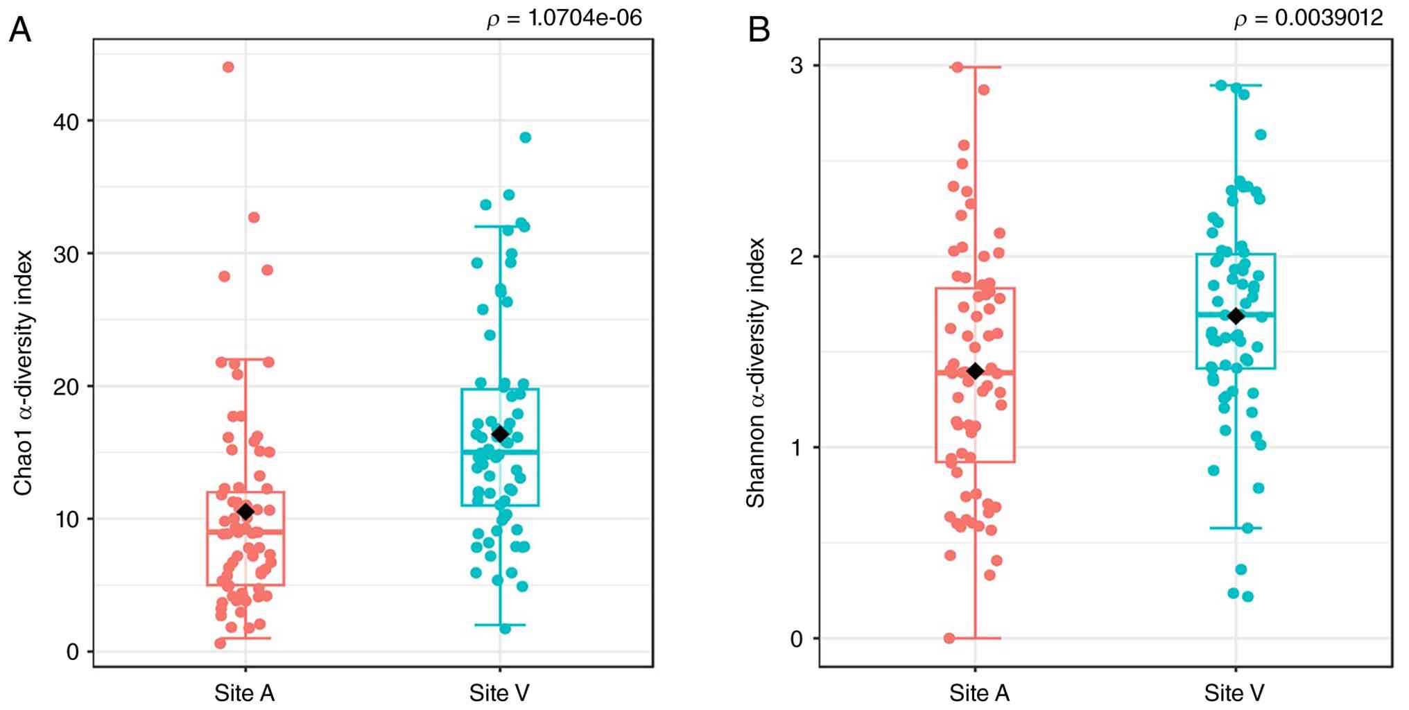
13,040±5,528 at site V. Using the Chao1 and Shannon indices,
α-diversity was determined (Fig.
1). Both mean and median values were found to be higher at site
V compared with site A, implying greater microbial diversity and a
lower proportion of samples with limited diversity. Although the
distribution at site V appeared less variable, with fewer outliers,
the results consistently suggested higher diversity compared with
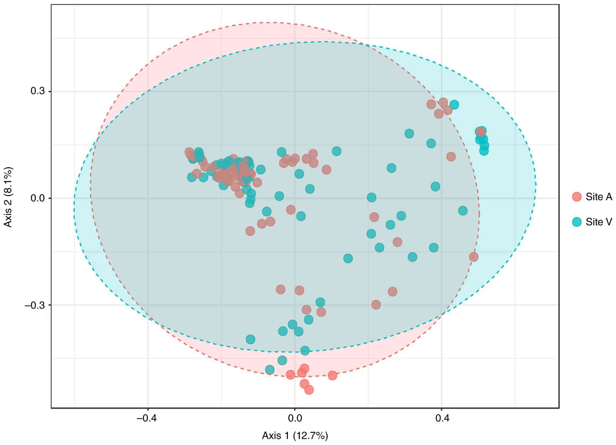
site A. Subsequent β-diversity analysis further supported these
findings (Fig. 2). Although a
substantial overlap between the two sites indicated shared
bacterial taxa, significant differences were observed (PERMANOVA,
P=0.025). Moreover, catheter samples from site V demonstrated
partial separation along axis 1, with a more scattered
distribution, suggesting the presence of bacterial taxa unique to
this site.
Relative abundance of bacteria in HD
catheter samples
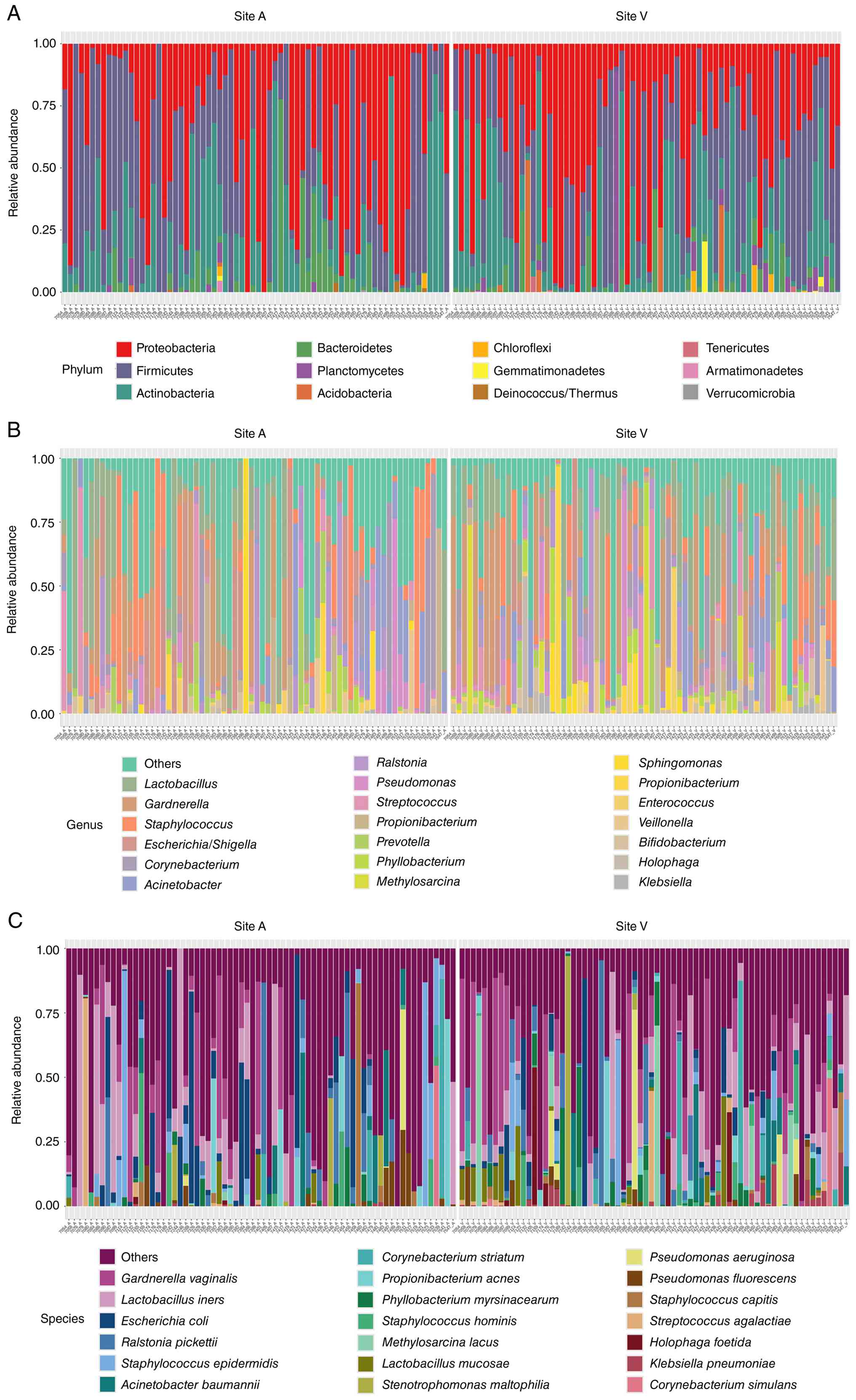
Relative abundances of bacteria from site A and site
V were visualized at the phylum, genus and species levels using
stacked bar charts (Fig. 3). At the
phylum level, Proteobacteria, Actinobacteria and Firmicutes
accounted for the majority of organisms found at both sites. At the
genus level, Lactobacillus, Gardnerella,
Escherichia, Shigella, Streptococcus and
Staphylococcus were the most prevalent genera. The 10 most
frequently identified bacterial species included Lactobacillus
iners, Gardnerella vaginalis, Escherichia coli,
Staphylococcus epidermidis, Acinetobacter baumannii,
Ralstonia pickettii, Corynebacterium striatum,
Stenotrophomonas maltophilia, Lactobacillus mucosae
and Methylosarcina lacus. Comparative analysis revealed
site-specific differences, with several known pathogens
(Pseudomonas aeruginosa, S. maltophilia and
Klebsiella pneumoniae) being notably enriched at site V.
Given their pathogenic potential and association with
catheter-associated bloodstream infections, sepsis and mortality,
these findings further highlighted the importance of strict hygiene
in patient care, healthcare personnel practices and dialysis
machine maintenance.
LEfSe of the bacterial profiles
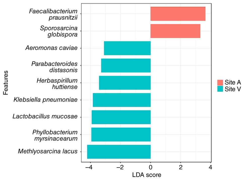
Based on the LEfSe presented in Fig. 4, analysis of bacterial profiles
between catheter sites [P<0.05; log LDA score ≥3.0] revealed
site-specific taxa. Faecalibacterium prausnitzii and
Sporosarcina globispora were primarily found at site A,
whereas Aeromonas caviae, Parabacteroides distasonis,
Herbaspirillum huttiense, K. pneumoniae, L.
mucosae, Phyllobacterium myrsinacearum and
Methylosarcina lacus were primarily observed at site V,
showing distinct microbial composition between catheter sites.
Association between clinical variables
and microbial species
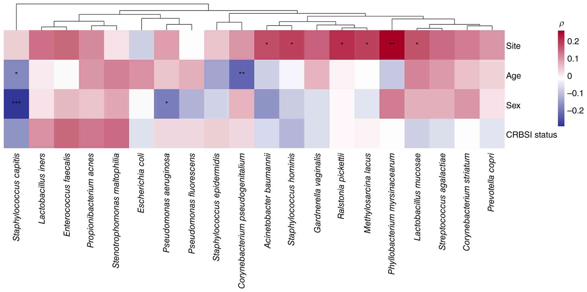
Spearman correlation analysis was subsequently
carried out to evaluate associations between clinical variables and
the top 20 bacterial species (Fig.
5). The catheter site demonstrated predominantly positive
correlations with several species. The strongest significant
association was observed for P. myrsinacearum (r=0.26;
P<0.01). Significant positive correlations with P<0.05 were
also identified for R. pickettii, Staphylococcus
hominis, A. baumannii, Methylosarcina lacus and
L. mucosae. These findings indicated enrichment of these
taxa at site V. However, age, as a clinical variable, showed a
small number of significant correlations. A significant negative
correlation was observed for Staphylococcus capitis
(r=-0.18; P<0.05) and Corynebacterium pseudogenitalium
(r=-0.22; P<0.01), suggesting a decreased abundance with
increasing age. However, no other species demonstrated
statistically significant associations with age; moreover,
sex-specific differences were observed for a limited number of
species. A strong negative correlation was identified for S.
capitis (r=-0.29; P<0.001), indicating enrichment in female
patients. In addition, P. aeruginosa showed a significant
negative correlation with the male sex (r=-0.19, P<0.05), also
suggesting a higher abundance in female patients. No species
demonstrated a statistically significant positive correlation with
the male sex. Neither were any statistically significant
correlations observed between CRBSI and non-CRBSI for any of the
evaluated species (all P>0.05), although weak trends were noted
for several taxa.
Discussion
The growing prevalence of antibiotic-resistant
bacteria highlights the limitations of conventional diagnostic
methods, and the necessity for more accurate strategies in pathogen
identification. Molecular approaches serve to provide a rapid and
reliable detection of bacterial strains to facilitate the
implementation of targeted therapeutic interventions (11). Among these techniques, 16S rDNA
amplicon sequencing, a widely utilized tool in metagenomic studies,
offers enhanced specificity for the identification of genes or
genomic regions of interest, while maintaining cost-effectiveness
(16,17). This sequencing technique is used to
characterize microbial communities across a variety of sample
types, including clinical specimens (such as blood and stool),
environmental samples (such as soil, wastewater and freshwater),
and host-associated microbiomes, such as those of humans, animals
and plants (2).
In the present study, full-length (V1-V9) 16S rDNA
amplification of representative catheter samples provided limited
information. This may be attributable to factors associated with
sample handling and preservation. Multiple freeze-thaw cycles and
prolonged storage at -20˚C may have resulted in compromised DNA
integrity, reducing the suitability of the material for full-length
amplification. Consequently, partial 16S rDNA amplification
targeting the V1-V4 region was selected in subsequent analyses to
ensure sufficient DNA for further amplification. Another potential
limitation was the choice of lysis buffer used during DNA
extraction. Although mammalian cells are readily disrupted due to
their relatively fragile plasma membranes, bacterial and fungal
cells are encapsulated within structurally resilient cell walls.
Mild lysis conditions can selectively lyse mammalian cells, while
sparing microbial populations. Nevertheless, certain microorganisms
may remain vulnerable under these conditions, leading to a
suboptimal DNA quantity. Such variability in DNA yield may
negatively influence the efficiency of downstream library
preparation and sequencing.
A previous study demonstrates the applicability of
16S rDNA amplicon sequencing in characterizing bacterial and fungal
communities in peritoneal dialysis fluid samples (30). However, to the best of our
knowledge, the present study is the first to use such an approach
to identify the bacterial composition within HD catheter samples.
Differences in bacterial communities between the catheter insertion
site (site A) and the vascular lumen (site V) were evident at each
of the phylum, genus and species levels.
The predominant phyla identified across the samples
were Proteobacteria, Actinobacteria and Firmicutes, findings which
are consistent with those of another study (31). At the genus level,
Lactobacillus, Gardnerella, Escherichia,
Shigella, Streptococcus and Staphylococcus
emerged as the most abundant genera. Moreover, species-level
analysis revealed that L. iners, G. vaginalis, E.
coli, S. epidermidis, A. baumannii, R.
pickettii, C. striatum, S. maltophilia, L.
mucosae and Methylosarcina lacus were among the 10 most
prevalent species. Furthermore, several of these organisms are
recognized either as established pathogens or as opportunistic
species with clinical relevance, suggesting the potential relevance
of these findings to catheter-associated infections.
The 10 most frequently identified bacterial species
in the present study may be broadly categorized into three groups,
namely environmental organisms, potential pathogens and beneficial
commensals. A. baumannii, a species commonly associated with
hospital-acquired infections, was detected in samples from both
sites, despite its usual environmental reservoirs in soil and water
(32). Similarly, S.
maltophilia, typically regarded as an environmental organism,
has emerged as a clinically relevant opportunistic pathogen,
especially in immunocompromised patients (33). Finally, R. pickettii,
although generally considered a contaminant, has also occasionally
been identified as a rare opportunistic pathogen (34).
Among the identified enteric organisms, E.
coli represents a species with both commensal and pathogenic
strains, the latter capable of causing intestinal and
extraintestinal infections, including urinary tract infections and
septicemia (35). G.
vaginalis, detected in samples from both sites, is a urogenital
pathogen, and the principal organism associated with bacterial
vaginosis (36). Regarding
skin-associated organisms, S. epidermidis, an important
component of the skin microbiota and a contributor to skin
homeostasis, is implicated in CRBSIs based on its capacity in
biofilm formation (37).
Additionally, C. striatum, a skin commensal, is reported as
an emerging opportunistic pathogen causing severe bloodstream
infections (38). The genus
Lactobacillus demonstrates contrasting roles. L.
iners, often described as a vaginal symbiont, is associated
with vaginal dysbiosis and an increased susceptibility to sexually
transmitted infections, whereas L. mucosae is considered
beneficial, and has been proposed for probiotic applications
(39). Finally, Methylosarcina
lacus, a methanotrophic organism capable of survival in
methane-rich environments, is classified as an environmental
species with limited clinical significance.
When comparing the bacterial profiles between the
two catheter sites, it was identified from the relative abundances
that host-associated bacterial species, including L. iners,
G. vaginalis, E. coli and S. epidermidis were
the most common at site A. Although these species were also present
in site V, additional environmental species were also found to be
increasingly abundant at the location. Such species included R.
pickettii and P. myrsinacearum, which both act as human
pathogens, and may also serve as indicators of water contamination
(40-42).
According to the relative abundance and LEfSe analyses, there was a
significantly increased abundance of G. vaginalis, as well
as the persistent presence of environmental species at site V.
These findings indicated the possible introduction of such species
via the water system that was used in the procedure. In addition,
several of the detected bacterial species were likely to have
translocated from other body sites, including the skin, the
intestines and the female reproductive tract. Examples of such
organisms included G. vaginalis, E. coli, S.
epidermidis and C. striatum. This also implicated their
possible introduction during HD, catheterization and catheter
removal. Given that dialysis machines are expected to be cleaned
and disinfected, these findings highlighted the potential need to
re-evaluate the cleaning and disinfection protocols used in
dialysis centers, as well as the quality control of dialysate and
water used in the dialysis system. Given the presence of known
human pathogens (such as A. baumannii and K.
pneumoniae) among the samples, an emphasis on enhancement of
the cleaning and disinfection protocols of the dialysis equipment,
along with improving personal hygiene among healthcare personnel
and patients with CKD, may be essential in preventing such
infections.
Spearman correlation analysis was also carried out
in the present study, which revealed that the catheter site had the
strongest influence on species-level microbial distribution,
whereas age and sex demonstrated limited, taxa-specific
associations. Furthermore, several species, including A.
baumannii, S. hominis and R. pickettii, were
positively associated with site V. These organisms are commonly
implicated in opportunistic infections and biofilm formation,
suggesting that the venous segment of the catheter may provide a
microenvironment conducive to colonization by potentially
pathogenic taxa (41,43). The enrichment of these species at
the venous site may reflect differences in blood flow dynamics,
nutrient availability or host-device interface characteristics that
facilitate microbial persistence (44-46).
The sex-specific associations were modest in comparison; however,
the populations of S. capitis and P. aeruginosa were
significantly enriched in female patients compared with male
patients. Both species are known to be skin- or
environment-associated organisms that have documented roles in
device-associated colonization (47-49).
Taken together, these findings suggested that host biological
differences, including hormonal influences or variations in skin
microbiota composition, may affect catheter-associated microbial
communities (47,50). The age-associated correlations were
also limited and primarily negative, indicating a potential decline
in certain commensal or skin-associated taxa with increasing age
(51). These observations may
reflect age-dependent alterations in skin barrier integrity, immune
function or microbiome composition (52). In addition, CRBSI status was not
significantly correlated with the abundance of the top 20 species.
This suggested that overt bloodstream infection may not be caused
by dominant taxa abundance, but rather by complex ecological
interactions, virulence factors or biofilm dynamics (53). Overall, the findings generated from
the Spearman correlation analysis highlighted the catheter site as
the principal determinant of microbial structuring at the species
level, whereas host demographic factors appeared to exert more
subtle modulatory effects (54).
Understanding these spatial and host-associated microbial patterns
may aid in the identification of high-risk colonization profiles,
and in informing targeted infection prevention strategies in
patients on HD (55,56).
In the clinical context, among all 70 participants
whose HD catheters were used in the study, 27 cases were confirmed
as cases of CRBSI. A total of 36 patients received antibiotic
treatment, including 9 patients with negative culture results, but
who demonstrated relevant symptoms (for example, fever,
nausea/vomiting and diarrhea). The management of CRBSI in patients
on HD relies on the prompt administration of antibiotics. Based on
the clinical data, a diverse correlation was identified between
specific antibiotic regimens and clinical outcomes, with
fourth-generation cephalosporins emerging as a main contributor for
positive outcomes, frequently associated with patients being able
to continue with HD. Vancomycin was often used in combination with
carbapenems or cephalosporins to support the maintenance of HD.
Meropenem was also used in several patients, allowing them to
either continue HD or to resume PD. An observation that concerned
the therapeutic limitations when treating multidrug-resistant (MDR)
pathogens was that patients with fatal outcomes often received the
most intensive antibiotic regimens, including combinations of
colistin, fosfomycin and meropenem, or sequences involving
piperacillin-tazobactam and vancomycin. These clinical data may
highlight the virulence of identified bacterial species, including
carbapenem-resistant P. aeruginosa, carbapenem-resistant
A. baumannii and MDR K. pneumoniae. Whereas
antibiotics such as ceftazidime and vancomycin are instrumental in
maintaining the treatment for numerous patients on HD, their
efficacy is limited by the presence of MDR gram-negative bacteria.
The relative abundance of these pathogens in site V samples
highlights the potential need for a rapid pathogen identification
to transition from empiric to precision therapy earlier in the
clinical course, which may potentially reduce the high mortality
rates associated with these resistant organisms.
However, the present study focused on characterizing
the taxonomic composition of catheter-associated microbiota, and
antibiotic-associated outcomes were not evaluated. Future
investigation is required in future studies, which constitutes one
of the limitations of the present study. Furthermore, studies on
the differences in the fungal, as well as the viral, microbiome
should also be carried out. Additionally, the DNA that was
sequenced from the biofilm may have resulted from both current and
past infections, highlighting that culture results may need to be
used as references, and that further validation is required.
Furthermore, since data regarding the catheter insertion location
and relevant previous infections were not obtained, these results
could not be used to determine what the direct causative effects
were. Further studies, such as investigations incorporating shotgun
metagenomic sequencing, may provide comprehensive functional
profiling, including the detection of antibiotic resistance genes,
virulence determinants, biofilm-associated bacterial pathways and
the symptoms or clinical outcome of the patients. Such an approach
would provide additional in-depth insights into the clinical
significance of catheter microbiomes and their potential
association with CRBSI.
In conclusion, the present study demonstrated
differences in the bacterial composition present at two distinct HD
catheter sites. Through the application of 16S rDNA amplicon
sequencing, the identification and characterization of bacterial
profiles were carried out in patients with CKD who were undergoing
HD. This molecular approach offers potential clinical utility by
enabling the precise and prompt identification of pathogens, which
may facilitate the delivery of optimal therapeutic interventions.
Therefore, its integration into clinical practice may improve
patient outcomes, potentially by reducing the morbidity and
mortality associated with CRBSI in this vulnerable population.
Acknowledgements
The authors would like to thank Mr. Chainarong
Bunma, Center of Excellence in Systems Microbiology, Department of
Biochemistry, Faculty of Medicine, Chulalongkorn University for
technical support and assistance with DNA extraction, and Dr
Trairak Pisitkun, Head of the Chula Excellence Center of Systems
Biology (CUSB), Faculty of Medicine, Chulalongkorn University, for
kindly providing access to the server used for data analysis in the
present study. The authors also thank Miss Piyaporn Towannang,
(King Chulalongkorn Memorial Hospital, Bangkok, Thailand) and Dr
Chanchana Boonyakrai, (Taksin Hospital, Bangkok, Thailand) for
their contributions to the provision of HD catheter samples.
Funding
Funding: The present study was supported by Genomics Thailand,
the Health Systems Research Institute (HSRI), Thailand (grant no.
68-109) and the Thailand Science Research and Innovation Fund,
Chulalongkorn University, Thailand (grant no.
HEA_FF_69_123_3000_012).
Availability of data and materials
The data generated in the present study may be found
in the NCBI database under the BioProject accession number
PRJNA1346282 and Sequence Read Archive accession numbers
SRR35811925-SRR35812066 or at the following URL: https://www.ncbi.nlm.nih.gov/bioproject/PRJNA1346282.
Authors' contributions
SP and TK designed the study. BS and SV performed
the experiments, interpreted the results, and contributed to
drafting and revising the manuscript. SV contributed to data
analysis. WW contributed to the acquisition and preparation of
clinical samples and the collection of patient data. TS and PP
contributed to the handling and transport of clinical samples and
the collection and compilation of patient data. SP and TK
supervised the study, critically revised the manuscript and confirm
the authenticity of all raw data. All authors critically reviewed
the manuscript, approved the final version for publication, and
agree to be accountable for all aspects of the work.
Ethics approval and consent to
participate
The present study was performed in line with the
Declaration of Helsinki (2013), and the protocol of the present
study was approved by the Institutional Review Board of the Faculty
of Medicine, Chulalongkorn University (approval no. 0834/2025; IRB
no. 0282/68; Bangkok, Thailand). Informed consent was obtained from
all of the included patients.
Patient consent for publication
Not applicable.
Competing interests
The authors declare that they have no competing
interests.
References
|
1
|
Kovesdy CP: Epidemiology of chronic kidney
disease: An update 2022. Kidney Int Suppl (2011). 12:7–11.
2022.PubMed/NCBI View Article : Google Scholar
|
|
2
|
Jin J, Liu X and Shiroguchi K: Long
journey of 16S rRNA-amplicon sequencing toward cell-based
functional bacterial microbiota characterization. IMetaOmics.
1(e9)2024.PubMed/NCBI View
Article : Google Scholar
|
|
3
|
GBD 2023 Chronic Kidney Disease
Collaborators. Global, regional, and national burden of chronic
kidney disease in adults, 1990-2023, and its attributable risk
factors: A systematic analysis for the global burden of disease
study 2023. Lancet. 406:2461–2482. 2025.PubMed/NCBI View Article : Google Scholar
|
|
4
|
Chen TK, Knicely DH and Grams ME: Chronic
kidney disease diagnosis and management: A review. JAMA.
322:1294–1304. 2019.PubMed/NCBI View Article : Google Scholar
|
|
5
|
Tzanakaki E, Boudouri V, Stavropoulou A,
Stylianou K, Rovithis M and Zidianakis Z: Causes and complications
of chronic kidney disease in patients on dialysis. Health Sci J.
8:343–349. 2014.
|
|
6
|
Schamroth Pravda M, Maor Y, Brodsky K,
Katkov A, Cernes R, Schamroth Pravda N, Tocut M, Zohar I, Soroksky
A and Feldman L: Blood stream infections in chronic hemodialysis
patients-characteristics and outcomes. BMC Nephrol.
25(3)2024.PubMed/NCBI View Article : Google Scholar
|
|
7
|
Soi V, Moore CL, Kumbar L and Yee J:
Prevention of catheter-related bloodstream infections in patients
on hemodialysis: Challenges and management strategies. Int J
Nephrol Renovasc Dis. 9:95–103. 2016.PubMed/NCBI View Article : Google Scholar
|
|
8
|
Onder AM, Cuff CF, Liang X, Billings AA,
Onder S, Yu JJ and King JA: Detecting the prevalence of bacterial
colonization on tunneled cuffed hemodialysis catheters using
quantitative PCR targeting 16S rRNA and scanning electron
microscopy. J Vasc Access. 23:743–753. 2022.PubMed/NCBI View Article : Google Scholar
|
|
9
|
Bjarnsholt T: The role of bacterial
biofilms in chronic infections. APMIS. Suppl:1–51. 2013.PubMed/NCBI View Article : Google Scholar
|
|
10
|
Franco-Duarte R, Černáková L, Kadam S,
Kaushik KS, Salehi B, Bevilacqua A, Corbo MR, Antolak H,
Dybka-Stępień K, Leszczewicz M, et al: Advances in chemical and
biological methods to identify microorganisms-from past to present.
Microorganisms. 7(130)2019.PubMed/NCBI View Article : Google Scholar
|
|
11
|
Batool M and Galloway-Peña J: Clinical
metagenomics-challenges and future prospects. Front Microbiol.
14(1186424)2023.PubMed/NCBI View Article : Google Scholar
|
|
12
|
Ranjan R, Rani A, Metwally A, McGee HS and
Perkins DL: Analysis of the microbiome: Advantages of whole genome
shotgun versus 16S amplicon sequencing. Biochem Biophys Res Commun.
469:967–977. 2016.PubMed/NCBI View Article : Google Scholar
|
|
13
|
Chiu CY and Miller SA: Clinical
metagenomics. Nat Rev Genet. 20:341–355. 2019.PubMed/NCBI View Article : Google Scholar
|
|
14
|
Kuczynski J, Lauber CL, Walters WA,
Parfrey LW, Clemente JC, Gevers D and Knight R: Experimental and
analytical tools for studying the human microbiome. Nat Rev Genet.
13:47–58. 2011.PubMed/NCBI View Article : Google Scholar
|
|
15
|
Sims D, Sudbery I, Ilott NE, Heger A and
Ponting CP: Sequencing depth and coverage: Key considerations in
genomic analyses. Nat Rev Genet. 15:121–132. 2014.PubMed/NCBI View Article : Google Scholar
|
|
16
|
Gołębiewski M and Tretyn A: Generating
amplicon reads for microbial community assessment with
next-generation sequencing. J Appl Microbiol. 128:330–354.
2020.PubMed/NCBI View Article : Google Scholar
|
|
17
|
Matsuo Y, Komiya S, Yasumizu Y, Yasuoka Y,
Mizushima K, Takagi T, Kryukov K, Fukuda A, Morimoto Y, Naito Y, et
al: Full-length 16S rRNA gene amplicon analysis of human gut
microbiota using MinION™ nanopore sequencing confers
species-level resolution. BMC Microbiol. 21(35)2021.PubMed/NCBI View Article : Google Scholar
|
|
18
|
Woo PCY, Lau SKP, Teng JLL, Tse H and Yuen
KY: Then and now: Use of 16S rDNA gene sequencing for bacterial
identification and discovery of novel bacteria in clinical
microbiology laboratories. Clin Microbiol Infect. 14:908–934.
2008.PubMed/NCBI View Article : Google Scholar
|
|
19
|
Charalampous T, Kay GL, Richardson H,
Aydin A, Baldan R, Jeanes C, Rae D, Grundy S, Turner DJ, Wain J, et
al: Nanopore metagenomics enables rapid clinical diagnosis of
bacterial lower respiratory infection. Nat Biotechnol. 37:783–792.
2019.PubMed/NCBI View Article : Google Scholar
|
|
20
|
Santos R, Lee H, Williams A, Baffour-Kyei
A, Lee SH, Troakes C, Al-Chalabi A, Breen G and Iacoangeli A:
Investigating the performance of oxford nanopore long-read
sequencing with respect to illumina microarrays and short-read
sequencing. Int J Mol Sci. 26(4492)2025.PubMed/NCBI View Article : Google Scholar
|
|
21
|
De Maio N, Shaw LP, Hubbard A, George S,
Sanderson ND, Swann J, Wick R, AbuOun M, Stubberfield E, Hoosdally
SJ, et al: Comparison of long-read sequencing technologies in the
hybrid assembly of complex bacterial genomes. Microb Genom.
5(e000294)2019.PubMed/NCBI View Article : Google Scholar
|
|
22
|
Ferdous T, Jiang L, Dinu I, Groizeleau J,
Kozyrskyj AL, Greenwood CMT and Arrieta MC: The rise to power of
the microbiome: Power and sample size calculation for microbiome
studies. Mucosal Immunol. 15:1060–1070. 2022.PubMed/NCBI View Article : Google Scholar
|
|
23
|
Cohen J: Statistical Power Analysis for
the Behavioral Sciences. 2nd edition. L. Erlbaum Associates,
Hillsdale, NJ, p567, 1988.
|
|
24
|
Wick RR, Judd LM and Holt KE: Performance
of neural network basecalling tools for Oxford Nanopore sequencing.
Genome Biol. 20(129)2019.PubMed/NCBI View Article : Google Scholar
|
|
25
|
Lanfear R, Schalamun M, Kainer D, Wang W
and Schwessinger B: MinIONQC: Fast and simple quality control for
MinION sequencing data. Bioinformatics. 35:523–525. 2019.PubMed/NCBI View Article : Google Scholar
|
|
26
|
Rodríguez-Pérez H, Ciuffreda L and Flores
C: NanoCLUST: A species-level analysis of 16S rRNA nanopore
sequencing data. Bioinformatics. 37:1600–1601. 2021.PubMed/NCBI View Article : Google Scholar
|
|
27
|
Cole JR, Wang Q, Fish JA, Chai B,
McGarrell DM, Sun Y, Brown CT, Porras-Alfaro A, Kuske CR and Tiedje
JM: Ribosomal database project: Data and tools for high throughput
rRNA analysis. Nucleic Acids Res. 42:D633–D642. 2014.PubMed/NCBI View Article : Google Scholar
|
|
28
|
Bolyen E, Rideout JR, Dillon MR, Bokulich
NA, Abnet CC, Al-Ghalith GA, Alexander H, Alm EJ, Arumugam M,
Asnicar F, et al: Reproducible, interactive, scalable and
extensible microbiome data science using QIIME 2. Nat Biotechnol.
37:852–857. 2019.PubMed/NCBI View Article : Google Scholar
|
|
29
|
Chong J, Liu P, Zhou G and Xia J: Using
MicrobiomeAnalyst for comprehensive statistical, functional, and
meta-analysis of microbiome data. Nat Protoc. 15:799–821.
2020.PubMed/NCBI View Article : Google Scholar
|
|
30
|
Visedthorn S, Klomkliew P, Sawaswong V,
Sivapornnukul P, Chanchaem P, Saejew T, Pavatung P, Kanjanabuch T
and Payungporn S: Bacterial classification based on metagenomic
analysis in peritoneal dialysis effluent of patients with chronic
kidney disease. Biomed Rep. 21(102)2024.PubMed/NCBI View Article : Google Scholar
|
|
31
|
Yan Z, Wang Y, Zeng W, Xia R, Liu Y, Wu Z,
Deng W, Zhu M, Xu J, Deng H and Miao Y: Microbiota of long-term
indwelling hemodialysis catheters during renal transplantation
perioperative period: A cross-sectional metagenomic microbial
community analysis. Ren Fail. 45(2256421)2023.PubMed/NCBI View Article : Google Scholar
|
|
32
|
Ayoub Moubareck C and Hammoudi Halat D:
Insights into Acinetobacter baumannii: A review of
microbiological, virulence, and resistance traits in a threatening
nosocomial pathogen. Antibiotics (Basel). 9(119)2020.PubMed/NCBI View Article : Google Scholar
|
|
33
|
Adegoke AA, Stenström TA and Okoh AI:
Stenotrophomonas maltophilia as an emerging ubiquitous
pathogen: Looking beyond contemporary antibiotic therapy. Front
Microbiol. 8(2276)2017.PubMed/NCBI View Article : Google Scholar
|
|
34
|
Nasir N, Sayeed MA and Jamil B:
Ralstonia pickettii bacteremia: An emerging infection in a
tertiary care hospital setting. Cureus. 11(e5084)2019.PubMed/NCBI View Article : Google Scholar
|
|
35
|
Braz VS, Melchior K and Moreira CG:
Escherichia coli as a multifaceted pathogenic and versatile
bacterium. Front Cell Infect Microbiol. 10(548492)2020.PubMed/NCBI View Article : Google Scholar
|
|
36
|
Morrill S, Gilbert NM and Lewis AL:
Gardnerella vaginalis as a cause of bacterial vaginosis:
Appraisal of the evidence from in vivo models. Front Cell Infect
Microbiol. 10(168)2020.PubMed/NCBI View Article : Google Scholar
|
|
37
|
Severn MM and Horswill AR:
Staphylococcus epidermidis and its dual lifestyle in skin
health and infection. Nat Rev Microbiol. 21:97–111. 2023.PubMed/NCBI View Article : Google Scholar
|
|
38
|
Chen FL, Hsueh PR, Teng SO, Ou TY and Lee
WS: Corynebacterium striatum bacteremia associated with
central venous catheter infection. J Microbiol Immunol Infect.
45:255–258. 2012.PubMed/NCBI View Article : Google Scholar
|
|
39
|
de Moraes GMD, de Abreu LR, do Egito AS,
Salles HO, da Silva LMF, Nero LA, Todorov SD and Dos Santos KMO:
Functional properties of Lactobacillus mucosae strains
isolated from Brazilian goat milk. Probiotics Antimicrob Proteins.
9:235–245. 2017.PubMed/NCBI View Article : Google Scholar
|
|
40
|
Ryan MP and Adley CC: Ralstonia spp.:
Emerging global opportunistic pathogens. Eur J Clin Microbiol
Infect Dis. 33:291–304. 2014.PubMed/NCBI View Article : Google Scholar
|
|
41
|
K Thet M, Pelobello MLF, Das M, Alhaji MM,
Chong VH, Khalil MAM, Chinniah T and Tan J: Outbreak of
nonfermentative Gram-negative bacteria (Ralstonia pickettii
and Stenotrophomonas maltophilia) in a hemodialysis center.
Hemodial Int. 23:E83–E89. 2019.PubMed/NCBI View Article : Google Scholar
|
|
42
|
Hughes G, Pollard C, Chavda S and David
MD: The first case report of human infection with
Phyllobacterium myrsinacearum causing spondylodiscitis. Clin
Infect Pract. 7-8(100029)2020.
|
|
43
|
Xiao BL, Hu XQ and Li M: Dysbiosis and
Staphylococcus species over representation in the exit site
skin microbiota of hemodialysis patients carrying tunneled cuffed
central venous catheter. Ren Fail. 46(2363417)2024.PubMed/NCBI View Article : Google Scholar
|
|
44
|
Tejera D, Limongi G, Bertullo M and
Cancela M: Ralstonia pickettii bacteremia in hemodialysis
patients: A report of two cases. Rev Bras Ter Intensiva.
28:195–198. 2016.PubMed/NCBI View Article : Google Scholar
|
|
45
|
Bowler PG, Duerden BI and Armstrong DG:
Wound microbiology and associated approaches to wound management.
Clin Microbiol Rev. 14:244–269. 2001.PubMed/NCBI View Article : Google Scholar
|
|
46
|
Hall-Stoodley L, Costerton JW and Stoodley
P: Bacterial biofilms: From the natural environment to infectious
diseases. Nat Rev Microbiol. 2:95–108. 2004.PubMed/NCBI View Article : Google Scholar
|
|
47
|
Lowes MA, Kikuchi T, Fuentes-Duculan J,
Cardinale I, Zaba LC, Haider AS, Bowman EP and Krueger JG:
Psoriasis vulgaris lesions contain discrete populations of Th1 and
Th17 T cells. J Invest Dermatol. 128:1207–1211. 2008.PubMed/NCBI View Article : Google Scholar
|
|
48
|
Ebenezer A, Noutcha AEM, Agi PI, Okiwelu
SN and Commander T: Spatial distribution of the sibling species of
Anopheles gambiae sensu lato (Diptera: Culicidae) and malaria
prevalence in Bayelsa State, Nigeria. Parasit Vectors.
7(32)2014.PubMed/NCBI View Article : Google Scholar
|
|
49
|
Leece EA, Girard NM and Maddern K:
Alfaxalone in cyclodextrin for induction and maintenance of
anaesthesia in ponies undergoing field castration. Vet Anaesth
Analg. 36:480–484. 2009.PubMed/NCBI View Article : Google Scholar
|
|
50
|
Chen X, Cui T, Fu J, Peng J and Shan J:
Trend-residual dual modeling for detection of outliers in low-cost
GPS trajectories. Sensors (Basel). 16(2036)2016.PubMed/NCBI View Article : Google Scholar
|
|
51
|
Shahar OD, Gabizon R, Feine O, Alhadeff R,
Ganoth A, Argaman L, Shimshoni E, Friedler A and Goldberg M:
Acetylation of lysine 382 and phosphorylation of serine 392 in p53
modulate the interaction between p53 and MDC1 in vitro. PLoS One.
8(e78472)2013.PubMed/NCBI View Article : Google Scholar
|
|
52
|
Grice EA and Segre JA: The skin
microbiome. Nat Rev Microbiol. 9:244–253. 2011.PubMed/NCBI View Article : Google Scholar
|
|
53
|
Whitehead TR, Spence C and Cotta MA:
Inhibition of hydrogen sulfide, methane, and total gas production
and sulfate-reducing bacteria in in vitro swine manure by tannins,
with focus on condensed quebracho tannins. Appl Microbiol
Biotechnol. 97:8403–8409. 2013.PubMed/NCBI View Article : Google Scholar
|
|
54
|
Ferrier A, Garin D and Crance JM: Rapid
inactivation of vaccinia virus in suspension and dried on surfaces.
J Hosp Infect. 57:73–79. 2004.PubMed/NCBI View Article : Google Scholar
|
|
55
|
Darouiche RO: Device-associated
infections: A macroproblem that starts with microadherence. Clin
Infect Dis. 33:1567–1572. 2001.PubMed/NCBI View
Article : Google Scholar
|
|
56
|
Donlan RM: Biofilms and device-associated
infections. Emerg Infect Dis. 7:277–281. 2001.PubMed/NCBI View Article : Google Scholar
|