Introduction
With the transformation of lifestyles and the
increase in the obesity rate, non-alcoholic fatty liver disease
(NAFLD) has become the most common chronic liver disease worldwide
(1). According to a systematic
review and meta-analysis, ~25% of adults are affected by NAFLD
globally, and its incidence is still on the rise (1). The core pathological feature of NAFLD
is excessive lipid deposition in the liver, which not only impairs
liver function but is also closely related to disorders of glucose
and lipid metabolism (2). A number
of clinical studies have shown that patients with NAFLD often have
metabolic abnormalities, such as insulin resistance, hyperglycemia,
and hyperlipidemia (3,4). These metabolic disorders are
intertwined, further increasing the risk of serious complications
such as cardiovascular diseases and type 2 diabetes, posing a
notable threat to patient health and quality of life.
Bile acids are important products of liver
metabolism and serve key roles in lipid metabolism.
Chenodeoxycholic acid (CDCA), a notable component of bile acids,
has received considerable attention in previous years (5). Previous research has demonstrated that
CDCA can improve hepatic lipid metabolism, as evidenced by animal
experiments where triglyceride and cholesterol contents in animal
livers were markedly reduced and beneficial changes occurred in the
expression of lipid metabolism-related genes after CDCA
intervention (6). However, its
specific molecular mechanism has not been fully elucidated,
limiting its further application in the treatment of fatty
liver.
Pyruvate kinase (PK), a key rate-limiting enzyme in
the glycolysis pathway, catalyzes the conversion of
phosphoenolpyruvate to pyruvate and simultaneously generates ATP,
providing energy for cells (7). In
previous years, an increasing number of studies have shown that PK
serves an important role in the regulation of hepatic lipid
metabolism (8-10).
Changes in the activity and expression levels of PK can affect
fatty acid synthesis and oxidation, thereby affecting the balance
of hepatic lipid metabolism (7).
However, there are currently relatively few studies on whether
there is an interaction between CDCA and PK and how this
interaction affects hepatic lipid metabolism.
The present study aimed to explore whether CDCA
regulates hepatic lipid metabolism by interacting with PK. This not
only helps to reveal the potential molecular mechanism of CDCA in
improving hepatic lipid metabolism, and enriches the understanding
of the mechanism of bile acids in hepatic lipid metabolism, but
also provides novel targets and a theoretical basis for the
treatment of fatty liver, with important theoretical significance
and clinical application value.
Materials and methods
Cell culture
The human normal hepatocyte cell line THLE-2 was
purchased from American Type Culture Collection (ATCC; cat. no.
CRL-2706), and human hepatoma cells HepG2 were purchased from ATCC
(cat. no. HB-8065) and preserved in the laboratory. All cell lines
used in the present study were authenticated using short tandem
repeat (STR) profiling (Genewiz, Inc.) within 6 months prior to
experimentation, and the STR profiles of THLE-2 and HepG2 matched
the reference databases (ATCC), confirming no cross-contamination.
The THLE-2 cells were cultured in BEGM™ Bronchial
Epithelial Cell Growth Medium (cat. no. CC-3170; Lonza Group,
Ltd.), which included 5 ng/ml EGF, 0.5 µg/ml hydrocortisone, 0.5
µg/ml epinephrine, 10 µg/ml transferrin, 5 µg/ml insulin, 0.1 ng/ml
retinoic acid, 6.5 ng/ml triiodothyronine and 50 µg/ml
gentamicin/amphotericin B. HepG2 cells were cultured in DMEM (cat.
no. SH30243.01; HyClone™; Cytiva) containing 10% fetal
bovine serum (FBS; Gibco; cat. no. 10099-141; Thermo Fisher
Scientific, Inc.) and 1% penicillin-streptomycin (cat. no. P1400;
Beijing Solarbio Science & Technology Co., Ltd.), and both cell
lines were routinely cultured in an incubator at 37˚C and 5%
CO2.
High-fat (HF) cell model
When the confluence of THLE-2 and HepG2 cells
reached 70-80%, the medium was replaced with serum-free DMEM
containing oleic acid (OA; cat. no. P1145; Sigma-Adrich; Merck
KGaA) and palmitic acid (PA; cat. no. P5585; Sigma-Adrich; Merck
KGaA). Various OA/PA combinations were tested (0.25/0.125, 0.5/0.25
and 1.0/0.5 mM) and the cells were induced for 24 h. The
concentrations (0.5 mM OA + 0.25 mM PA) were selected because this
combination induced optimal lipid droplet accumulation (as detected
by BODIPY staining) without excessive cytotoxicity [viability
>80% via Cell Counting Kit-8 (CCK-8) assay (Beyotime Institute
of Biotechnology); data not shown]. For the CCK-8 assay, 10 µl of
CCK-8 solution was added to the cells after 48 h of culture. The
cells were then incubated for an additional 2 h, and the absorbance
at 450 nm (OD value) was measured using a microplate reader. Cells
were divided into the following groups for treatment: The control
group, normal cultured THLE-2 and HepG2 cells; the HF group, cells
induced with OA/PA without additional treatment; the CDCA group,
during HF induction, 10 and 50 µM CDCA (cat. no. HY-76847;
MedChemExpress) were added for intervention; and the CDCA +
Shikonin (cat. no. HY-N0822; MedChemExpress) group, 1 h before
adding 50 µM CDCA for intervention, 10 µM Shikonin was added for
pretreatment. Each group had 6 replicate wells and subsequent
detection was performed after 24 h of treatment.
BODIPY staining
THLE-2 and HepG2 cells were fixed in 4% ice-cold
paraformaldehyde (cat. no. P0099; Beyotime Institute of
Biotechnology) at 4˚C for 20 min, then treated with 0.1% Triton
X-100 (Beyotime Institute of Biotechnology) at room temperature for
5 min, and washed twice with phosphate-buffered saline (PBS) to
remove the residual 4% paraformaldehyde. A working fluid solution
containing 10 µM BODIPY 493/503 (cat. no. GC42959; GLPBIO
Technology LLC) was added to the cells, and the mixture was
incubated at 37˚C for 30 min in the dark. Subsequently, the cell
nuclei were stained with DAPI at room temperature for 5 min after
two washes with PBS. After three more washes with PBS, the cells
were immediately imaged using a fluorescence microscope (IX73;
Olympus Corporation) at a magnification of x200. The area and
fluorescence intensity of the lipid droplets were quantified using
ImageJ software (version 1.8.0; National Institutes of Health) by
analyzing five random fields per well (6).
Animal model
All animal experiments were performed in compliance
with all relevant ethical regulations for animal testing and
research and in accordance with the animal protocols approved
(approval no. SW-2020R0126-F03) by Pingxiang People's Hospital
(Pingxiang, China). A total of 24 male, 6-week-old C57BL/6J mice
were purchased from Beijing Vital River Laboratory Animal
Technology Co., Ltd. The mice were housed in SPF-grade animal rooms
with controlled temperature (22±2˚C), humidity (50±10%), and a 12-h
light/dark cycle. Each cage contained 6 mice (to ensure social
interaction). Sterile food and filtered water were provided ad
libitum. After 1 week of adaptive feeding in an SPF-grade
animal room, the experiment was performed. The NAFLD model was
established by feeding the mice a high-fat diet (HFD; cat. no.
D12492; 60% fat content Research Diets, Inc.) for 12 weeks.
Successful NAFLD modeling was determined based on body weight, with
mice in the HF group exhibiting a body weight ≥20% higher than the
normal diet group (average HF group weight ~38 g vs. normal group
~31 g).
After successful NAFLD modeling, 18 mice were
randomly divided into three groups (n=6): i) A high-fat diet group
(HF); ii) HF + CDCA (50 mg/kg/d); and iii) HF + CDCA (100 mg/kg/d).
A normal diet group (fed with normal feed) was used as a control
(n=6). During the entire study period, all the mice included at the
start were monitored closely. There were no exclusions or losses;
all mice completed the experimental procedures as scheduled, which
ensured the integrity and reliability of the experimental data. The
normal feed (cat. no. D12450B; Research Diets, Inc.) contained 10%
fat, 70% carbohydrate and 20% protein, which was used as a direct
contrast to the HFD (60% fat). CDCA (purity ≥98%; MedChemExpress)
was dissolved in 0.5% carboxymethylcellulose sodium solution (cat.
no. HY-Y0703; MedChemExpress) and administered by oral gavage at
the corresponding dose once a day for 8 weeks. The total experiment
duration (21 weeks) included 1 week of adaptation, 12 weeks of HFD
feeding, and 8 weeks of CDCA intervention.
After the intervention, the mice were weighed and
fasted for 12 h without water deprivation. For blood collection,
50-60 mg/kg sodium pentobarbital was administered intraperitoneally
(anesthetic route, intraperitoneal injection) to induce temporary
anesthesia, and 0.2 ml of blood was collected by eyeball
enucleation. The animals were euthanized using an intraperitoneal
overdose of sodium pentobarbital (100 mg/kg body weight; euthanasia
method, intraperitoneal injection of anesthetic overdose). Death
was verified by: i) No spontaneous breathing (chest movement) or
heartbeat (chest palpation) for ≥5 min; and ii) no hind paw
withdrawal reflex when pinched with forceps.
During the experiment, mice were monitored daily for
health (coat condition, wounds, jaundice) and behavior (activity,
feeding/drinking); body weight was measured weekly. Humane
endpoints for euthanasia included: Severe weight loss (>20% of
initial weight in 1 week), persistent lethargy/refusal to eat/drink
for >24 h, visible organ damage (abdominal distension, and
jaundice), or abnormal behaviors (trembling, convulsions). No mice
met these endpoints or died unexpectedly; all 24 mice were
euthanized at the end of the intervention.
The serum was separated, and the liver was quickly
removed and fixed in 4% paraformaldehyde at 4˚C for 24 h. After
fixation, the tissues were dehydrated and then cut into 8-10
µm-thick frozen sections using a cryostat. The sections were
air-dried at room temperature for 10 min, followed by staining with
freshly prepared Oil Red O (Thermo Fisher Scientific, Inc.) working
solution at room temperature for 20 min in the dark. Finally, the
stained sections were mounted with glycerol gelatin, and lipid
droplet morphology was observed under a bright-field light
microscope (Nikon E100) at a magnification of x400 (scale bar, 10
µm) and the remaining tissue was frozen at -80˚C for biochemical
and molecular biology detection.
Determination of total cholesterol
(TC), triglyceride (TG), malondialdehyde (MDA) and superoxide
dismutase (SOD) contents
After cell collection, the cell lysate [2% Triton
X-100 (Beyotime Institute of Biotechnology) lysate] was added to
the cell pellet and incubated for 30 min to completely lyse the
cells. Experiments were performed according to the instructions of
the following kits: TC and TG detection kits (cat. nos. A111-1-1
and A110-1-1; Nanjing Jiancheng Bioengineering Institute), and SOD
(cat. no. BC0170) and MDA (cat. no. BC0020) detection kits (Beijing
Solarbio Science & Technology Co., Ltd.).
Reverse transcription-quantitative PCR
(RT-qPCR)
Total RNA from HepG2 cells or mouse liver tissues
was extracted using TRIzol® reagent (cat. no. 15596026;
Inivtrogen; Thermo Fisher Scientific, Inc.), reverse-transcribed
into cDNA (cat. no. RR037A; Takara Bio, Inc.), and qPCR reactions
of lipid synthesis [fatty acid synthase (FASN) and sterol
regulatory element-binding transcription factor 1 (SREBP-1c)] and
β-oxidation [CTP synthase 1 (CPT1α) and peroxisome
proliferator-activated receptor α (PPARα)]-related genes were
performed with SYBR Green Master Mix (cat. no. 04707516001; Roche
Diagnostics), according to the manufacturer's instructions. The
thermocycling conditions were as follows: An initial denaturation
step was performed at 95˚C for 10 min. This was followed by 40-45
cycles consisting of three sequential steps of denaturation at 95˚C
for 10 sec, annealing at 60˚C for 20 sec, and elongation at 72˚C
for 20 sec. After the cycling stage, a final extension step was
conducted at 72˚C for 3 min. GAPDH was used as an internal
reference gene, and the relative gene expression was calculated
using the 2-ΔΔC method (11). The primer sequences for the target
and reference genes are listed in Table
I.
 | Table IPrimer sequences for reverse
transcription-quantitative PCR. |
Table I
Primer sequences for reverse
transcription-quantitative PCR.
| Genes | Forward primer
(5'→3') | Reverse primer
(5'→3') | Organisms |
|---|
| FASN |
CACAGTGCTCAAAGGACATGCC |
CACCAGGTGTAGTGCCTTCCTC | Mus musculus |
| |
TTCTACGGCTCCACGCTCTTCC |
GAAGAGTCTTCGTCAGCCAGGA | Homo sapiens |
| SREBP-1c |
CCGTAGAGAAGCTTGGCGAT |
CACTAGAGGTCGGCATGGTC | Mus musculus |
| |
ACTTCTGGAGGCATCGCAAGCA |
AGGTTCCAGAGGAGGCTACAAG | Homo sapiens |
| CPT1α |
GACCGCGCTCTTAGGACTAC |
GAGCAGCAAGGAAGGACACT | Mus musculus |
| |
GATCCTGGACAATACCTCGGAG |
CTCCACAGCATCAAGAGACTGC | Homo sapiens |
| PPARα |
AGAGGGCTGAGCGTAGGTAA |
ATTGGGCCGGTTAAGACCAG | Mus musculus |
| |
TCGGCGAGGATAGTTCTGGAAG |
GACCACAGGATAAGTCACCGAG | Homo sapiens |
| GAPDH |
CATCACTGCCACCCAGAAGACTG |
ATGCCAGTGAGCTTCCCGTTCAG | Mus musculus |
| |
GTCTCCTCTGACTTCAACAGCG |
ACCACCCTGTTGCTGTAGCCAA | Homo sapiens |
Detection of PK activity and
pyruvate
Experiments were performed according to the
instructions of the PK activity detection kit and pyruvate
detection kit (cat. nos. BC0540 and BC2200; Beijing Solarbio
Science & Technology Co., Ltd.).
Co-immunoprecipitation (co-IP) and
high-performance liquid chromatography (HPLC) detection
To determine the binding site of CDCA and PK, after
treating cells with CDCA, the cells were lysed in
Pierce™ RIPA Buffer (cat. no. 89900; Thermo Fisher
Scientific, Inc.) to extract the total protein, and a PK antibody
was added for IP. For each IP reaction, 500 µl of normalized cell
lysate (containing 1,000 µg total protein) was incubated with 2 µg
of anti-PK polyclonal antibody (cat. no. ab137852; Abcam), while
normal rabbit IgG (cat. no. ab37415; Abcam) at the same amount was
used as the negative control. Prior to use, 20 µl of settled
Protein A agarose beads (cat. no. 20334; Thermo Fisher Scientific,
Inc.) were pre-washed three times with cold PBS to remove the
storage buffer, and then added to the antibody-lysate mixture for
binding. After eluting the bound protein, the CDCA content was
determined using HPLC. A Waters-C18 chromatographic column (250x4.6
mm; 5 µm) was used, with a volume flow rate of 1 ml/min, an
injection volume of 20 µl, a column temperature of 30˚C and a
mobile phase consisting of 0.1% formic acid aqueous solution (A
phase) and acetonitrile (B phase) at a ratio of 40:60. Under these
chromatographic conditions, the retention time of CDCA was 8.5±0.2
min, which was used to confirm the specificity of CDCA
detection.
Molecular docking to simulate the
spatial structure interaction
Discovery Studio software (version 2023; BIOVIA;
Dassault Systèmes) was used to dock the small-molecule structure of
CDCA with the crystal structure of PK, and to analyze the key amino
acid residues and binding modes of their interaction.
Statistical analysis
Experimental data are expressed as the mean ±
standard deviation (SD) with the number of independent replicates
indicated in the figure legends or corresponding table notes. The
experimental data were analyzed using one-way analysis of variance
using SPSS Statistics software (Version 21.0; IBM Corp.), followed
by Tukey's honestly significant difference (HSD) test for post hoc
multiple comparisons. P<0.05 was considered to indicate a
statistically significant difference. Origin 2024b software
(OriginLab Corporation) was used to create the graphs.
Results
CDCA ameliorates hepatic lipid
deposition
The effect of CDCA on hepatic lipid deposition was
evaluated with both in vitro and in vivo experiments.
Screening of OA/PA combinations (0.25/0.125, 0.5/0.25 and 1.0/0.5
mM) revealed that 0.5 mM OA combined with 0.25 mM PA induced
optimal lipid droplet accumulation without significant cytotoxicity
(cell viability >80% as determined using a Cell Counting Kit-8
assay; data not shown). Following the establishment of the cell
model using 0.5 mM OA combined with 0.25 mM PA, 10 and 50 µM CDCA
were applied for 24-h intervention. The results demonstrated that
compared with the HF group, intracellular TG content was
significantly reduced in the CDCA-treated groups (P<0.05), with
a comparable trend observed in HepG2 cells (Fig. 1A). TC content also exhibited a
significant reduction (Fig. 1A),
indicating that CDCA effectively mitigated intracellular lipid
accumulation.
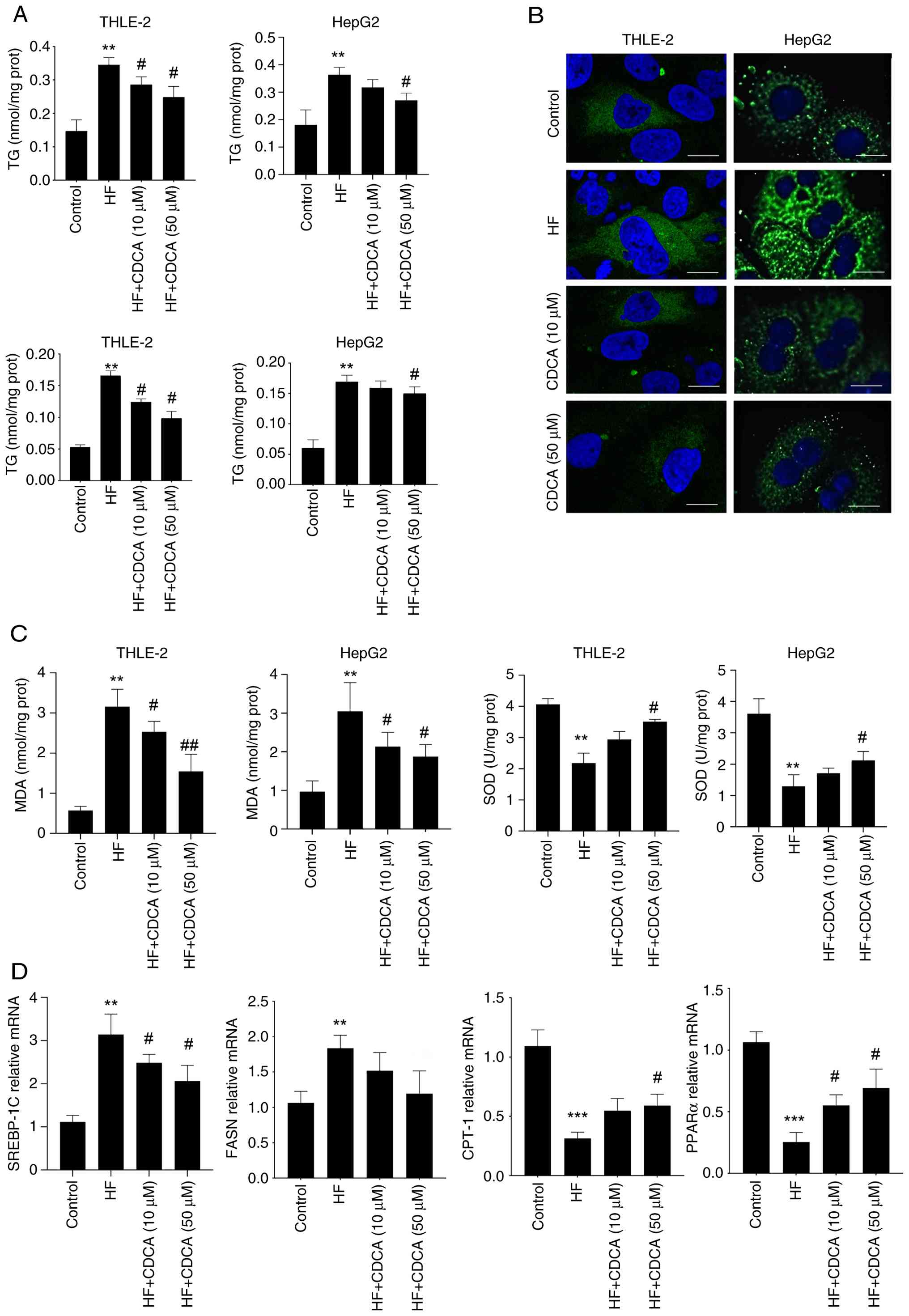 | Figure 1CDCA alleviates lipid deposition at
the cellular level (n=4). (A) Intracellular TG and TC contents. (B)
BODIPY staining of intracellular lipid droplets (magnification,
x400; scale bar, 10 µm). (C) Intracellular MDA contents and SOD
activities. (D) Reverse transcription-quantitative PCR detection of
FASN, SREBP-1c, CTP-1 and PPARα. **P<0.01 and
***P<0.001 vs. control; #P<0.05 and
##P<0.01 vs. HF. Error bars represent the standard
deviation. CDCA, chenodeoxycholic acid; TG, triglycerides; TC,
total cholesterol; MDA, malondialdehyde; SOD, superoxide dismutase;
FASN, fatty acid synthase; SREBP-1c, sterol regulatory
element-binding transcription factor 1; CTP1α, CTP synthase 1;
PPARα, peroxisome proliferator-activated receptor α; HF, high
fat. |
BODIPY staining visually illustrated intracellular
lipid deposition. Abundant lipid droplets were observed in the HF
group cells, whereas both the number and volume of lipid droplets
were markedly decreased in the CDCA-treated groups, further
confirming the inhibitory effect of CDCA on cellular lipid
deposition (Fig. 1B). Additionally,
cellular oxidative stress indices were assessed. Results indicated
that, compared with the HF group, intracellular MDA content was
significantly decreased in the CDCA-treated groups (P<0.05),
while SOD activity was significantly elevated (Fig. 1C). To further investigate the
molecular mechanisms underlying CDCA-mediated regulation of hepatic
lipid metabolism, RT-qPCR analysis was performed. Results revealed
that, in cell-based experiments, mRNA expression levels of lipid
synthesis-related genes (FASN and SREBP-1c) were significantly
downregulated following CDCA treatment compared with the HF group,
whereas expression of lipid metabolism-related genes (CTP1α and
PPARα) exhibited an opposite trend (Fig. 1D).
In the in vivo experiments, for the NAFLD
mouse model established by 12-week HFD feeding, 50 and 100 mg/kg/d
of CDCA were administered via gavage for 8-week intervention. The
biochemical detection results showed that, compared with the HF
group, the TC in the high-dose CDCA-treated group and TG in the
high- and low-dose CDCA-treated groups were significantly reduced
(P<0.01; Fig. 2A). Oxidative
stress analysis in the liver showed that, compared with the HF
group, the MDA content in the liver tissues of the CDCA-treated
groups was significantly reduced (P<0.05), and the SOD activity
was significantly increased (Fig.
2B). Oil Red O staining of liver tissue sections showed that
the livers of mice in the HF group were filled with numerous red
lipid droplets, whereas the lipid droplets in the CDCA-treated
groups were notably reduced, especially in the high-dose group.
Hepatic lipid deposition was markedly improved, which was
consistent with the biochemical results, indicating that CDCA also
reduced hepatic lipid deposition in vivo (Fig. 2C). In the liver tissues of mice, the
mRNA expression levels in the high-dose CDCA-treated group of lipid
synthesis genes, such as FASN and SREBP-1c, were significantly
decreased compared with those in the HF group, and the expression
of fatty acid β-oxidation-related genes (CPT1α and PPARα) were
significantly upregulated (Fig.
2D), indicating that CDCA improved hepatic lipid deposition by
inhibiting fatty acid synthesis and promoting fatty acid
β-oxidation.
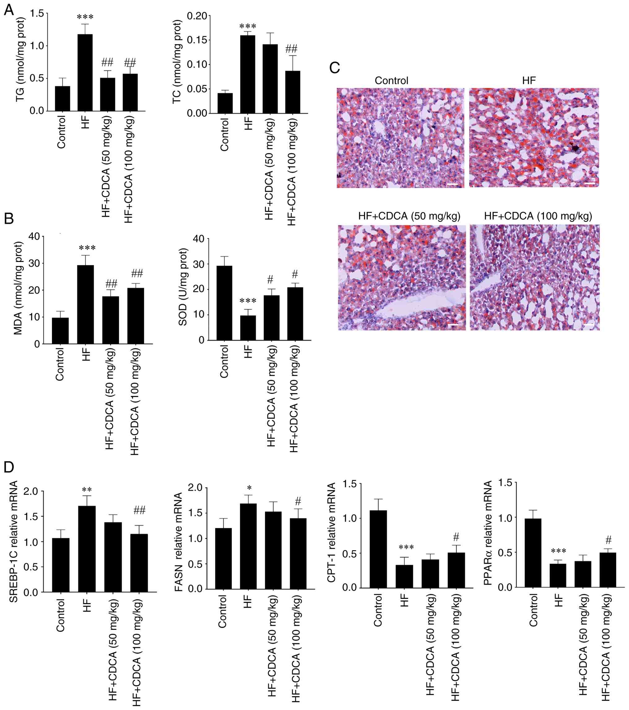 | Figure 2CDCA alleviates lipid deposition in
vivo (n=6). (A) TG content and TC contents in the liver. (B)
MDA content and SOD activity in liver tissues. Reverse
transcription-quantitative PCR detection of FASN, SREBP-1c, CTP-1
and PPARα. (C) Oil Red O staining of lipid droplets in liver
tissues (magnification, x200; scale bar, 20 µm). (D) Reverse
transcription-quantitative PCR detection of FASN, SREBP-1c, CTP-1
and PPARα. *P<0.05, **P<0.01 and
***P<0.001 vs. control; #P<0.05 and
##P<0.01 vs. HF. Error bars represent the standard
deviation. CDCA, chenodeoxycholic acid; TG, triglycerides; TC,
total cholesterol; MDA, malondialdehyde; SOD, superoxide dismutase;
SREBP-1c, sterol regulatory element-binding transcription factor 1;
CTP1α, CTP synthase 1; PPARα, peroxisome proliferator-activated
receptor α; HF, high fat. |
CDCA directly binds to PK and
regulates its activity
To explore whether there was a direct interaction
between CDCA and PK, a co-IP experiment was performed. After
treating HepG2 cells with CDCA, the cells were lysed to extract the
total protein, and a PK antibody was added for co-IP. The results
showed that in the CDCA-treated group, CDCA bound to PK was
successfully detected, while no obvious binding signal was detected
in the control group, indicating specific binding between CDCA and
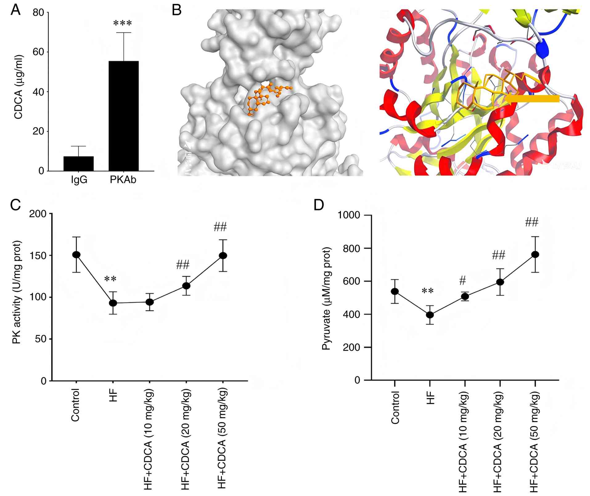
PK (Fig. 3A). Furthermore, the
results of molecular docking showed that CDCA specifically binds to
the PK-M2 subtype. The binding site was mainly located in the
catalytic domain of PK-M2, and the key amino acid sites were
Arg246, Lys247, Glu275, Arg278 and Arg279 in PKM2. The binding free
energy score between CDCA and PK-M2 was -10.33 kcal/mol, indicating
a strong binding stability between the two molecules (Fig. 3B). To study the regulatory effect of
CDCA on PK activity, intracellular PK activity was measured. The
results showed that CDCA enhanced PK activity in a
concentration-dependent manner. When the CDCA concentration was in
the range of 10-50 µM, the PK activity gradually increased with
increasing CDCA concentration, and the difference was statistically
significant (P<0.05; Fig. 3C).
The enhancement of PK activity promoted the production of pyruvate,
a product of glycolysis process (Fig.
3D), indicating that CDCA affects glycolysis by regulating PK
activity.
PK mediates the regulation of hepatic
lipid metabolism by CDCA
To verify the role of PK in the CDCA-mediated
improvement of hepatic lipid metabolism, a functional rescue
experiment was performed. HepG2 cells were pretreated with 10 µM of
the PK inhibitor (Shikonin) 1 h before intervention with 50 µM
CDCA. These results showed that Shikonin pretreatment completely
reversed the inhibitory effect of CDCA on lipid synthesis and
enhanced β-oxidation. Compared with the CDCA-alone treatment group,
the intracellular TG and TC levels in the CDCA + Shikonin group
were significantly increased (P<0.01; Fig. 4A).
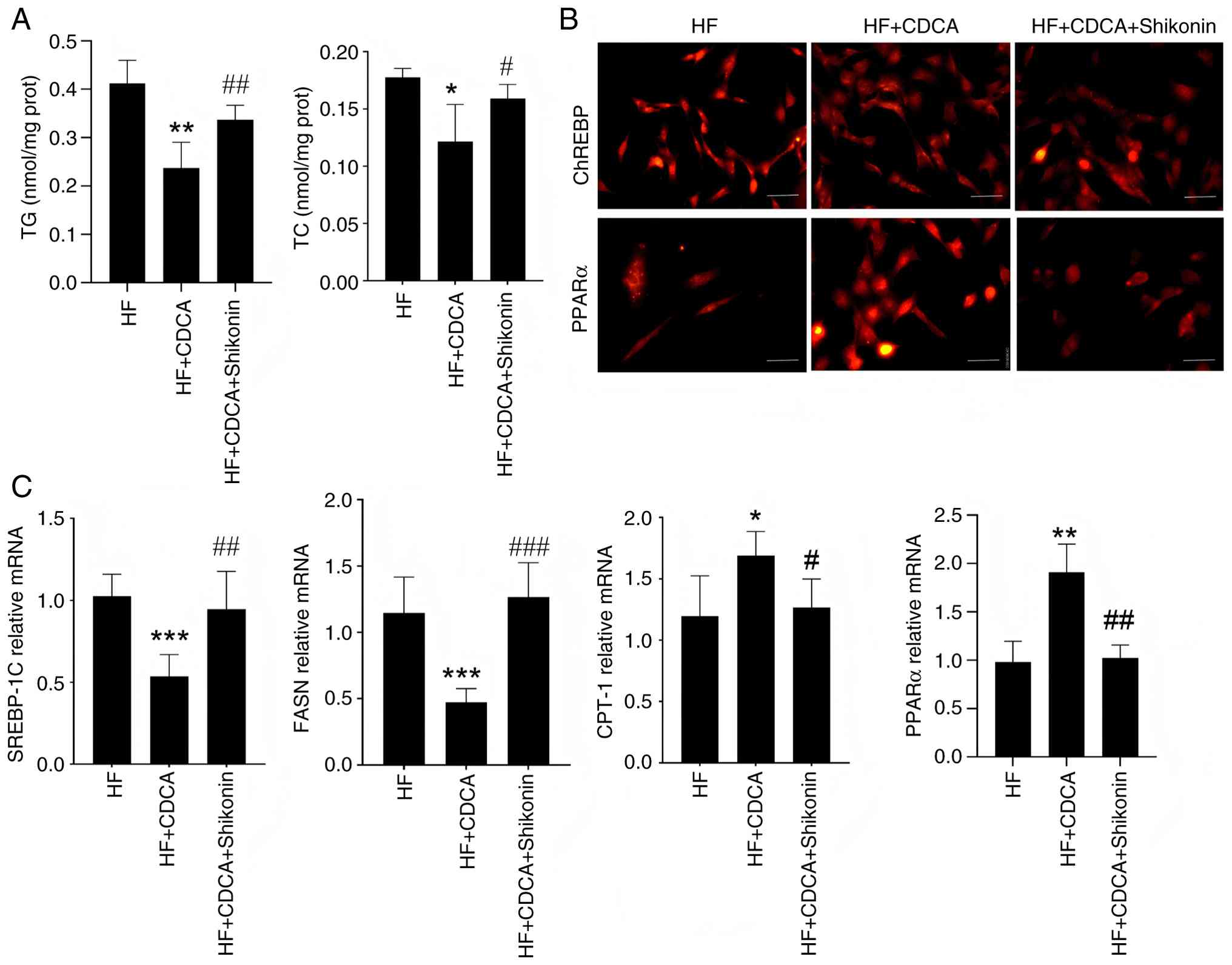 | Figure 4PK mediates the regulation of hepatic
lipid metabolism by CDCA (n=4). (A) Intracellular TG and TC
contents. (B) Immunofluorescence detection of ChREBP and PPARα
nuclear translocation (magnification, x200; scale bar, 50 µm). (C)
Reverse transcription-quantitative PCR detection of FASN, SREBP-1c,
CTP1α and PPARα. *P<0.05, **P<0.01 and
***P<0.001 vs. control; #P<0.05,
##P<0.01 and ###P<0.001 vs. HF. Error
bars represent the standard deviation. CDCA, chenodeoxycholic acid;
TG, triglycerides; TC, total cholesterol; ChREBP,
carbohydrate-responsive element-binding protein; SREBP-1c, sterol
regulatory element-binding transcription factor 1; CTP1α, CTP
synthase 1; PPARα, peroxisome proliferator-activated receptor α;
HF, high fat. |
Immunofluorescence results showed that the
fluorescence intensity of carbohydrate-responsive element-binding
protein (ChREBP) in the nucleus was markedly reduced in the
CDCA-treated group, whereas the nuclear translocation of ChREBP was
restored in the CDCA + Shikonin group, and the fluorescence
intensity in the nucleus was enhanced (Fig. 4B). Meanwhile, the CDCA-PK
interaction promoted the nuclear localization of PPARα (Fig. 4B). Concurrently, 50 µM CDCA
treatment reduced the mRNA expression levels of FASN and SREBP-1c,
and these reduced levels returned to a state similar to the
baseline after Shikonin intervention. By contrast, the mRNA
expression levels of CPT1α and PPARα were increased following 50 µM
CDCA treatment, and this upregulatory effect was reversed by
Shikonin (Fig. 4C), indicating that
PK serves a key mediating role in the regulation of hepatic lipid
metabolism by CDCA.
Discussion
To the best of our knowledge, for the first time,
the present study revealed a direct interaction between CDCA and PK
and demonstrated that this interaction promotes PK activation and
pyruvate production. A previous study has shown that CDCA mainly
improves hepatic lipid metabolism by activating the farnesoid X
receptor (FXR) (12). After FXR
activation, on the one hand, CDCA was demonstrated to inhibit the
expression of cholesterol 7α-hydroxylase, reducing the synthesis of
bile acids. By contrast, it was also shown to promote the
expression of fibroblast growth factor 15 (FGF15). FGF15, in turn
inhibited hepatic fatty acid synthesis and cholesterol uptake
through a negative feedback mechanism, thereby reducing hepatic
lipid content (13). However, the
present study found that the interaction between CDCA and PK
provides a novel pathway for improving hepatic lipid
metabolism.
CDCA specifically binds to the PK-M2 subtype and
notably enhances PK activity by stabilizing enzyme conformation. As
a key rate-limiting enzyme in glycolysis, enhanced activity of PK
accelerates glycolysis, resulting in a marked increase in the
production of pyruvate, a glycolytic product. As an important
metabolic intermediate, pyruvate not only provides more substrate
acetyl-CoA for fatty acid oxidation, promoting the β-oxidation
decomposition of fatty acids and reducing the accumulation of
hepatic fatty acids, but also, reduces de novo fatty acid
synthesis by activating the PPARα pathway and inhibiting the
expression of key lipid-synthesis genes and proteins such as FAS
and SREBP-1c (14). This two-way
regulatory mechanism acts simultaneously at the level of fatty acid
synthesis and decomposition, effectively improving hepatic lipid
metabolism and forming a novel glycolipid metabolism regulatory
axis that complements the classical pathway of CDCA activation of
FXR, to jointly maintain the homeostasis of hepatic lipid
metabolism. PK catalyzes the formation of pyruvate from
phosphoenolpyruvate and generates ATP during glycolysis, which is a
key enzyme in maintaining cellular energy metabolism (15). The present study further found that
PK not only serves a key role in glycolysis but also serves as a
central hub in the regulation of hepatic lipid metabolism. The
interaction between CDCA and PK endows PK with a novel function in
lipid metabolism regulation and reveals the important role of PK in
the cross-pathway of glycolipid metabolism.
Previous research has demonstrated that changes in
PK activity can directly affect the activities of transcription
factors such as ChREBP and PPARα, which are associated with
carbohydrates (16). When PK
activity is enhanced, on the one hand, it has been shown to inhibit
the nuclear translocation of ChREBP, reducing the activation of the
FASN promoter by ChREBP and thus inhibiting fatty acid synthesis.
By contrast, it has also been shown to promote the nuclear
localization of PPARα, enhance the expression of fatty acid
oxidation-associated genes and promote the β-oxidation of fatty
acids. This indicates that PK may act as a ‘molecular switch’ in
the cross-pathway of glycolipid metabolism. By regulating the
activities of key transcription factors, a coordinated regulation
of the two major pathways of carbohydrate and lipid metabolism can
be achieved. This regulatory mechanism helps to maintain the
balance of carbohydrate and lipid metabolism in the liver and
ensures normal physiological functions of the liver. Once the
activity of PK or its interaction with CDCA is disrupted, this
balance may be disturbed, leading to disorders of hepatic lipid
metabolism and further triggering metabolic diseases, such as
NAFLD.
Currently, the treatment of NAFLD mainly focuses on
lifestyle interventions and there is a lack of effective drug
treatment methods. The discovery of the CDCA-PK axis opens a new
direction for the development of novel anti-fatty liver drugs. By
designing and synthesizing small-molecule compounds that can
specifically enhance the interaction between CDCA and PK or mimic
its effects, more effective and safer drugs for NAFLD treatment may
be developed. Follow-up studies can further explore the specific
regulatory roles of different PK subtypes in hepatic lipid
metabolism and the synergistic mechanism of CDCA in the gut-liver
axis. The gut microbiota serves an important role in bile acid and
hepatic lipid metabolism. The metabolic transformation of CDCA in
the intestine and its interaction with gut microbiota may affect
its regulatory effect on hepatic lipid metabolism. In-depth
research on these mechanisms will help to understand the regulatory
network of hepatic lipid metabolism more comprehensively and
provide a more solid theoretical basis for the clinical precision
treatment of NAFLD.
Despite the novel findings and theoretical
contributions, the present study has several limitations that
should be acknowledged. First, the current research was mainly
based on in vitro cell models and/or animal models of NAFLD;
lack of clinical data from patients with NAFLD limits the
translational value of the findings. Whether the CDCA-PK
interaction and its regulatory role in hepatic lipid metabolism are
conserved in humans, and the correlation between CDCA/PK levels and
NAFLD severity in clinical populations, remain to be verified.
Second, the molecular details of the direct binding between CDCA
and PK-M2 were not fully elucidated. For instance, the specific
binding sites on PK-M2 for CDCA, and whether this binding induces
conformational changes in PK-M2 through specific amino acid
residues, require further verification using techniques such as
X-ray crystallography or cryo-electron microscopy. Third, the study
focused on the overall regulatory effect of CDCA on PK, but did not
systematically explore the potential crosstalk between the CDCA-PK
axis and other bile acid-related signaling pathways (such as FXR
and TGR5) in hepatic lipid metabolism. The synergistic or
antagonistic effects between these pathways remain unclear. Fourth,
the present study did not consider the influence of individual
differences such as sex, age, and genetic background on the CDCA-PK
regulatory axis. These factors may affect the response of hepatic
lipid metabolism to CDCA-PK interaction, which needs to be
clarified in subsequent studies. Finally, the study lacked
long-term intervention experiments to evaluate the sustained
effects and potential side effects of targeting the CDCA-PK axis in
the treatment of NAFLD, which is essential for the development of
clinical therapeutic strategies.
In conclusion, CDCA markedly improved hepatic lipid
deposition, and its mechanism was shown to be closely associated
with the direct binding of CDCA to PK and the enhancement of its
activity. The present study not only expands the target network of
bile acid-regulated metabolism, but also provides novel potential
drug targets and treatment strategies for NAFLD, which has
important theoretical and clinical significance.
Acknowledgements
Not applicable.
Funding
Funding: No funding was received.
Availability of data and materials
The data generated in the present study may be
requested from the corresponding author.
Authors' contributions
XZhang designed and conceived the study, and was
responsible for original manuscript preparation as well as review
and editing. XZhang, ST and XZhou undertook data collection and
analyzed the data. All authors confirm the authenticity of all the
raw data. All authors read and approved the final manuscript.
Ethics approval and consent to
participate
The study was approved (approval no.
SW-2020R0126-F03) by the Ethics Board of Pingxiang People's
Hospital (Pingxiang, China). This study was conducted in compliance
with the principles of the Declaration of Helsinki.
Patient consent for publication
Not applicable.
Competing interests
The authors declare that they have no competing
interests.
References
|
1
|
Riazi K, Azhari H, Charette JH, Underwood
FE, King JA, Afshar EE, Swain MG, Congly SE, Kaplan GG and Shaheen
AA: The prevalence and incidence of NAFLD worldwide: A systematic
review and meta-analysis. Lancet Gastroenterol Hepatol. 7:851–861.
2022.PubMed/NCBI View Article : Google Scholar
|
|
2
|
Yki-Järvinen H, Luukkonen PK, Hodson L and
Moore JB: Dietary carbohydrates and fats in nonalcoholic fatty
liver disease. Nat Rev Gastroenterol Hepatol. 18:770–786.
2021.PubMed/NCBI View Article : Google Scholar
|
|
3
|
Chang TC, Chiou WC, Lai WH, Huang HC,
Huang YL, Liu HK, Liang YC and Huang C: Ugonin J improves metabolic
disorder and ameliorates nonalcoholic fatty liver disease by
regulating the AMPK/AKT signaling pathway. Pharmacol Res.
163(105298)2021.PubMed/NCBI View Article : Google Scholar
|
|
4
|
Hüttl M, Markova I, Miklankova D,
Zapletalova I, Poruba M, Haluzik M, Vaněčkova I and Malinska H: In
a prediabetic model, empagliflozin improves hepatic lipid
metabolism independently of obesity and before onset of
hyperglycemia. Int J Mol Sci. 22(11513)2021.PubMed/NCBI View Article : Google Scholar
|
|
5
|
Murakami K, Tenge VR, Karandikar UC, Lin
SC, Ramani S, Ettayebi K, Crawford SE, Zeng XL, Neill FH, Ayyar BV,
et al: Bile acids and ceramide overcome the entry restriction for
GII.3 human norovirus replication in human intestinal enteroids.
Proc Natl Acad Sci USA. 117:1700–1710. 2020.PubMed/NCBI View Article : Google Scholar
|
|
6
|
Xu Y, Zhu Y, Hu S, Xu Y, Stroup D, Pan X,
Bawa FC, Chen S, Gopoju R, Yin L and Zhang Y: Hepatocyte nuclear
factor 4α prevents the steatosis-to-NASH progression by regulating
p53 and bile acid signaling (in mice). Hepatology. 73:2251–2265.
2021.PubMed/NCBI View Article : Google Scholar
|
|
7
|
Du D, Liu C, Qin M, Zhang X, Xi T, Yuan S,
Hao H and Xiong J: Metabolic dysregulation and emerging
therapeutical targets for hepatocellular carcinoma. Acta Pharm Sin
B. 12:558–580. 2022.PubMed/NCBI View Article : Google Scholar
|
|
8
|
Dai P, Tang Z, Qi M, Liu D, Bajinka O and
Tan Y: Dispersion and utilization of lipid droplets mediates
respiratory syncytial virus-induced airway hyperresponsiveness.
Pediatr Allergy Immunol. 33(e13651)2022.PubMed/NCBI View Article : Google Scholar
|
|
9
|
Fan N, Zhang X, Zhao W, Zhao J, Luo D, Sun
Y, Li D, Zhao C, Wang Y, Zhang H and Rong J: Covalent inhibition of
pyruvate kinase m2 reprograms metabolic and inflammatory pathways
in hepatic macrophages against non-alcoholic fatty liver disease.
Int J Biol Sci. 18:5260–5275. 2022.PubMed/NCBI View Article : Google Scholar
|
|
10
|
Liu Z, Zhang C, Lee S, Kim W, Klevstig M,
Harzandi AM, Sikanic N, Arif M, Ståhlman M, Nielsen J, et al:
Pyruvate kinase L/R is a regulator of lipid metabolism and
mitochondrial function. Metab Eng. 52:263–272. 2019.PubMed/NCBI View Article : Google Scholar
|
|
11
|
Bajinka O, Tang Z, Mao Y, Qiu X, Darboe A
and Tan Y: Respiratory syncytial virus infection disrupts pulmonary
microbiota to induce microglia phenotype shift. J Med Virol.
95(e28976)2023.PubMed/NCBI View Article : Google Scholar
|
|
12
|
Deng W, Fan W, Tang T, Wan H, Zhao S, Tan
Y, Oware KA, Tan J, Li J and Qu S: Farnesoid X receptor deficiency
induces hepatic lipid and glucose metabolism disorder via
regulation of pyruvate dehydrogenase kinase 4. Oxid Med Cell
Longev. 2022(3589525)2022.PubMed/NCBI View Article : Google Scholar
|
|
13
|
Song L, Hou Y, Xu D, Dai X, Luo J, Liu Y,
Huang Z, Yang M, Chen J, Hu Y, et al: Hepatic FXR-FGF4 is required
for bile acid homeostasis via an FGFR4-LRH-1 signal node under
cholestatic stress. Cell Metab. 37:104–120.e9. 2025.PubMed/NCBI View Article : Google Scholar
|
|
14
|
Qiu X, Ye Q, Sun M, Wang L, Tan Y and Wu
G: Saturated hydrogen improves lipid metabolism disorders and
dysbacteriosis induced by a high-fat diet. Exp Biol Med (Maywood).
245:512–521. 2020.PubMed/NCBI View Article : Google Scholar
|
|
15
|
Li Z, Peng Y, Li J, Chen Z, Chen F, Tu J,
Lin S and Wang H: N6-methyladenosine regulates
glycolysis of cancer cells through PDK4. Nat Commun.
11(2578)2020.PubMed/NCBI View Article : Google Scholar
|
|
16
|
Mzhel'skaya KV, Trusov NV, Guseva GN,
Aksenov IV, Kravchenko LV and Tutelyan VA: Effects of quercetin on
expression of genes of carbohydrate and lipid metabolism enzymes in
the liver of rats receiving high-fructose ration. Bull Exp Biol
Med. 167:263–266. 2019.PubMed/NCBI View Article : Google Scholar
|