Introduction
KBG syndrome (KBGS, Online Mendelian Inheritance in
Man no.148050) is a rare autosomal dominant disorder caused by
heterozygous disease-causing variants in the ankyrin repeat domain
11 (ANKRD11) gene or by microdeletions of the 16q24.3 region
encompassing this gene (1,2-6).
ANKRD11-related KBG syndrome is increasingly recognized, with
>375 individuals reported in the literature (7,8).
ANKRD11 encodes a transcriptional coregulator involved in chromatin
remodeling and regulation of gene expression through interactions
with histone-modifying enzymes, transcriptional cofactors and the
cohesin complex (5,6,8,9,10).
ANKRD11 expression is key for nervous system development and
function, influencing neural progenitor proliferation, neuronal
generation, positioning and plasticity and dendritic
differentiation (6,8). Structurally, the protein contains five
N-terminal ankyrin repeats, two repression domains, one activation
domain and C-terminal D-box motifs that regulate proteasomal
degradation during the cell cycle (5,6). KBGS
is primarily caused by heterozygous loss-of-function ANKRD11
variants, including truncating frameshift and nonsense variants, or
16q24.3 microdeletions involving ANKRD11, resulting in
haploinsufficiency (2-6).
Pathogenic mechanisms include impaired chromatin regulation, and
altered transcriptional control of developmental genes, supporting
the classification of KBGS as a chromatinopathy (8-10).
These molecular alterations are consistent with the
neurodevelopmental, skeletal and craniofacial manifestations
observed in affected individuals, including developmental delay,
intellectual disability, short stature, facial dysmorphism and
dental anomalies such as macrodontia (1,2,5,9,10).
Currently, the prevalence of KBGS is hypothesized to
be underestimated due to its wide phenotypic variability, variable
severity and mild presentations in some individuals. Numerous
pathogenic ANKRD11 variants and 16q24.3 deletions have been
described (2-4,8,11,12).
Although no consensus clinical diagnostic criteria has been
published, KBGS should be suspected in individuals with
developmental delay and/or intellectual disability,
attention-deficit/hyperactivity disorder or autism spectrum
disorder, together with characteristic phenotypic features or
associated comorbidities. The primary clinical features include
postnatal short stature, hand and costovertebral anomalies,
macrodontia of the upper central incisors as a key feature and a
triangular facial shape (1-4,8,13,14).
Considering the substantial clinical heterogeneity,
other disorders with overlapping clinical features with KBGS have
been considered in the differential diagnosis, such as Cornelia de
Lange syndrome (OMIM No. 122470), Silver-Russell syndrome (OMIM No.
180860), Aarskog-Scott syndrome (OMIM No. 305400) and Coffin-Siris
syndrome (OMIM No. 135900) (4,9,10,15,16).
For this reason, KBGS has recently been classified as part of a new
class of disorder, known as chromatinopathies. These disorders
result from variants in proteins involved in chromatin remodeling
and transcriptional regulation (6,8-10,16).
The present study aimed to describe the clinical and
molecular findings of 10 individuals with ANKRD11 variants,
highlighting the broad spectrum of associated phenotypes, and to
establish genotype-phenotype associations to refine the
characterization of this syndrome and compare its phenotypic
features with those of classical chromatinopathy (2-4,13,17-19).
The present study conducted a systematic review of
the literature addressing the clinical and molecular aspects,
providing a broader framework for interpreting our findings
(2-4,11,13,17,19,20).
Materials and methods
Study design and participants
The present retrospective observational case series
included 10 individuals with molecularly confirmed KBGS evaluated
at the Clinical Genetics Service of Fundación Valle del Lili (Cali,
Colombia) between January 2017 and February 2025. Inclusion
criteria comprised a compatible clinical phenotype and the
identification of a pathogenic or likely pathogenic ANKRD11
variant or a chromosomal rearrangement involving the 16q24.3
region. Patients without molecular confirmation of KBGS or with
insufficient clinical or photographic data for analysis were
excluded. Clinical, demographic and molecular data were obtained
from medical records and standardized clinical genetic evaluations.
The patient group comprised six males and four females, with an age
range of 6-37 years at the time of evaluation.
To assess facial dysmorphology in KBGS, the present
study included a comparative sample of 10 sex- and age-matched
controls recruited during data collection sessions at Universidad
Icesi and the Clinical Research Center of Fundación Valle del Lili,
Cali, Colombia, between January and March 2021. The control group
comprised six males and four females, with an age range of 6-37
years. Control individuals were included only for the 2D facial
morphometric analysis if they were from the same geographic region,
had no known history of neurodevelopmental disorder, craniofacial
anomaly or genetic syndrome, and had standardized frontal facial
photographs of sufficient quality for landmark annotation. No
genetic testing or screening was performed in the control
group.
The present study was approved by the Ethics
Committee of Hospital Universitario Fundación Valle del Lili
(approval no. 2025.140). Written informed consent was obtained from
all patients or their legal guardians for the publication of
clinical data and photographic images. The present study was
conducted in accordance with the ethical principles of the
Declaration of Helsinki, ensuring confidentiality and appropriate
use of identifiable information for research and publication.
Clinical evaluation
Phenotypic assessment included growth parameters,
neurodevelopmental profile, behavioral manifestations, craniofacial
dysmorphism, skeletal anomaly and involvement of other organ
systems. Clinical features were described using standardized
dysmorphology terminology. Developmental delay, intellectual
disability and neuropsychiatric features were recorded based on
multidisciplinary clinical and neuropsychological evaluation.
Molecular analysis
Molecular diagnosis and evaluation of clinically
relevant differential diagnoses were performed using clinical exome
sequencing. In total, four exome analyses were performed
institutionally, whereas the remaining studies were performed by
external diagnostic laboratories as part of the patient clinical
diagnostic workflow. For externally performed tests, sequencing and
bioinformatic procedures were performed out according to validated
protocols, and the results were reviewed based on the clinical
reports available.
For institutionally performed exome analyses,
genomic DNA was extracted and purified from peripheral blood
samples, followed by preparation of genomic fragment libraries
using the KeyExome-GeneSGKit X. 48 Reactions kit, reference LV4283
(Genomic Systems). DNA quantity and purity were assessed using
NanoDrop spectrophotometry, including the 260/280 nm ratio, and DNA
quantification was performed using Qubit and Quantus fluorometers.
Library quality was evaluated using TapeStation capillary
electrophoresis, with library fragments showing a Gaussian
distribution and a peak of approximately 320 bp. Clonal
amplification and sequencing of the selected regions were performed
on an Illumina NextSeq 500 platform using Illumina bridge
sequencing technology and a paired-end sequencing strategy.
Sequencing was performed using the NextSeq 500/550 High Output kit
v2.5, 300 cycles, supporting 2x150 bp reads (cat. no. 20024908;
Illumina Inc.). Mean sequencing coverage was recorded according to
the clinical laboratory report. Bioinformatic analysis of the DNA
sequences was performed by comparison with the reference genome
sequence GRCh38 using the GeneSystems® platform v4.0.1
(IVD-CE; https://platform.genesystm.com/services) (21). Variants were considered for analysis
when they showed a read depth ≥20X and a variant allele
fraction/read ratio ≥0.2.
ANKRD11 variants were described according to
the Human Genome Variation Society (HGVS) nomenclature using the
NM_013275.6 reference transcript and classified following the
American College of Medical Genetics and Genomics/Association for
Molecular Pathology (ACMG/AMP) guidelines (22,23).
Variant interpretation was supported by variant interpretation
platforms, including VarSome (varsome.com/)
and Franklin (Genoox/QIAGEN; franklin.genoox.com/clinical-db/home), together with
genotype-phenotype association and available clinical evidence.
In silico predictive analyses were performed for variants of
uncertain significance using VarSome (24,25).
Variants classified as pathogenic or likely
pathogenic were confirmed by Sanger sequencing in four patients for
whom confirmation was available as part of the clinical diagnostic
workflow. For the remaining six patients, Sanger sequencing
chromatograms were generated by external diagnostic laboratories as
part of the clinical diagnostic workflow (data not available).
Parental segregation studies were performed when
parental samples were available. Novelty was assessed based on the
absence of previous reports in VarSome, Franklin and the available
literature. Novel variants are currently in the process of being
submitted to ClinVar (26).
Two-dimensional facial
morphometry
Two-dimensional facial morphometric analysis was
performed in patients and controls using standardized 2D frontal
photographs. Images were acquired with participants in an upright
head position and neutral expression, with open eyes and closed
mouth.
A set of 21 anatomical facial landmarks were
manually annotated using the tpsDig2 software version 2.32, 64-bit
executable (27) and quantitative
shape analyses were conducted to identify craniofacial patterns
associated with KBGS. Facial shape variation was assessed using
geometric morphometrics, a robust set of statistical tools designed
for measuring and comparing 2D/3D shapes with high precision and
efficiency (27,28-31).
Facial landmark configurations were aligned in a common morphospace
using generalized Procrustes analysis, removing non-shape variance
due to translation, rotation and scale. Multivariate linear
regression was performed to correct for age-associated facial shape
differences. The regression residuals were analyzed using principal
component analysis (PCA) to explore facial differences between
patients with KBGS and matched controls. Morphological variation
was visualized in the morphospace defined by the first two
principal components. Group differentiation was assessed using
Procrustes distances between mean shapes, calculated as the square
root of the sum of squared differences between homologous
landmarks. All statistical analyses were performed in MorphoJ
1.08.01(32) and R 4.4.2 using the
geomorph package v4.0.10(33).
Statistical significance was determined using 1,000-permutation
tests, with P<0.05 considered statistically significant.
Results
Cohort characteristics
A total of 10 individuals with molecularly confirmed
KBGS were included, comprising six males and four females. Age at
molecular diagnosis ranged from 4 to 36 years, with a mean age of
16.4 years. All individuals were followed at Fundación Valle del
Lili (Cali, Colombia) and underwent comprehensive clinical genetic
evaluation.
Developmental delay was present in all patients
(10/10, 100%) and all met criteria for intellectual disability,
with severity ranging from mild to severe. Growth parameters were
variable. Current height ranged from 99 to 171 cm, and birth length
data were available for 4/10 individuals, ranging from 47 to 52
cm.
All phenotypic and molecular characteristics are
described in Tables I and II.
 | Table IClinical and molecular features of
patients with KBG Syndrome. |
Table I
Clinical and molecular features of
patients with KBG Syndrome.
| Patient no. | 1 | 2 | 3 | 4 | 5 | 6 | 7 | 8 | 9 | 10 |
|---|
| Sex | M | F | M | M | M | M | M | F | F | F |
| Age at diagnosis
years | 36 | 27 | 15 | 17 | 15 | 10 | 7 | 4 | 16 | 17 |
| Age at last
evaluation, years (March 2025) | 37 | 29 | 19 | 19 | 21 | 16 | 11 | 6 | 18 | 20 |
| Exon | 9 | 12 | 7 | 9 | 12 | 9 | 9 | 9 | 9 | 9 |
| Heterozygous
variant | c.3600dup (p.
Val1201 SerfsTer3) | c.7753C>T (p.
Arg2585 Cys) | c.741C>A
(p.Tyr247Ter) | c.4218C>A (p.
Tyr1406 Ter) | c.7744_7745 dup (p.
Asn2583 Serfs*20) | c.6517delG (p.
Val2173 Serfs*2) | c.1903_1907del (p.
Lys635 Glnfs*26) | c.5397dup (p.
Glu1800 ArgfsTer150) | c.3499dup
(p.Ser1167 PhefsTer) | c.3055_ 3059del
(p.Arg1019 GlyfsTer14) |
| Type of
mutation | Frameshift | Missense | Nonsense | Nonsense | Frameshift | Frameshift | Frameshift | Frameshift | Frameshift | Frameshift |
| Novel variant | + | + | + | - | + | + | - | + | + | + |
| Inheritance | NI | NI | De novo | De novo | NI | De novo | De novo | De novo | NI | NI |
| ACMG/AMP
classification | LP | LP | LP | P | LP | LP | P | LP | LP | P |
| Height at birth,
cm | NI | NI | NI | 50 | 52 | NI | 47 | NI | 50 | NI |
| Height at at last
evaluatio, cm (March 2025) Craniofacial features | 147 | 141 | 162 | 149 | 171 | 154 | 153 | 99 | 149 | 150 |
|
Brachycephaly
HP:0000248 | + | - | - | - | + | + | - | - | + | - |
|
Microcephaly
HP:0000252 | - | + | + | - | - | - | - | + | - | - |
|
Low hair
implantation HP:0002162 | + | + | + | + | + | + | + | - | + | - |
|
Coarse
facies HP:0000280 | + | + | - | + | - | + | + | - | - | - |
|
Facial
asymmetry HP:0000324 | + | + | - | + | + | + | + | - | - | + |
|
Triangular
face HP:0000325 | - | - | + | - | - | + | + | - | + | + |
|
Prominent
forehead HP:0011220 | - | - | + | - | - | + | - | - | - | - |
|
Synophrys
HP:0000664 | + | + | - | + | - | - | + | - | + | + |
|
Full, arched
eyebrows HP:0002553 | - | + | + | + | + | - | + | - | + | + |
|
Hypertrichosis
HP:0000998 | - | - | + | + | - | - | - | - | + | - |
|
Hypertelorism
HP:0000316 | - | - | + | - | + | + | - | - | + | - |
|
Downslanting
palpebral fissures HP:0000494 | - | - | - | - | +; left | + | + | - | - | - |
|
Upslanting
palpebral fissures HP:0000582 | + | - | - | - | - | - | - | + | - | + |
|
Prominent
nasal bridge HP:0000426 | - | + | + | + | - | + | + | - | + | + |
|
Bulbous nose
HP:0000414 | + | + | + | + | + | + | + | - | + | + |
|
Long
philtrum HP:0000343 | - | + | + | + | - | - | - | - | + | - |
|
Tented upper
lip HP:0010804 | - | - | - | - | - | - | - | - | + | - |
|
Thin upper
lip HP:0000219 | - | + | - | - | - | - | - | - | - | - |
|
Thick lips
HP:0012471 | - | - | - | + | - | - | + | - | - | - |
|
Macrostomy
HP:0000154 | + | - | - | - | - | + | + | - | - | - |
|
Macrodontia
HP:0001572 | - | - | + | + | + | - | + | - | + | - |
|
Wide-spaced
teeth HP:0000687 | - | - | - | + | - | + | + | - | - | - |
|
Prominent
ears HP:0000411 Cleft palate/lip | + | - | + | + | + | + | - | - | - | + |
|
HP:0000175,
HP:0410030 | - | + | - | - | - | - | - | - | - | - |
|
Prognathism
HP:0000303 | - | + | - | - | - | - | - | - | - | - |
|
Micrognathia
HP:0000347 | - | - | + | + | + | - | + | - | + | - |
|
Low-set
posteriorly rotated ears HP:0000358 | - | - | - | + | + | + | + | - | - | - |
|
Cupped ears
HP:0000378 | - | - | - | - | + | + | - | - | - | - |
|
Preauricular
skin tags HP:0000384 | - | - | - | - | - | - | - | - | - | + |
| Skeletal
features | | | | | | | | | | |
|
Scoliosis
HP:00026 50 | - | + | - | - | + | + | + | - | + | + |
|
Short neck
HP:0000470 | - | - | - | - | - | - | - | - | + | + |
|
Cubitus
valgus HP:0002967 | + | - | - | + | - | - | - | - | - | - |
|
Single
transverse line HP:0000954 | - | - | + | - | - | - | + | - | - | - |
|
Clinodactyly
and camptodactyly HP:0030084 and HP:0012385 | - | + | + | + | - | + | + | - | + | + |
|
Broad thumbs
HP:0011304 | - | - | - | + | - | - | + | - | - | - |
|
Broad hallux
HP:0010055 | - | - | - | + | - | - | - | - | - | - |
|
Short 4th
and 5th fingers HP:0008093, HP:0009237 | + | - | - | + | + | - | + | - | + | + |
|
Persistence
of the fingerpads HP:0001212 | - | - | - | + | + | + | + | - | + | - |
| Neurological
findings | | | | | | | | | | |
|
Developmental
delay HP:0001263 | + | + | + | + | + | + | + | + | + | + |
|
Intellectual
disability HP:0001249 | + | +; mild | +;moderate to
severe | +; severe | +; mild | +; mild | +; moderate | + | +; moderate | +; moderate |
|
Hypotonia
HP:0001252 | + | - | - | + | + | + | - | + | - | - |
|
Attention-deficit
hyperactivity disorder HP:0007018 | - | - | - | + | - | + | + | + | - | - |
|
Autistic
features HP:0000729 | - | - | + | + | - | - | + | + | - | - |
|
Behavioral
disorder HP:0000708 | - | + | - | + | - | + | + | + | - | - |
|
Seizures
HP:0001250 | + | + | - | + | - | - | + | - | + | - |
|
Abnormal
brain MRI HP:0410263 | - | - | - | NI | - | - | - | Periventricular
hyperintensity compatible with mild leukoencephalomalacia | - | - |
| Other | | | | | | | | | | |
|
Cardiovascular
congenital disease HP:0030680 | - | - | - | +; ASD and VSD | - | - | - | - | - | +; persistent vena
cava and dilated pulmonary trunk |
|
Renal
defects HP:0000077 | - | - | - | +; left
hydronephrosis | - | - | - | - | - | - |
|
Sensorineural
hearing loss HP:0000407 | + | + | - | +; bilateral
conductive deafness | - | +; bilateral
sensorineural hearing loss | +; mild to moderate
bilateral conductive hearing loss | +; right mild
conductive hearing loss | +; mild conductive
hearing loss | +; severe mixed
conductive hearing impairment |
| Cryptorchidism
HP:0000028 | - | - | - | - | - | - | + | - | - | - |
| Gastrointestinal
disorder | | | | | | | | | | |
|
Feeding
difficulties HP:0011968 | + | + | - | + | - | + | - | + | - | + |
|
Low weight
HP:0004325 | - | + | - | - | - | + | - | + | - | - |
|
Gastric
reflux HP:0002020 | + | - | - | + | - | - | - | + | - | - |
|
Anal
anomalies HP:0002023 | - | - | - | - | - | - | - | - | - | +; imperforate
anus |
| Intestinal
abnormalities HP:0002242 | - | - | - | +; intestinal
malrotation | - | - | - | - | - | - |
| Table IIFrequency of the main clinical
features observed in the KBG syndrome cohort (n=10). |
Table II
Frequency of the main clinical
features observed in the KBG syndrome cohort (n=10).
| Clinical
feature | n (%) |
|---|
| Craniofacial
features | |
|
Brachycephaly | 4(40) |
|
Microcephaly | 3(30) |
|
Low hair
implantation | 8(80) |
|
Coarse
facies | 4(40) |
|
Facial
asymmetry | 7(70) |
|
Triangular
face | 4(40) |
|
Prominent
forehead | 2(20) |
|
Synophrys | 6(60) |
|
Full, arched
eyebrows | 7(70) |
|
Hypertrichosis | 3(30) |
|
Hypertelorism | 4(40) |
|
Downslanting
palpebral fissures | 2(20) |
|
Upslanting
palpebral fissures | 3(30) |
|
Prominent
nasal bridge | 6(60) |
|
Bulbous
nose | 9(90) |
|
Long
philtrum | 4(40) |
|
Tented upper
lip | 1(10) |
|
Thin upper
lip | 1(10) |
|
Thick
lips | 1(10) |
|
Macrostomy | 2(20) |
|
Macrodontia | 5(50) |
|
Wide-spaced
teeth | 2(20) |
|
Prominent
ears | 6(60) |
|
Cleft
palate/lip | 1(10) |
|
Prognathism | 1(10) |
|
Micrognathia | 4(40) |
|
Low-set
posteriorly rotated ears | 3(30) |
|
Cupped
ears | 2(20) |
|
Preauricular
skin tags | 2(20) |
| Skeletal
features | |
|
Scoliosis | 6(60) |
|
Short
neck | 2(20) |
|
Cubitus
valgus | 2(20) |
|
Single
transverse line | 1(10) |
|
Clinodactyly
and campodactyly | 7(70) |
|
Broad
thumbs | 1(10) |
|
Broad
hallux | 1(10) |
|
Short 4th
and 5th fingers | 5(50) |
|
Persistence
of the fingerpads | 4(40) |
| Neurological
features | |
|
Developmental
delay | 10(100) |
|
Intellectual
disability | 10(100) |
|
Hypotonia | 5(50) |
|
Attention-deficit
hyperactivity disorder | 4(40) |
|
Autistic
features | 4(40) |
|
Behavioral
disorder | 5(50) |
|
Seizures | 4(40) |
| Gastrointestinal
features | |
|
Feeding
difficulties | 6(60) |
|
Low
weight | 3(30) |
|
Gastric
reflux | 3(30) |
|
Anal
anomalies | 1(10) |
|
Intestinal
abnormality | 1(10) |
| Other features | |
|
Cardiovascular
congenital disease | 2(20) |
|
Renal
defects | 1(10) |
|
Sensorineural
hearing loss | 8(80) |
Clinical features
Semiological findings and phenotypic abnormalities
were defined using terms from Human Phenotype Ontology (34).
Craniofacial findings. Craniofacial
dysmorphism was identified in all individuals, although the
specific combination and severity of features varied (Fig. 1). The most frequent findings
included low anterior hairline (8/10, 80%), bulbous nasal tip
(8/10, 80%), synophrys (6/10, 60%), facial asymmetry (7/10, 70%)
and prominent ears (6/10, 60%). Additional recurrent features
included full or arched eyebrows (7/10, 70%), triangular face,
brachycephaly, coarse facial features, hypertelorism, long philtrum
and macrodontia of the permanent upper central incisors (all 5/10,
50%) (Fig. 2). Less frequent
findings included microcephaly (3/10, 30%), downslanting palpebral
fissures (2/10, 20%), upslanting palpebral fissures (3/10, 30%),
macrostomy (2/10, 20%), cleft lip and/or palate (1/10, 10%),
prognathism (1/10, 10%) and micrognathia (4/10, 40%).
Skeletal manifestation. Skeletal anomalies
were commonly observed. Scoliosis was the most frequent finding
(6/10, 60%). Digital anomalies were prominent, including
clinodactyly and/or campodactyly (7/10, 70%), short fourth and
fifth fingers (5/10, 50%) and persistent fetal fingertip pads
(4/10, 40%). Less frequent skeletal features included cubitus
valgus (2/10, 20%), short neck (2/10, 20%), single transverse
palmar crease (1/10, 10%), broad thumbs (1/10, 10%), and broad
hallux (1/10, 10%).
Neurological and behavioral features. All
individuals exhibited developmental delay and intellectual
disability. Hypotonia was observed in half of the cohort (5/10,
50%). Behavioral and neuropsychiatric manifestations were frequent,
including behavioral disorder (5/10, 50%), attention-deficit
hyperactivity disorder (4/10, 40%) and autistic features (4/10,
40%). Seizures were reported in four individuals (40%). Brain
magnetic resonance imaging, when available, demonstrated variable
findings, including periventricular white matter signal
abnormalities compatible with mild leukoencephalomalacia in one
patient.
Additional systemic findings
Sensorineural or conductive hearing loss was
identified in eight individuals (80%), making it one of the most
prevalent non-neurological manifestations in the cohort.
Gastrointestinal involvement was frequent, including feeding
difficulties (6/10, 60%), gastroesophageal reflux (3/10, 30%), low
weight (3/10, 30%) and isolated cases of anal anomaly (1/10, 10%)
and intestinal abnormality (1/10, 10%). Congenital cardiovascular
defects were observed in two individuals (20%) and renal anomalies
were identified in one patient. Cryptorchidism was documented in
one male individual.
Molecular findings
All 10 individuals carried heterozygous
ANKRD11 variants, consistent with autosomal dominant KBGS.
The molecular spectrum was predominantly composed of
loss-of-function variants, primarily frameshift and nonsense
mutations, supporting ANKRD11 haploinsufficiency as the
underlying pathogenic mechanism (Table
I; Fig. 3). In addition to the
ANKRD11 variants reported, no other variants were identified
during exome analysis that were considered likely to contribute to
the clinical conditions.
 | Figure 3Schematic overview of the
ANKRD11 gene (13 exons) and its protein product (2,663 amino
acids), showing the distribution of reported pathogenic variants.
Exon 9 is highlighted as a mutational hotspot. Variants identified
in the present cohort (n=10) are mapped alongside previously
reported variants across key functional domains of the protein
(ANK, RD1, AD, and RD2). ANKRD11, ankyrin repeat
domain-containing protein 11; RD1, repression domain 1; AD,
activation domain; N-term, N-terminal region; C-term, C-terminal
region; aa, amino acids. |
Most variants were located in exon 9, a known
mutational hotspot, and a single missense variant was identified in
one patient (2,6). There were no recurrent variants or
16q24.3 chromosomal rearrangements. Eight variants were considered
novel based on the absence of previous reports in variant
interpretation platforms, including VarSome and Franklin (24,25),
and available literature: c.3600dup (p.Val1201SerfsTer3),
c.7753C>T (p.Arg2585Cys), c.741C>A (p.Tyr247Ter),
c.7744_7745dup (p.Asn2583Serfs20), c.6517delG
(p.Val2173Serfs2), c.5397dup (p.Glu1800ArgfsTer150), c.3499dup
(p.Ser1167PhefsTer) and c.3055_3059del (p.Arg1019GlyfsTer14)
(Table I; Fig. 3). These variants are currently in
the process of being submitted to ClinVar (26).
According to ACMG/AMP guidelines, three variants
were classified as pathogenic and seven as likely pathogenic, with
strong phenotypic concordance (23). Pathogenic or likely pathogenic were
confirmed by Sanger sequencing in four patients for whom
confirmation was available as part of the clinical diagnostic
workflow (Figs. S1, S2, S3
and S4). Parental studies, when
available, confirmed multiple de novo occurrences. Detailed
molecular data are summarized in Table
I and Fig. 3.
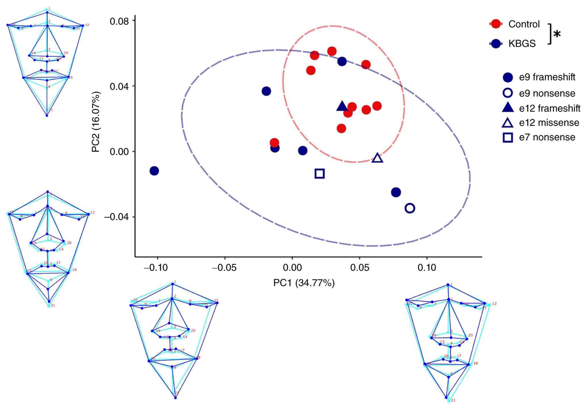
2D facial morphometric analysis
The PCA of 2D facial landmarks revealed partial
overlap among diagnostic groups. In total, two patients fell within
the range of variation of control individuals, whereas the
remaining eight exhibited broad dispersion along PC1, which
accounted for 34.7% of the total variance. PC2 accounted for 16.1%
of variance and revealed a distinct separation between patients and
controls (Fig. 4). Permutation
tests based on Procrustes distances confirmed that facial shape
differences between controls and patients with KBGS were
statistically significant (Procrustes distance=0.045;
P=0.0288).
At the extremes of PC1, individuals with the most
severe dysmorphic features exhibited pronounced facial asymmetries,
affecting either the left or the right side of the face (Fig. 4). At the positive extreme, patients
had narrower faces, upward displacement of the right orbit,
downward nasal tip displacement, increased lower lip thickness and
lateral chin deviation to the right. At the negative extreme,
patients exhibited wider faces, downward displacement of the right
orbit, upward nasal tip displacement, increased upper lip thickness
and lateral chin deviation to the left (Fig. 4).
PC2 highlighted characteristic KBGS facial features,
including increased forehead height, nasal prominence, ticker lower
lip and reduced chins (Fig. 4).
Patients displayed heterogeneous craniofacial
phenotypes (Fig. 4), but no
significant associations were detected between facial variation and
mutation type (frameshift, nonsense, missense) or exon location
(11,14,20).
Discussion
KBGS is a rare autosomal dominant neurodevelopmental
disorder caused by haploinsufficiency of ANKRD11, a gene
encoding a chromatin regulator involved in transcriptional control,
neuronal development and skeletal formation (2,5,9,10).
Since its initial description, the phenotypic spectrum of KBGS has
expanded to include a wide range of neurodevelopmental,
craniofacial, skeletal, behavioral and systemic manifestations
(1-4,11,13,17,20).
To date, >300 molecularly confirmed cases have been reported
worldwide, with >1,000 distinct ANKRD11 variants
described, the majority of which are truncating and cluster within
exon 9, a well-established mutational hotspot (2-4,11,12,19).
Despite increasing recognition, KBGS remains
underdiagnosed and typically misclassified due to its marked
phenotypic variability and overlap with other genetic conditions
(4,9,10,13,15,16,35).
The syndrome is referred to as a ‘great imitator’ because of its
clinical resemblance to other chromatinopathies, such as Cornelia
de Lange and Coffin-Siris syndromes, as well as multisystem
developmental conditions including VACTERL association (vertebral
defects, anal atresia, cardiac defects, tracheoesophageal
fistula/esophageal atresia, renal anomalies and limb abnormality
(VACTERL) and Turner syndrome (4,9,10,15,16).
Genotype-phenotype associations in KBGS are weak or absent, with
variant type and location providing little predictive value for
clinical severity (2,3,13,18).
Consequently, molecular studies are essential to establish a
definitive diagnosis, as clinical features alone are not
pathognomonic and genotype-phenotype associations in KBGS remain
weak or absent (5,12,36,37).
The present study presents a comprehensive
characterization of 10 individuals with KBGS, integrating detailed
clinical evaluation, molecular findings, 2D facial morphometric
analysis and a structured comparison with previously published
large cohorts. The present findings reinforce the concept of KBGS
as a highly heterogeneous chromatinopathy and demonstrate the
diagnostic challenges associated with its broad phenotypic
spectrum.
Clinically, all individuals in the present cohort
exhibited neurodevelopmental involvement, confirming developmental
delay and intellectual disability as key features of KBGS.
Craniofacial dysmorphism was universal, although the specific
combination and severity of features varied. Notably, the most
frequent craniofacial findings included facial asymmetry, coarse
facial features, low anterior hairline, bulbous nasal tip and
prominent ears. These features appeared at higher frequencies than
in previously reported cohorts (2,3,6,11,19,20),
suggesting they may be under-recognized or inconsistently
documented in previous studies. This underscores the importance of
detailed and systematic dysmorphological assessment in individuals
with suspected KBGS.
Comparison with six previously published cohorts
with detailed phenotypic data showed that the present cohort shares
the core clinical features of KBGS, including intellectual
disability/developmental delay, craniofacial dysmorphism, skeletal
anomaly and neuropsychiatric manifestations (2,3,6,11,19,20).
Certain features, such as low anterior hairline, facial asymmetry,
hearing loss, and hypotonia, were observed at higher frequencies in
the present series, potentially reflecting more systematic
phenotypic assessment or cohort-specific differences (Table III) (2,3,6,11,19,20).
| Table IIIComparative summary of the primary
clinical features reported in six previously published large
cohorts of individuals with KBG syndrome (2,3,6,11,19,20)
and the present study. |
Table III
Comparative summary of the primary
clinical features reported in six previously published large
cohorts of individuals with KBG syndrome (2,3,6,11,19,20)
and the present study.
| | Study |
|---|
| Feature | Serra et al,
2025 | Choi et al,
2022 | Parenti et
al, 2021 | Low et al,
2016 | Goldenberg et
al, 2016 | Ockeloen et
al, 2016 | Present study | Overall |
|---|
| Number of
patients | 3 | 8 | 23 | 32 | 39 | 19 | 10 | 134 |
| Country | Italy | South Korea | Multiple,
including: Ger, Ita, Can, Ire, USA and Col | United Kingdom | France | Netherlands | Colombia | Multiple
countries |
| Sex
(female/male) | 2/1 | 5/3 | 11/12 | 20/12 | 23/16 | 9/10 | 4/6 | 74/60 |
| Age at diagnosis,
years | 0.25-18.00 (mean,
6.44) | 0.25-37.00 (mean,
8.95) | 4.00-23.00 (mean,
6.40) | 2.00-47.00 (mean,
13.30) | 1.00-66.00 (mean,
14.26) | 6.00-47.00 (mean,
20.74) | 4.00-36.00 (mean,
16.40) | 0.25-66.00 (mean,
12.36) |
| Intellectual
disability/developmental delay | 3/3 (100.00%) | 8/8 (100.00%) | 16/23 (69.57%) | 31/32 (96.88%) | 28/39 (71.79%) | 18/19 (94.74%) | 10/10
(100.00%) | 114/134
(85.07%) |
| Low hair
implantation | ND | ND | 15/23 (65.22%) | ND | ND | ND | 8/10 (80.00%) | 23/33 (69.7%) |
| Facial
asymmetry | ND | ND | ND | ND | ND | ND | 7/10 (70.00%) | 7/10 (70%) |
| Clinodactyly and
camptodactyly | 3/3 (100.00%) | ND | 15/23 (65.22%) | 15/32 (46.88%) | 23/39 (58.97%) | 18/19 (94.74%) | 7/10 (70.00%) | 81/126
(64.28%) |
| Bulbous nose | 2/3 (66.67%) | 1/8 (12.50%) | 16/23 (69.57%) | ND | ND | ND | 9/10 (90.00%) | 28/44 (63.64%) |
| Macrodontia | 1/3 (33.33%) | 6/8 (75.00%) | 15/23 (65.22%) | 23/32 (71.88%) | 18/39 (46.15%) | 17/19 (89.47%) | 5/10 (50.00%) | 85/134
(63.43%) |
| Synophrys | 1/3 (33.33%) | 4/8 (50.00%) | 15/23 (65.22%) | ND | ND | ND | 6/10 (60.00%) | 26/44 (59.1%) |
| Prominent ears | 1/3 (33.33%) | 5/8 (62.50%) | ND | ND | ND | ND | 6/10 (60.00%) | 12/21 (57.14%) |
| Full, arched
eyebrows | 1/3 (33.33%) | ND | 11/23 (47.83%) | ND | ND | ND | 7/10 (70.00%) | 19/36 (52,78%) |
| Psychiatric
features | 1/3 (33.33%) | ND | 15/23 (65.22%) | 12/32 (37.50%) | 17/39 (43.59%) | 17/19 (89.47%) | 4/10 (40.00%) | 66/126
(52.38%) |
| Hypotonia | 1/3 (33.33%) | ND | ND | ND | ND | ND | 5/10 (50.00%) | 6/13 (46,14%) |
| Prominent nasal
bridge | 1/3 (33.33%) | 1/8 (12.50%) | ND | ND | ND | ND | 7/10 (70.00%) | 9/21 (42.85%) |
| Scoliosis | 0/3 (0.00%) | 1/8 (12.50%) | ND | ND | ND | 9/19 | 6/10 (60.00%) | 16/40 (40%) |
| Feeding
difficulties | ND | ND | 7/23 (30.43%) | 10/32 (31.25%) | ND | ND | 6/10 (60.00%) | 23/65 (35.38%) |
| Seizures | 1/3 (33.33%) | 3/8 (37.50%) | 6/23 (26.09%) | 13/32 (40.62%) | 12/39 (30.77%) | 5/19 (26.32%) | 5/10 (50.00%) | 45/134
(33.58%) |
| Sensorineural
hearing loss | 1/3 (33.33%) | ND | 6/23 (26.09%) | 8/32 (25.00%) | 11/39 (28.21%) | 6/19 (31.58%) | 8/10 (80.00%) | 40/126
(31.74%) |
| Hypertrichosis | 0/3 (0.00%) | ND | 5/23 (21.74%) | ND | ND | ND | 3/10 (30.00%) | 8/36 (22.22%) |
| Coarse facies | ND | ND | 2/23 (8.70%) | ND | ND | ND | 5/10 (50.00%) | 7/33 (21.21%) |
Skeletal anomaly and neurological manifestations
were also common, including scoliosis, digital anomaly, hypotonia,
seizures and behavioral disturbances. Hearing impairment was
identified in a high proportion of patients, representing one of
the most prevalent extracranial manifestations in the present
series and reinforcing the need for routine auditory screening in
the clinical evaluation of individuals with KBGS.
At the molecular level, the present findings support
ANKRD11 haploinsufficiency as the primary pathogenic
mechanism underlying KBGS. The predominance of truncating variants
and their clustering within exon 9 align with previous reports,
confirming this region as a global mutational hotspot (2,6).
Previously reported ANKRD11 variants and the 10 variants
identified in the present cohort are summarized in Fig. 5. Importantly, several variants in
the present cohort were novel, expanding the ANKRD11
mutational spectrum and contributing data from an under-represented
Latin American population. Despite this detailed molecular
characterization, no association between variant type, location, or
novelty and phenotypic severity was established, reinforcing the
lack of robust genotype-phenotype association in KBGS.
 | Figure 5Schematic representation of
ANKRD11, its functional domains and the distribution of
pathogenic variants associated with KBG syndrome. (A) Exonic
structure of ANKRD11 and major protein functional domains,
including ANK, RD1 and RD2 and AD. Exon 9 is highlighted as a
mutational hotspot. Distribution of pathogenic variants located (B)
within and (C) outside exon 9. Variants identified in the present
study are highlighted in red, whereas previously reported variants
are shown in yellow. ANKRD11, ankyrin repeat
domain-containing protein 11; RD1, repression domain 1; AD,
activation domain; N-term, N-terminal region; C-term, C-terminal
region; aa, amino acids. |
A novel aspect of the present study is the
incorporation of 2D facial morphometric analysis. Quantitative
assessment demonstrated consistent alterations in facial shape,
especially asymmetry and dysmorphologies in the upper face, midface
and nasal regions, supporting the existence of a craniofacial
signature associated with ANKRD11 haploinsufficiency.
However, substantial interindividual variability persisted,
reinforcing that genotype-phenotype associations cannot yet be
reliably inferred from facial features alone. Future research
should include larger cohorts to explore potential genetic
associations between facial dysmorphology, the impact of
developmental canalization on varying degrees of asymmetry and the
influence of genetic ancestry. This is particularly relevant in
populations with high ancestral diversity and admixture, where
phenotypic expression may be modulated by complex genetic
backgrounds that are under-represented in existing reference
datasets. Longitudinal studies employing 3D facial modeling may
clarify whether KBGS facial phenotype changes over time, as facial
dysmorphology may become more pronounced during postnatal
development (38). Rather than
defining molecular subgroups, facial morphometry serves as a
complementary tool that enhances phenotypic recognition and
diagnostic confidence.
Comparison with six large published cohorts
(2,3,6,11,19,20)
revealed substantial overlap in key clinical features but also
highlighted differences that may reflect cohort-specific
characteristics or improved phenotypic recognition. By contrast
with the literature, macrodontia, typically described as a cardinal
feature of KBGS, was not prominent in the present cohort. This
emphasizes that its absence should not preclude diagnosis and
supports the need for comprehensive phenotypic evaluation rather
than relying on isolated hallmark features.
Overall, the present findings reinforce KBGS as a
complex and heterogeneous disorder that frequently overlaps with
other genetic conditions, making clinical diagnosis challenging.
Molecular testing remains essential for accurate diagnosis,
appropriate classification and the implementation of targeted
clinical interventions.
The present study expanded the clinical and
molecular spectrum of KBGS through the detailed characterization of
a Colombian case series, contributing novel data regarding
ANKRD11-associated disorders. The present findings confirm
the marked phenotypic variability of KBGS and demonstrated that
variant type and location are not directly associated with
phenotypic severity.
Despite the use of advanced approaches, such as 2D
facial morphometric analysis, no robust genotype-phenotype
association was identified. Instead, KBGS emerges as a highly
heterogeneous condition that frequently mimics other genetic
syndromes, particularly chromatinopathy, VACTERL association and
Turner syndrome, making clinical diagnosis challenging.
The prominence of facial asymmetry, coarse facial
features, low anterior hairline, bulbous nasal tip and prominent
ears in the present cohort, together with the limited relevance of
macrodontia, underscores the importance of detailed and systematic
dysmorphological evaluation. Ultimately, the present study
highlights the critical role of molecular testing in confirming the
diagnosis, enabling appropriate intervention and improving clinical
care of individuals with KBGS.
Supplementary Material
Sanger sequencing confirmation of the
ANKRD11 variant in patient 1. Sanger sequencing chromatogram
showing (A) reference and (B) patient 1 carrying the ANKRD11
c.3600dup (p.Val1201SerfsTer3) variant. The arrow indicates the
position of the nucleotide duplication. ANKRD11, ankyrin
repeat domain-containing protein 11.
Sanger sequencing confirmation of the
ANKRD11 variant in patient 2. Sanger sequencing chromatogram
showing patient 2 carrying the ANKRD11: c.7753C>T
(p.Arg2585Cys) variant. The arrow indicates the position of the
nucleotide substitution. ANKRD11, ankyrin repeat
domain-containing protein 11.
Sanger sequencing confirmation of the
ANKRD11 variant in patient 4. Sanger sequencing chromatogram
showing (A) patient 4 carrying the ANKRD11: c.4218C>A
(p.Tyr1406Ter) variant and (B) reference sequence. The arrow
indicates the position of the nucleotide substitution.
ANKRD11, ankyrin repeat domain-containing protein 11.
Sanger sequencing confirmation of the
ANKRD11 variant in patient 9. Sanger sequencing chromatogram
for (A) patient 9 carrying the ANKRD11: c.3499dup
(p.Ser1167PhefsTer) variant and (B) referencesequence. The arrow
indicates the position of the nucleotide duplication.
ANKRD11, ankyrin repeat domain-containing protein 11.
Acknowledgements
Not applicable.
Funding
Funding: No funding was received.
Availability of data and materials
The data generated in the present study are not
publicly available due to ethical, legal, consent-related and
identifiability restrictions but may be requested from the
corresponding author.
Authors' contributions
SBN, JANC and HP conceived and designed the study
and analyzed and interpreted data. SBN, LEP and LVCC interpreted
data and wrote the manuscript. MAM and NMA performed the
two-dimensional facial morphometric analysis and wrote the
manuscript. NMA revised the manuscript. All authors have read and
approved the final manuscript. JANC and HP confirm the authenticity
of all the raw data.
Ethics approval and consent to
participate
The present study was approved by the ethics
committee of the Hospital Universitario Fundación Valle del Lili,
Cali, Colombia; approval no. 2025.140). Written informed consent to
participate was obtained from all adult participants, including
healthy controls and individuals with KBGS, who were capable of
providing consent. For minors and individuals with intellectual
disability impairing autonomous consent capacity, written informed
consent was obtained from parents or legal guardians.
Patient consent for publication
Written informed consent for publication of clinical
data and unredacted photographs was obtained from all adult
participants, including healthy controls and individuals with KBGS,
who were capable of providing consent, and from parents or legal
guardians of minors and individuals with intellectual disability
impairing autonomous consent capacity. This consent was obtained
specifically for this study.
Competing interests
The authors declare that they have no competing
interests.
References
|
1
|
Skjei KL, Martin MM and Slavotinek AM: KBG
syndrome: Report of twins, expansion of the phenotype, and review
of the literature. Am J Med Genet. 143A (Part A):292–300.
2007.PubMed/NCBI View Article : Google Scholar
|
|
2
|
Low K, Ashraf T, Canham N, Clayton-Smith
J, Deshpande C, Donaldson A, Fisher R, Flinter F, Foulds N, Fryer
A, et al: Clinical and genetic aspects of KBG syndrome. Am J Med
Genet A. 170:2835–2846. 2016.PubMed/NCBI View Article : Google Scholar
|
|
3
|
Goldenberg A, Riccardi F, Tessier A,
Pfundt R, Busa T, Cacciagli P, Capri Y, Coutton C, Delahaye-Duriez
A, Frebourg T, et al: Clinical and molecular findings in 39
patients with KBG syndrome caused by deletion or mutation of
ANKRD11. Am J Med Genet A. 170:2847–2859. 2016.PubMed/NCBI View Article : Google Scholar
|
|
4
|
Brancati F, D'Avanzo MG, Digilio MC,
Sarkozy A, Biondi M, De Brasi D, Mingarelli R and Dallapiccola B:
KBG syndrome in a cohort of Italian patients. Am J Med Genet A.
131A:144–149. 2004.PubMed/NCBI View Article : Google Scholar
|
|
5
|
Sirmaci A, Spiliopoulos M, Brancati F,
Powell E, Duman D, Abrams A, Bademci G, Agolini E, Guo S, Konuk B,
et al: Mutations in ANKRD11 cause KBG syndrome, characterized by
intellectual disability, skeletal malformations, and macrodontia.
Am J Hum Genet. 89:289–294. 2011.PubMed/NCBI View Article : Google Scholar
|
|
6
|
Parenti I, Mallozzi MB, Hüning I,
Gervasini C, Kuechler A, Agolini E, Albrecht B, Baquero-Montoya C,
Bohring A, Bramswig NC, et al: ANKRD11 variants: KBG syndrome and
beyond. Clin Genet. 100:187–200. 2021.PubMed/NCBI View Article : Google Scholar
|
|
7
|
Orphanet: KBG syndrome. Orphanet.
https://www.orpha.net/en/disease/detail/2332.
|
|
8
|
Swols DM and Tekin M: ANKRD11-related KBG
syndrome. 2018 Mar 22 [Updated 2026 Jun 11]. In: Adam MP, Bick S,
Mirzaa GM, Pagon RA, Wallace SE and Amemiya A (eds).
GeneReviews® [Internet]. Seattle (WA): University of
Washington, Seattle; 1993-2026.
|
|
9
|
Fahrner JA and Bjornsson HT: Mendelian
disorders of the epigenetic machinery: Tipping the balance of
chromatin states. Annu Rev Genomics Hum Genet. 15:269–293.
2014.PubMed/NCBI View Article : Google Scholar
|
|
10
|
Bjornsson HT: The Mendelian disorders of
the epigenetic machinery. Genome Res. 25:1473–1481. 2015.PubMed/NCBI View Article : Google Scholar
|
|
11
|
Serra G, Elefante P, Gazzitano Y, Memo L,
Mineo V, Morando C, Nardello R, Piro E, Travan L and Corsello G:
KBG syndrome: Report and follow-up on three unrelated patients
observed at different ages. Ital J Pediatr. 51(54)2025.PubMed/NCBI View Article : Google Scholar
|
|
12
|
Willemsen MH, Fernandez BA, Bacino CA,
Gerkes E, de Brouwer AP, Pfundt R, Sikkema-Raddatz B, Scherer SW,
Marshall CR, Potocki L, et al: Identification of ANKRD11 and ZNF778
as candidate genes for autism and variable cognitive impairment in
the novel 16q24.3 microdeletion syndrome. Eur J Hum Genet.
18:429–435. 2010.PubMed/NCBI View Article : Google Scholar
|
|
13
|
Novara F, Rinaldi B, Sisodiya SM, Coppola
A, Giglio S, Stanzial F, Benedicenti F, Donaldson A, Andrieux J,
Stapleton R, et al: Haploinsufficiency for ANKRD11-flanking genes
makes the difference between KBG and 16q24.3 microdeletion
syndromes: 12 new cases. Eur J Hum Genet. 25:694–701.
2017.PubMed/NCBI View Article : Google Scholar
|
|
14
|
Herrmann J, Pallister PD, Tiddy W and
Opitz JM: The KBG syndrome-a syndrome of short stature,
characteristic facies, mental retardation, macrodontia and skeletal
anomalies. Birth Defects Orig Artic Ser. 11:7–18. 1975.PubMed/NCBI
|
|
15
|
Kleefstra T, Kramer JM, Neveling K,
Willemsen MH, Koemans TS, Vissers LE, Wissink-Lindhout W, Fenckova
M, van den Akker WM, Kasri NN, et al: Disruption of an
EHMT1-associated chromatin-modification module causes intellectual
disability. Am J Hum Genet. 91:73–82. 2012.PubMed/NCBI View Article : Google Scholar
|
|
16
|
Bögershausen N and Wollnik B: Mutational
landscapes and phenotypic spectrum of SWI/SNF-related intellectual
disability disorders. Front Mol Neurosci. 11(252)2018.PubMed/NCBI View Article : Google Scholar
|
|
17
|
Peluso F, Caraffi SG, Contrò G, Valeri L,
Napoli M, Carboni G, Seth A, Zuntini R, Coccia E, Astrea G, et al:
Deep phenotyping of the neuroimaging and skeletal features in KBG
syndrome: A study of 53 patients and review of the literature. J
Med Genet. 60:1224–1234. 2023.PubMed/NCBI View Article : Google Scholar
|
|
18
|
Chanes NM, Wong J and Lacassie Y: Further
delineation of DDX3X syndrome. Clin Dysmorphol. 28:149–151.
2019.PubMed/NCBI View Article : Google Scholar
|
|
19
|
Choi Y, Choi J, Do H, Hwang S, Seo GH,
Choi IH, Keum C, Choi JH, Kang M, Kim GH, et al: KBG syndrome:
Clinical features and molecular findings in seven unrelated Korean
families with a review of the literature. Mol Genet Genomic Med.
11(e2127)2023.PubMed/NCBI View Article : Google Scholar
|
|
20
|
Ockeloen CW, Willemsen MH, de Munnik S,
van Bon BW, de Leeuw N, Verrips A, Kant SG, Jones EA, Brunner HG,
van Loon RL, et al: Further delineation of the KBG syndrome
phenotype caused by ANKRD11 aberrations. Eur J Hum Genet.
23:1176–1185. 2015.PubMed/NCBI View Article : Google Scholar
|
|
21
|
GeneSystems: GeneSystems®
platform platform v4.0.1. https://platform.genesystm.com.
|
|
22
|
den Dunnen JT, Dalgleish R, Maglott DR,
Hart RK, Greenblatt MS, McGowan-Jordan J, Roux AF, Smith T,
Antonarakis SE and Taschner PE: HGVS recommendations for the
description of sequence variants: 2016 update. Hum Mutat.
37:564–569. 2016.PubMed/NCBI View Article : Google Scholar
|
|
23
|
Richards S, Aziz N, Bale S, Bick D, Das S,
Gastier-Foster J, Grody WW, Hegde M, Lyon E, Spector E, et al:
Standards and guidelines for the interpretation of sequence
variants: A joint consensus recommendation of the American college
of medical genetics and genomics and the association for molecular
pathology. Genet Med. 17:405–424. 2015.PubMed/NCBI View Article : Google Scholar
|
|
24
|
Saphetor SA: VarSome. https://varsome.com/.
|
|
25
|
Genoox/QIAGEN: Franklin clinical database.
https://franklin.genoox.com/clinical-db/home.
|
|
26
|
National Center for Biotechnology
Information: ClinVar. https://www.ncbi.nlm.nih.gov/clinvar/.
|
|
27
|
Rohlf FJ: tpsDig2, version 2.32, 64-bit
executable [computer software]. Stony Brook University, 2021.
Available from: https://www.sbmorphometrics.org/soft-dataacq.html.
|
|
28
|
Echeverry-Quiceno LM, Candelo E, Gómez E,
Solís P, Ramírez D, Ortiz D, González A, Sevillano X, Cuéllar JC,
Pachajoa H and Martínez-Abadías N: Population-specific facial
traits and diagnosis accuracy of genetic and rare diseases in an
admixed Colombian population. Sci Rep. 13(6869)2023.PubMed/NCBI View Article : Google Scholar
|
|
29
|
Bookstein FL: Morphometric tools for
landmark data: Geometry and biology. Cambridge University Press,
1991.
|
|
30
|
Dryden IL and Mardia KV: Statistical shape
analysis with applications in R. 2nd edition. John Wiley &
Sons, 2016.
|
|
31
|
Hallgrimsson B, Percival CJ, Green R,
Young NM, Mio W and Marcucio R: Morphometrics, 3D imaging, and
craniofacial development. Curr Top Dev Biol. 115:561–597.
2015.PubMed/NCBI View Article : Google Scholar
|
|
32
|
Klingenberg CP: MorphoJ: An integrated
software package for geometric morphometrics. Mol Ecol Resour.
11:353–357. 2011.PubMed/NCBI View Article : Google Scholar
|
|
33
|
Adams D, Collyer M, Kaliontzopoulou A and
Baken E: geomorph: Geometric Morphometric Analyses of 2D and 3D
Landmark Data. Version 4.0.10. https://cran.r-project.org/package=geomorph.
|
|
34
|
Gargano MA, Matentzoglu N, Coleman B,
Addo-Lartey EB, Anagnostopoulos AV, Anderton J, Avillach P, Bagley
AM, Bakštein E, Balhoff JP, et al: The human phenotype ontology in
2024: Phenotypes around the world. Nucleic Acids Res. 52
(D1):D1333–D1346. 2024.PubMed/NCBI View Article : Google Scholar
|
|
35
|
Wright CF, McRae JF, Clayton S, Gallone G,
Aitken S, FitzGerald TW, Jones P, Prigmore E, Rajan D, Lord J, et
al: Making new genetic diagnoses with old data: Iterative
reanalysis and reporting from genome-wide data in 1,133 families
with developmental disorders. Genet Med. 20:1216–1223.
2018.PubMed/NCBI View Article : Google Scholar
|
|
36
|
Deciphering Developmental Disorders Study.
Prevalence and architecture of de novo mutations in developmental
disorders. Nature. 542:433–438. 2017.PubMed/NCBI View Article : Google Scholar
|
|
37
|
Wright CF, FitzPatrick DR and Firth HV:
Paediatric genomics: Diagnosing rare disease in children. Nat Rev
Genet. 19:253–268. 2018.PubMed/NCBI View Article : Google Scholar
|
|
38
|
Brancati F, Sarkozy A and Dallapiccola B:
KBG syndrome. Orphanet J Rare Dis. 1(50)2006.PubMed/NCBI View Article : Google Scholar
|