Introduction
Type 2 diabetes is the most common type of diabetes.
It is one of the non-communicable diseases with an increasing
incidence worldwide; global data indicates that approximately 589
million adults (aged 20-79) are living with diabetes (1). The pathophysiology of diabetes is
caused by insulin resistance and/or insulin deficiency.
Contributing factors include eating a diet high in fat and
carbohydrates, lack of exercise, stress and obesity. The result of
prolonged high blood sugar levels causes complications in various
organs, such as neuropathy, retinopathy and nephropathy (2).
Normally, the progression of clinical diabetes 2
will be insulin resistance or decreased insulin function first due
to various contributing factors such as daily behavior, eating
foods high in starch and fat, stress and lack of exercise. In
addition, there are abnormalities of some genes related to insulin
signaling in target cells, resulting in high blood sugar levels,
which will stimulate pancreatic β-cells to respond by working more
to produce insulin sufficient for the body's needs. If blood sugar
levels are continuously high, the cells will work hard until
exhaustion occurs, producing less insulin, resulting in continuous
high blood sugar levels, which will create free radicals reactive
oxygen species (ROS) and stimulate endoplasmic reticulum (ER)
stress (3) and mitochondrial stress
(4), which are toxic to pancreatic
β-cells, called glucotoxicity, resulting in apoptosis and insulin
deficiency in the later stages and eventually diabetes.
Currently, medicinal plants that have medicinal
properties for treating diabetes are another alternative that has
gained much interest. In the present study, the researchers were
interested in the medicinal properties of Justicia
gendarussa Burm.f. (JG; Acanthaceae) leaf extract. JG is a
local medicinal plant that is easy to find and is popularly grown
in the eastern region of Thailand. The important substances found
in leaves that have antioxidant properties include phenolic
compounds and flavonoids, which are found in high quantities
(5,6). Anti-inflammatory studies have shown
that flavonoids (vitexin and apigenin) can reduce inflammation as
well as nonsteroidal anti-inflammatory drugs (7). Studies have demonstrated that JG
extracts possess significant anti-inflammatory and antioxidant
activities, primarily through the inhibition of pro-inflammatory
cytokines and the scavenging of free radicals (8,9), while
their potential in treating asthma and hyperuricemia conditions has
been explored (10,11). Another interesting property is the
anti-diabetic effect. It was found that the JG leaf extract at
concentrations of 50 and 100 mg/kg/bw can reduce blood sugar and
lipid levels and reduce urine sugar in diabetic rats (12). Moreover, the 12 h treatment period
of the methanolic leaf extract of JG at 400 mg/kg bw reduced blood
glucose level to 52.75% (12).
However, the mechanism used to reduce the blood sugar of JG leaf
extract is still unknown. Therefore, the aims of the present study
are to develop an extract from JG leaf and to test the
anti-diabetic mechanism of JG leaf extract through preventing the
death of pancreatic β-cells and increasing the efficiency of
pancreatic β-cells in producing and secreting insulin in INS-1 rat
insulinoma cell line.
Materials and methods
Plant collection and extraction
JG leaves were collected from Ban Bueng District,
Chonburi Province, Thailand. The plant was identified by Associated
Professor Dr Watchara Damjuti (Faculty of Integrative Medicine,
Rajamangala University of Technology Thanyaburi (RMUTT),
Pathumtani, Thailand) and the voucher specimen (identification no.
CR0011/25) is kept at herbarium of the Faculty of Integrative
Medicine, RMUTT. Plants were cleaned and dried in an oven at
50-60˚C. A total of 366 g of dried plant powder was subjected to
extraction using ethanol as the solvent via the Soxhlet extraction
method, with a plant-to-solvent ratio of 20 g:300 ml. The
extraction was continued until the active compounds were completely
extracted from the plant material (~6 h). The resulting extract
solution was then concentrated and dried at 50-55˚C under vacuum
conditions using a rotary evaporator.
Determination of flavonoid content in
the extract
The JG extract has been previously reported to
possess a high content of flavonoids. To quantify this, the total
flavonoid content was determined using the aluminium chloride
colorimetric method, employing quercetin as a reference standard.
For each standard solution, 200 µl was mixed with 100 µl of 10%
(w/v) aluminium chloride and 100 µl of 0.1 mM potassium acetate in
a test tube. The mixtures were incubated at room temperature for 30
min to allow for color development. Absorbance was subsequently
measured at 415 nm using a microplate reader and a standard
calibration curve was constructed by plotting absorbance values
against quercetin concentrations. The JG extract was prepared at a
single concentration and subjected to the same assay conditions as
the standard solutions. All measurements were performed in
triplicate. The absorbance of the extract was interpolated from the
standard curve to calculate the flavonoid content, which was
expressed as milligrams of quercetin equivalents per gram of
extract (mg QE/g).
Determination of total phenolic
content (TPC)
The total phenolic content of the JG extract was
measured using the Folin-Ciocalteu colorimetric method, as
described by Singleton et al in 1999(13). Briefly, 25 µl of the JG extract (at
concentrations ranging from 0.0156-1 mg/ml) was mixed with 25 µl of
0.2 N Folin-Ciocalteu reagent. The plate was incubated in the dark
at room temperature for 30 min. Then, 100 µl of a 7% sodium
carbonate solution was added and the reaction mixture was incubated
for 5 min in the dark at room temperature. The absorbance was
measured at 730 nm using a microplate reader (FLUOstar
Omega®; BMG Labtech GmbH). The total phenolic content
was calculated as gallic acid equivalents (GAE) in mg per gram of
extract (mg GAE/g dry wt.), using a standard curve prepared with
gallic acid. The experiment was conducted in triplicate.
Phytochemical analysis by
high-performance liquid chromatography (HPLC)
According to a previously report, vitexin,
naringenin, apigenin and kaempferol are found in JG leaves
(14). In the present study,
vitexin was used as a chemical marker and its quantitative analysis
in JG extract was measured using the HPLC following a previous
report (15). HPLC analysis was
performed on the Agilent-1260 Infinity system (Agilent
Technologies, Inc.) coupled with a photodiode array detector. In
the separation condition, reverse phase C18 column (Kromasil 100-5;
250x4.6 mm; Agilent Technologies, Inc.) was used as a stationary
phase and acetonitrile:0.1% ortho-phosphoric acid (20:80, v/v) was
used as a mobile phase. The flow rate was 1 ml/min. The vitexin
signal was detected at 335 nm. The calibration curve of vitexin was
conducted from vitexin concentrations and obtained area under the
curve. Vitexin content in JG extract was calculated from the
calibration curve.
Examination of antioxidant activity by
2,2-diphenyl-1-picrylhydrazyl (DPPH) radical scavenging assay
The ability to scavenge DPPH radicals of JG extract
was assessed by using a modified version of the Clarke method from
2013(16). Briefly, a 96-well plate
was filled with 50 µl of JG extract in 5% DMSO to the final
concentration range of 0.0156-1 mg/ml, followed by 150 µl of 0.3 M
DPPH reagent in absolute ethanol. After shaking for two min, the
plate was kept in the dark at room temperature for 30 min. At the
end of incubation, the absorbance was measured by a
spectrophotometer (FLUOstar Omega®; BMG Labtech GmbH) at
a wavelength 517 nm. The ability of the JG extract to scavenge free
radicals was compared with standard gallic acid. The experiment was
conducted in 3 dependent experiments. % DPPH scavenging potential
was calculated by the following formula:
% DPPH scavenging potential=[(Control
absorbance-Sample absorbance)/Control absorbance] x100
Examination of antioxidant activity by
2,2'-azino-bis(3-ethylbenzothiazoline-6-sulfonic acid (ABTS)
radical scavenging assay
The ABTS radical scavenging assay was conducted
using a modified version by a previous study (17). ABTS•+ radical cations were generated
by reacting 7 mM ABTS solution with 2.45 mM potassium persulfate in
distilled water and allowing the mixture to stand for 12-16 h in
the dark at room temperature. The resulting ABTS•+ solution was
diluted with methanol to an absorbance of 0.7±0.02 at 734 nm. To
100 µl of the JG extract at various concentrations (10, 50, 100 and
200 µg/ml), 100 µl of ABTS•+ solution was added and the mixture was
incubated for 6 min at room temperature. Absorbance was then
measured at 734 nm using a microplate reader (FLUOstar
Omega®; BMG Labtech GmbH). % ABTS scavenging potential
was calculated by the following formula:
% ABTS scavenging potential=[(Control
absorbance-Sample absorbance)/Control absorbance] x100
INS-1-cell culture
INS-1 cells (INS1 832/13 rat insulinoma cell line;
MilliporeSigma) were cultivated in RPMI-1640 media (Gibco; Thermo
Fisher Scientific, Inc.) supplemented with 10% fetal calf serum
(FCS) (Gibco; Thermo Fisher Scientific, Inc.), 100 U/ml penicillin
and 100 g/ml streptomycin at 37˚C in humidified air containing 5%
CO2. The culture medium was changed every 2 days. INS-1
cells were maintained normally in a solution containing 11.1 mM
glucose (their basal level) and they were cultured in an
approximately fourfold (40 mM) glucose to induce glucotoxicity.
Cytotoxicity and cytoprotective effect
assessment by MTT assay
The MTT assay was performed to determine toxicity as
previously described (18).
Briefly, INS-1 cells, ~10,000 cells per well, were seeded into
96-well plates. Cells were cultured in RPMI-1640 medium containing
10% fetal bovine serum (FBS; Gibco; Thermo Fisher Scientific, Inc.)
and 0.05 mM β-mercaptoethanol at 37˚C with 5% CO2
incubator. To examine cytotoxicity of JG extract, cells were
exposed to varying concentrations of the JG extract ranging from
31.2-2,000 µg/ml for 72 h. To evaluate the cytoprotective effect of
the JG extract, cells were divided into several groups: a negative
control (11.1 mM glucose), a glucotoxic/positive control (40 mM
glucose) and treatment groups (40 mM glucose + 50 or 100 µg/ml JG
extract, as well as 10, 20, or 40 µM vitexin). After 72 h of
incubation, a 100 µl of MTT solution (final concentration 0.5
mg/ml) was added to each well. After incubation at 37˚C with 5%
CO2 for 90 min, MTT solution were removed, then, the
formazan crystals were dissolved in 150 µl DMSO. The absorbance was
determined using a microplate scanning spectrophotometer (FLUOstar
Omega®; BMG Labtech GmbH) at 570 nm. All experiments
were conducted in triplicate in three independent experiments. The
following calculation was used to calculate the proportion of
viable cells using the averaged 570-nm absorbance values.
Cell viability (%)=Absorbance of sample/Absorbance
of control x100(1).
Measurement of intracellular ROS
production by 2'-7'dichlorofluorescin diacetate (DCFH-DA)
assay
The DCFH-DA probe was used to assay the ROS in
cells. The cells were cultured with 40 mM glucose alone or with 40
mM glucose plus various concentrations of JG leaf extract or
vitexin. The cells were grown in a 5% CO2 incubator at
37˚C for 48 h. The cells were then aspirated from 96-well plates
and the cells were washed with 100 µl of Sterile PBS. 100 µM of
DCFH-DA solution was added. The cells were incubated in 5%
CO2 incubator at 37˚C for 30 min. The fluorescence
absorbance was measured at the wavelengths of Excitation 495 nm and
Emission 529 nm using a spectrophotometer (FLUOstar
Omega®; BMG Labtech GmbH). All experiments were
conducted in triplicate in three independent experiments.
Analysis of cell apoptosis by Annexin
V-fluorescein isothiocyanate (FITC)/propidium iodide (PI) staining,
followed by flow cytometry
Apoptosis was assessed using FITC/PI staining
followed by flow cytometric analysis, in accordance with the
manufacturer's protocol and the MIFlowCyt guidelines for flow
cytometry experiments by Lee et al 2008(19). INS-1 pancreatic β-cells were
cultured under standard conditions and exposed to high-glucose
stress (40 mM glucose) in the presence or absence of JG leaf
extract (50 µg/ml) or vitexin (10 µM). Untreated cells cultured
under normal glucose conditions served as controls. Cells were
incubated for 72 h prior to analysis. After treatment, cells were
harvested, washed twice with cold phosphate-buffered saline (PBS)
and resuspended in binding buffer. Cells were then stained with 5
µl of Annexin V-FITC and 5 µl of PI for 15 min at 4˚C in
the dark. Following incubation, samples were immediately analyzed
by flow cytometry using a FACSort flow cytometer (Becton, Dickinson
and Company). Forward scatter and side scatter parameters were used
to exclude debris and cell aggregates. A minimum of 10,000 events
per sample were collected. Apoptotic cells were quantified as the
percentage of Annexin V-positive cells within the gated population.
Data analysis was performed using appropriate flow cytometry
analysis software (CellQuest Pro software version 5.2.1; BD
Biosciences). Axis labels in all flow cytometry plots indicate the
fluorochrome-marker combinations, with Annexin V-FITC displayed on
the X-axis and PI on the Y-axis. Cells were gated and classified
into four distinct quadrants representing different cellular
states: Viable cells (Annexin V negative + PI negative), Early
apoptotic cells (Annexin V Positive + PI negative), Late apoptotic
cells (Annexin V positive + PI positive), and Necrotic cells
(Annexin V positive + PI positive). The total apoptotic rate was
calculated using the following equation:
Total Apoptotic Rate (%)=[Percentage of Early
Apoptotic Cells] + [Percentage of Late Apoptotic Cells]. All
experiments were conducted in triplicate in three independent
experiments.
Measurement of insulin and insulin
transcription factors mRNA expression by reverse
transcription-quantitative (RT-q) PCR
Total RNA was isolated from 1x10⁶ cells
using the PureDireX® Total RNA Isolation Kit (Bio-Helix
Co., Ltd.) following a modified manufacturer's protocol. Briefly,
harvested cells were transferred to a sterile microcentrifuge tube
and lysed in 100 µl of Lysis Buffer supplemented with 4 µl of
β-mercaptoethanol. The mixture was homogenized by gentle pipetting
and incubated at room temperature for 5 min. The cell lysate was
then centrifuged at 16,000 x g for 10 min at 4˚C to pellet cellular
debris. The supernatant was carefully collected, transferred to a
new microcentrifuge tube, and mixed with 500 µl of 70% ethanol
prepared in RNase- and DNase-free water. The total volume was
loaded onto a spin column assembled with a collection tube and
centrifuged at 14,000 x g for 1 min at 4˚C. The flow-through was
discarded, and the column was placed into a new collection tube.
Next, the column was washed by adding 400 µl of Wash Buffer 1 and
centrifuged at 14,000 x g for 30 sec at 4˚C, followed by a second
wash step with 600 µl of Wash Buffer 2 and centrifugation at 14,000
x g for 2 min at 4˚C. To elute the bound RNA, the column was placed
into a fresh microcentrifuge tube, and a 50 µl of Elution Buffer
was applied directly to the membrane. After incubating at room
temperature for 2 min, the purified total RNA was recovered by
centrifugation at 14,000 x g for 2 min at 4˚C. The concentration
and purity of the isolated total RNA were determined using a
NanoDrop spectrophotometer. cDNA synthesis and qPCR complementary
DNA (cDNA) synthesis was performed using the RScript cDNA Synthesis
Kit (Bio-Helix Co., Ltd.) according to the manufacturer's
instructions. The resulting cDNA was subsequently used as a
template to measure the mRNA expression level of the target genes.
RT-qPCR was employed to assess the mRNA expression levels of the
insulin, Nrf2, and Trx genes. The reaction was performed using a
ready-to-use 5X HOT FIREPol EvaGreen® qPCR Mix Plus
(Solis BioDyne OÜ). Each PCR reaction contained 450 ng of cDNA
template, 0.5 µl of 10 ng/µl forward and reverse primers and 5 µl
of the qPCR master mix (Bio-Helix Co., Ltd.). The thermal cycling
conditions were as follows: Initial polymerase activation at 95˚C
for 15 min, followed by 45 cycles of denaturation at 95˚C for 15
sec, annealing at 58˚C for 60 sec and extension at 72˚C for 60 sec.
β-actin was used as the endogenous reference gene and the untreated
control group was used as the calibrator. Relative gene expression
levels were calculated using the comparative
2-ΔΔCq method based on
fluorescence signal detection. A melting curve analysis was
performed at the end of the amplification cycles to confirm the
specificity and presence of a single amplicon. All reactions were
conducted in triplicate. Primer sequences are following; Forward
primer for insulin: 5'-CTGCCCAGGCTTTTGTCAAA-3' Reverse primer for
insulin: 5'-CTTCCACCAAGTGAGAACCACA-3' Forward primer for pancreatic
and duodenal homeobox 1 (Pdx1): 5'-GGTGCCAGAGTTCAGTGCTAA-3' Reverse
primer for Pdx1: 5'-CCAGTCTCGGTTCCATTCG-3' Forward primer for Nrf2:
5'-AGGACATGGAGCAAGTTTGG-3' Reverse primer for Nrf2:
5'-TTCTTTTTCCAGCGAGGAGA-3' Forward primer for Trx:
5'-CCTTCTTTCATTCCCTCTGTGA-3' Reverse primer for Trx:
5'-CCCAACCTTTTGACCCTTTTTA-3'. Forward primer for β-actin:
5'-ATCCTGGCCTCACTGTCCAC-3' Reverse primer for β-actin:
5'-CTAGAAGCATTTGCGGTGCA-3'. RT-qPCR was performed to analyze
relative gene expression levels. The relative quantification of
target genes was calculated using the comparative
2-ΔΔCq method (20). Target gene expression levels were
normalized to the reference gene (β-actin) and expressed relative
to the control group (untreated cells). All experiments were
conducted in triplicate in three independent experiments.
Statistical analysis
All data are expressed as mean ± SEM. Statistical
significance was determined using one-way analysis of variance
(ANOVA) followed by Tukey's multiple comparison test and Dunnett
multiple comparison test. All analyses were performed using
GraphPad Prism 6 (Dotmatics). P<0.05 was considered to indicate
a statistically significant difference.
Results
Extraction and quantification of
phenolic and flavonoid compounds in JG leaf extract
The leaves of JG were extracted using ethanol as the
solvent via Soxhlet extraction. The process yielded 56.14 grams of
crude extract, representing an extraction yield of 15.06% based on
the dry powder weight. The resulting extract exhibited a viscous
consistency and a deep green color. Phytochemical analysis revealed
the presence of key bioactive constituents, particularly phenolic
and flavonoid compounds. Quantitative determination indicated that
flavonoids were the predominant group, present at a high
concentration of 172.10 mg QE/g of extract. By contrast, the total
phenolic content was measured at 23.47 mg GAE/g of extract
(Table I), substantially lower than
the flavonoid content. These findings highlighted the potential of
JG leaf as a rich natural source of antioxidant flavonoids and
support their application in the development of herbal therapeutics
and functional health products.
 | Table IYield of Justicia gendarussa
leaf extract and contents of phenolic and flavonoid compounds in
the extract. |
Table I
Yield of Justicia gendarussa
leaf extract and contents of phenolic and flavonoid compounds in
the extract.
| Plant dry
weight | Extract weight | % Yield | Total phenolic
content (quercetin equivalents/g extract) | Total flavonoid
content (gallic acid equivalents/g extract) |
|---|
| 386 g | 58.14 g | 15.06 | 172.10±71.50
mg | 23.47±4.51 mg |
Analysis of bioactive compounds by
HPLC
The vitexin content in JG extract was quantified by
HPLC. The calibration curve was conducted by measuring six
different concentrations of standard solution in the range of
6.25-200 µg/ml. The obtained linear equation was y=17.024x +
40.949, with a coefficient of determination (R2) of
0.9997. The chromatograms of vitexin standard and JG extract are
shown in Fig. 1. The vitexin
content was found to be 0.19±0.02 mg/g extract.
In vitro evaluation of the antioxidant
activity of JG extract
The antioxidant activity of JG leaf extract was
evaluated in vitro using DPPH and ABTS radical scavenging
assays. Gallic acid was employed as a standard reference
antioxidant. As presented in Table
II, the extract exhibited radical scavenging capacity against
both DPPH and ABTS•+ radicals. The extract demonstrated
IC50 values of 1.15±0.14 mg/ml for the DPPH assay and
0.42±0.07 mg/ml for the ABTS assay, respectively. These results
indicated that JG leaf extract possesses notable antioxidant
potential, suggesting its utility as a natural source of free
radical scavengers for pharmaceutical or nutraceutical
applications.
| Table IIIn vitro DPPH and ABTS•+
antioxidant activity of JG leaf extracts. |
Table II
In vitro DPPH and ABTS•+
antioxidant activity of JG leaf extracts.
| Sample | DPPH
IC50 (mg/ml) | ABTS•+
IC50 (mg/ml) |
|---|
| JG leaf
extract | 1.15±0.14 | 0.42±0.07 |
| Gallic acid | 0.006±0.01 | 0.0019±0.02 |
Cytotoxicity assessment of JG leaf
extract on pancreatic β-cells
The cytotoxicity of JG leaf extract was evaluated
in vitro using INS-1 cells, a rat-derived pancreatic β-cell
line. Cells were exposed to varying concentrations of the JG
extract ranging from 31.2-2,000 µg/ml for 72 h. The results
demonstrated that concentrations between 31.2-1,000 µg/ml did not
exert cytotoxic effects on INS-1 cells. The percentage of cell
viability in these treatment groups was not markedly different from
that of the untreated control group. However, at the highest tested
concentration (2,000 µg/ml), cell viability decreased markedly to
~70% compared with the control (Fig.
2). These findings indicated that JG leaf extract was
non-cytotoxic to pancreatic β-cells at concentrations up to 1,000
µg/ml and therefore, this concentration range was selected for
subsequent biological assays.
Protective effect of JG leaf extract
on pancreatic β-cell viability under high glucose-induced
glucotoxicity
Chronic exposure to high glucose concentrations
induces glucotoxicity, leading to pancreatic β-cell dysfunction and
reduced cell survival (18,21). In the present study, the protective
effect of JG leaf extract against glucotoxicity-induced
cytotoxicity in β-cells was evaluated using the MTT assay. INS-1
cells cultured under high-glucose conditions for 72 h exhibited a
marked decrease in viability to ≤50% compared with the
normoglycemic control group. However, co-treatment with JG extract
at concentrations of 50 and 100 µg/ml markedly improved cell
viability, restoring it to levels comparable to the normoglycemic
control. Additionally, vitexin, a flavonoid compound identified in
the extract, was evaluated independently at concentrations of 10,
20 and 40 µM. Vitexin markedly enhanced β-cell survival under
high-glucose conditions, with effects comparable to those observed
with the crude extract (Fig. 3).
These findings suggested that vitexin may play a key role in the
cytoprotective activity of JG leaf extract under glucotoxic
stress.
Antioxidant effect of JG leaf extract
on pancreatic β-cells under high glucose-induced oxidative stress
is mediated through the upregulation of key antioxidant genes, Nrf2
and Trx
The intracellular antioxidant activity of JG leaf
extract was evaluated in pancreatic β-cells subjected to high
glucose-induced oxidative stress. The level of intracellular ROS
was measured using the DCFH-DA assay after 48 h of treatment. The
results demonstrated that treatment with JG extract at
concentrations of 50 and 100 µg/ml markedly reduced intracellular
ROS levels compared with the high-glucose-only group. Notably, ROS
levels in extract-treated groups were comparable to those in the
normoglycemic control group. Furthermore, vitexin was tested at
concentrations of 10, 20 and 40 µM and showed a similar reduction
in intracellular ROS levels (Fig.
4A). High glucose induces oxidative stress by increasing ROS
production. Which corresponds to suppressing key antioxidant
regulators (Trx and Nrf2) (Fig. 4B
and C). JG extract and vitexin
effectively counteract these effects by upregulating
antioxidant-related gene expression, supporting their protective
effect against high glucose-induced oxidative damage.
Anti-apoptotic effect of JG leaf
extract on pancreatic β-cells under high glucose conditions
Based on prior findings, the most effective
concentrations for cellular protection and intracellular
antioxidant activity were determined to be 50 µg/ml for JG leaf
extract and 10 µM for the flavonoid standard, vitexin. These
concentrations were subsequently used to evaluate the extract's
effect on apoptosis in pancreatic β-cells under high-glucose
conditions over a 72-h period. Apoptotic cell death was assessed
using Annexin V/propidium iodide (PI) staining followed by flow
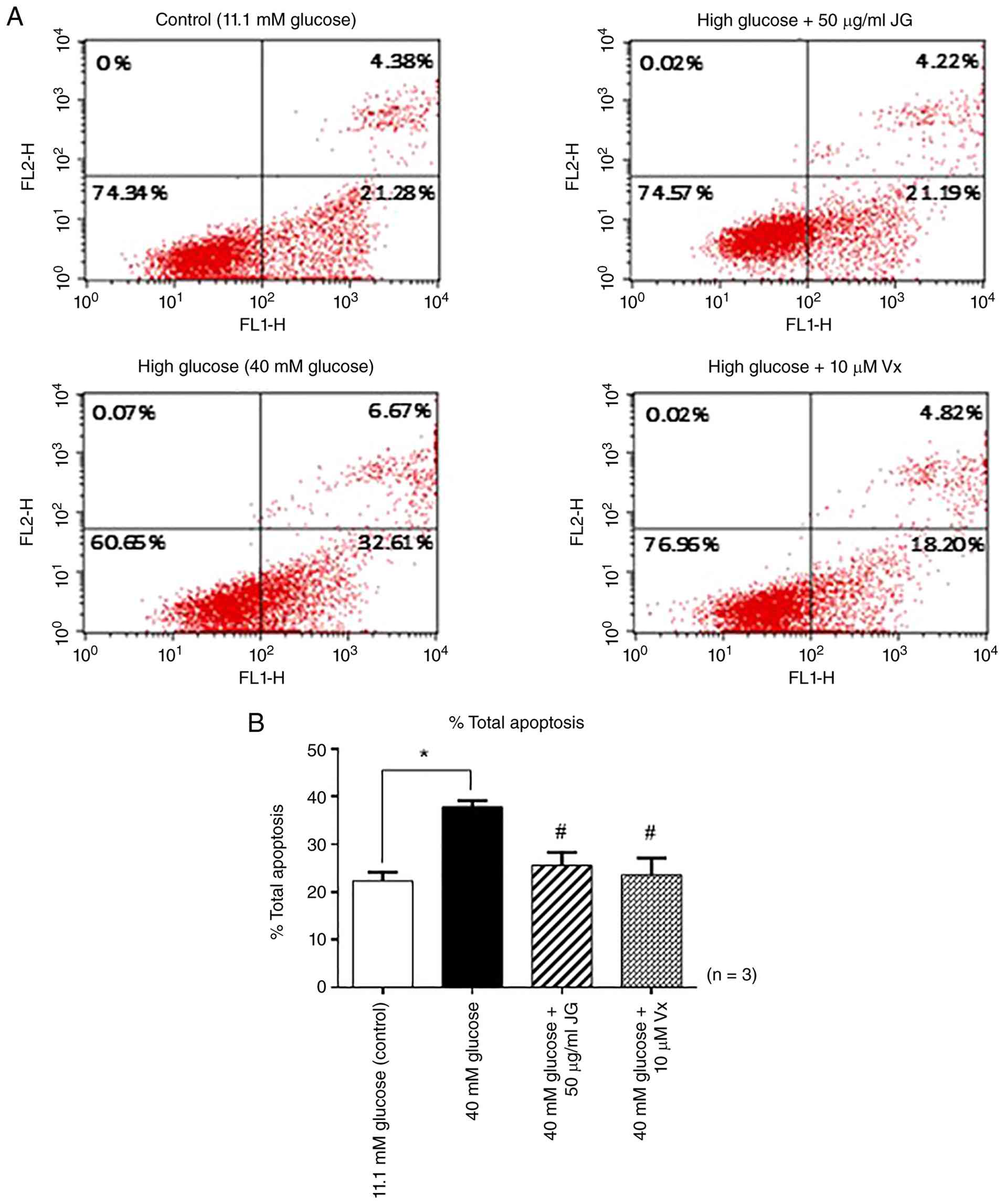
cytometric analysis. As shown in Fig.
5, cells exposed to high-glucose conditions exhibited a
significant increase in apoptosis, with 37.39% of cells undergoing
programmed cell death compared with the normoglycemic control.
Treatment with JG leaf extract at 50 µg/ml markedly reduced the
percentage of apoptotic cells to 25.59%, while vitexin at 10 µM
achieved a comparable reduction to 23.59%. These reductions were
statistically significant when compared with the high-glucose-only
group. These results highlight the potential of JG leaf extract and
vitexin to mitigate pancreatic β-cell apoptosis induced by
hyperglycemia. This cytoprotective effect suggests a promising
therapeutic role in preserving β-cell function under diabetic
conditions.
Effect of JG leaf extract on the
expression of insulin and the insulin transcription factor Pdx1
under high-glucose conditions
The effect of JG leaf extract on insulin
biosynthesis in pancreatic β-cells under high-glucose-induced
stress was investigated by measuring the mRNA expression levels of
the insulin gene and its key transcription factor, Pdx1. INS-1
cells were cultured under high-glucose conditions and treated with
either the extract (50 µg/ml) or the reference flavonoid compound,
vitexin (10 µM), for 48 h. Gene expression was quantified using
RTqPCR. The results demonstrated a marked downregulation of insulin
mRNA in the high-glucose-only group, with expression reduced to
0.109-fold relative to the normoglycemic control. Treatment with JG
leaf extract markedly restored insulin mRNA expression to
0.59-fold, while vitexin increased expression to 0.33-fold, both
compared with the high-glucose group. These data suggested that the
extract at 50 µg/ml was more effective in restoring insulin gene
expression than vitexin at 10 µM (Fig.
6A). Similarly, the expression pattern of Pdx1 mRNA mirrored
that of insulin, with significant restoration following treatment
with the extract (Fig. 6B). These
findings indicated that JG leaf extract enhanced insulin gene
expression under hyperglycemic conditions, at least in part,
through the upregulation of Pdx1, a master regulator of insulin
transcription. This suggested a potential mechanism by which the
extract may help preserve pancreatic β-cell function in
diabetes.
Discussion
The present study demonstrated that the extraction
of JG leaf using ethanol as a solvent via Soxhlet extraction
yielded a crude extract equivalent to 15.06% of the dry leaf powder
weight. The extract was characterized by a viscous consistency and
dark green coloration. Phytochemical analysis revealed that the
total flavonoid content (172.10 mg QE/g extract) was markedly
higher than the total phenolic content (23.47 mg GAE/g extract).
These findings are consistent with the report by Kuber et al
(22) in 2021, which documented
high flavonoid content (97.6±0.0342 mg Rutin equivalent/g) in
ethanolic extracts of JG leaf, while the total phenolic content was
relatively low (9.47±0.0216 mg GAE/g). However, the results differ
from those reported by Marliani et al (5) in 2022, who found that extraction using
varying ratios of water, ethanol and hexane yielded higher phenolic
content (112.076 mg GAE/g) than flavonoids (34.926 mg QE/g). Such
discrepancies may be attributed to differences in the choice of
extraction solvents, sample preparation techniques and
environmental factors affecting the plant materials used, such as
leaf maturity, seasonal variation and geographical origin. These
variables are known to influence the phytochemical composition of
herbal extracts.
The antioxidant capacity of JG leaf extract was
evaluated using DPPH and ABTS radical scavenging assays. The
extract demonstrated significant free radical scavenging activity,
with IC50 values of 1.15±0.14 mg/ml for the DPPH assay
and 0.42±0.07 mg/ml for the ABTS assay, indicating prominent
antioxidant potential. These findings are consistent with previous
studies reporting potent antioxidant properties of JG ethanol
extracts. For instance, a prior investigation found that the
ethanolic leaf extract exhibited strong DPPH radical scavenging
activity, with an IC50 value as low as 0.032 mg/ml
(22). Together, these results
support the conclusion that JG leaf possess substantial antioxidant
activity and may serve as a promising natural source of free
radical scavengers.
Vitexin and apigenin were shown to be the two major
flavonoids present in high content in the JG leaf extract (14,15).
However, vitexin was present in the extract at a relatively low
level (0.19±0.02 mg/g extract) in the present study. Despite its
low content, vitexin may still contribute to the observed
biological activity of the extract. This assumption is supported by
the result of vitexin standard that exhibits significant
cytoprotective effects against high glucose-induced pancreatic
β-cell apoptosis. Vitexin is a C-glycosylated flavone commonly
found in various plant species and is well documented for its
diverse biological activities, particularly antioxidant,
anti-inflammatory and anticancer properties (23). The cytoprotective effect of vitexin
was supported by Zhang et al (24) in 2017 and Raghu and Agrawal
(25) in 2016 reported that vitexin
plays a major role in the extract's antioxidant activity.
Nevertheless, the biological activity of JG extract is likely to
result from the combined effects of vitexin and other component in
the extract.
The cytotoxic effects of JG leaf extract on INS-1
cells, a rat pancreatic β-cell line, were evaluated across a
concentration range of 31.2-2,000 µg/ml. The extract exhibited no
statistically significant cytotoxicity on INS-1 cells at
concentrations between 31.2-1,000 µg/ml, with cell viability
percentages remaining comparable to the untreated control group.
However, at the highest tested concentration of 2,000 µg/ml, a
marked reduction in cell viability was observed, with survival
decreasing to ~70%, indicating cytotoxicity at this dose. These
findings corroborate previous reports demonstrating that ethanol
extracts of JG leaf at concentrations up to 1,000 µg/ml did not
induce toxicity in monkey kidney (Vero) cells or human breast
cancer (MCF-7) cells (26).
Additionally, no cytotoxic effects were noted in the mouse
macrophage cell line (RAW264) when exposed to 200 µg/ml of the
extract (8). Collectively, these
data supported that JG leaf extract concentrations ranging from
31.2-1,000 µg/ml are safe and appropriate for further bioactivity
evaluation. The observed decrease in cell viability at 2,000 µg/ml
may be attributable to the saturation of bioactive compounds
exerting intracellular homeostatic disturbances or the induction of
oxidative stress and apoptosis at high concentrations. A previous
study (27) reported that elevated
flavonoid levels can induce cell cycle arrest and apoptosis via
activation of the p53 signaling pathway, as demonstrated in bladder
cancer cell lines (T24 and EJ).
Prolonged exposure of cells to elevated glucose
levels induces oxidative stress, leading to glucotoxicity, a
critical mechanism underlying β-cell dysfunction and apoptosis. The
present study evaluated the protective effect of JG leaf extract
against high-glucose-induced damage in pancreatic β-cells. INS-1
cells cultured under hyperglycemic conditions (40 mM glucose) for
72 h exhibited a significant reduction in cell viability to <50%
compared with normoglycemic controls. Co-treatment with JG leaf
extract at concentrations of 50-100 µg/ml markedly improved cell
viability, restoring survival rates to levels comparable to the
normal glucose group. Furthermore, vitexin demonstrated a
dose-dependent protective effect on INS-1 cells under hyperglycemic
stress. Treatment with vitexin at 10, 20 and 40 µM concentrations
increased cell viability to levels comparable to those observed
with the crude extract, suggesting that vitexin may serve as a
principal bioactive constituent mediating a cytoprotective
mechanism against glucotoxicity. These findings are consistent with
previous reports by Ganesan et al (28) in 2020, which documented significant
enhancement of INS-1 cell survival under high glucose conditions
following treatment with vitexin at concentrations ranging from
20-40 µM.
The present study demonstrated that JG leaf extract
exhibits potent intracellular antioxidant activity in INS-1
pancreatic β-cells exposed to high glucose conditions. Treatment
with the extract at concentrations of 50 and 100 µg/ml markedly
reduced intracellular ROS levels compared with the high glucose
control group, with ROS levels approaching those observed under
normoglycemic conditions. Similarly, vitexin at concentrations of
10, 20 and 40 µM effectively attenuated ROS generation to levels
comparable to the crude extract. These findings are consistent with
previous reports indicating that vitexin restores antioxidant
system homeostasis by activating the Nrf2 signaling pathway, which
upregulates intracellular antioxidant molecules, while concurrently
inhibiting the NF-κB pathway involved in inflammation and oxidative
stress-induced apoptosis (28).
Supporting this, Tao et al (29) in 2023 reported that vitexin at 10-80
µM markedly reduced ROS accumulation and lipid peroxidation under
hyperglycemic stress. The result of intracellular antioxidant
activity showed that JG extract and vitexin upregulated
antioxidant-related gene, Nrf2 and Trx expression, supporting their
protective effect against high glucose-induced oxidative damage.
These findings are consistent with previous reports indicating that
vitexin restores antioxidant system homeostasis by activating the
Nrf2 signaling pathway, which upregulates intracellular antioxidant
molecules, while concurrently inhibiting the NF-κB pathway involved
in inflammation and oxidative stress-induced apoptosis (28). The activation of Nrf2 and Trx
pathway by JG extract is strongly supported by the known activities
of its bioactive components, vitexin and isovitexin. A previous
study (29) demonstrated that these
vitexin and isovitexin function as potent Nrf2 activators by
directly binding to the Keap1 protein pocket, thereby preventing
Nrf2 degradation and facilitating its translocation to the nucleus.
Furthermore, in C. elegans models, these compounds were
witnessed to increase the nuclear accumulation of SKN-1 (the
mammalian homolog of Nrf2), leading to enhanced stress resistance
and longevity. The present findings of upregulated Nrf2 and Trx
mRNA levels are highly consistent with these established
protein-level mechanisms, suggesting that the observed antioxidant
effects are indeed driven by the activation and translocation of
the Nrf2 signaling cascade (30).
Beyond its antioxidant properties, the present study further
revealed that JG leaf extract at 50 µg/ml and vitexin at 10 µM
markedly inhibited apoptosis in pancreatic β-cells under high
glucose conditions, compared with untreated hyperglycemic controls.
This protective effect corroborates previous evidence that vitexin
downregulates the pro-apoptotic protein Bax, upregulates the
anti-apoptotic protein Bcl-2 and mitigates mitochondrial stress in
pancreatic β-cells (30).
Collectively, these results substantiate the cytoprotective role of
JG leaf extract against glucotoxicity via attenuation of oxidative
stress and apoptosis, with vitexin likely serving as a key
bioactive constituent mediating these effects.
Prolonged exposure to high glucose levels is a
critical factor contributing to pancreatic β-cell dysfunction,
characterized by impaired insulin secretion and downregulation of
genes involved in insulin synthesis. A key mechanism underlying
glucotoxicity is the suppression of insulin gene expression and the
Pdx1 gene, the latter serving as a crucial insulin transcription
factor regulating insulin gene transcription in pancreatic β-cells.
The present study demonstrated that JG leaf extract at a
concentration of 50 µg/ml markedly upregulated the expression of
insulin and Pdx1 genes in INS-1 pancreatic β-cells cultured under
hyperglycemic conditions. Specifically, insulin mRNA levels, which
were reduced to 0.109-fold in the high glucose control group,
increased to 0.59-fold following treatment with the JG leaf extract
and to 0.33-fold with vitexin at 10 µM, indicating a greater
potential of the JG leaf extract compared with vitexin alone in
enhancing insulin synthesis. Similarly, Pdx1 mRNA expression showed
a parallel increase, reflecting a coordinated regulatory mechanism.
This finding aligns with the established role of Pdx1 as a direct
regulator of insulin gene transcription and a primary target
inhibited during glucotoxicity, resulting in diminished insulin
production. These results are consistent with previous reports by
Zhang et al (30) in 2023,
who demonstrated that vitexin prevents the decline in Pdx1 and
insulin mRNA expression induced by high glucose in INS-1 cells via
mechanisms involving the reduction of oxidative stress and
mitigation of mitochondrial dysfunction. Activation of Pdx1 thereby
restores the insulin-synthesizing capacity of β-cells under
oxidative stress (29). At present,
there is no evidence on the effect of JG leaf extract on insulin
and Pdx1 gene expression in glucotoxic conditions, particularly in
INS-1 cells. A previous study demonstrated that decreased protein
levels of PDX1 and MafA in pancreatic β-cell under
glucolipotoxicity results in the reduction of glucose-stimulated
insulin secretion (GSIS) (31).
Even though the present study did not perform the GSIS assay, it
demonstrated the regenerative and biosynthetic potential of the
extract on pancreatic β-cells, for which mRNA levels of Insulin and
Pdx1 are established primary indicators.
The protective effects of JG extract against
glucotoxicity in INS-1 cells compare favorably with other
established herbal antidiabetics. While a number of extracts, such
as those from apigenin, a plant-derived flavonoid, primarily act
through general ROS scavenging (32), JG extract demonstrated a specialized
capacity to upregulate the Pdx1/Insulin axis alongside the Nrf2/Trx
pathway. This dual regulatory role is critical for the improvement
of pancreatic β-cell mass and function. The present study
identified vitexin as the major bioactive in our JG extract, which
further distinguished JG from broader extracts. Specifically,
vitexin has the ability to bind directly to Keap1, thereby
disrupting the Keap1-Nrf2 interaction and subsequently promoting
Nrf2 activation. The present study showed that the flavonoid
concentration (29). The present
study showed that the flavonoid concentration in the extract
(172.10 mg QE/g) was markedly higher than that reported for several
other tropical medicinal plants, such as Curcuma longa and
Centella asiatica (33,34).
While Curcuma longa is known as a potent Nrf2 activator
(35), the markedly higher
flavonoid content in the present JG extract suggested a more robust
therapeutic potential for activation of the Nrf2 pathway, a
comprehensive shield for pancreatic β-cells against oxidative and
inflammatory insult in diabetes.
Regarding the translational potential of the present
findings, although in vivo experiments of JG extract were
not conducted in the present study, the physiological relevance of
the in vitro findings is supported by established
pharmacokinetic data for vitexin in rat models (36). Oral administration of vitexin at 30
mg/kg achieved a peak plasma concentration (Cmax)
of 0.51±0.015 µg/ml or 1.18 µM. This concentration was reached
rapidly, with a Tmax of ~15.82±0.172 min.
Although the concentration of 10 µM (4.32 µg/ml) used in the in
vitro model exceeds Cmax typically observed
in rat pharmacokinetic studies, the findings demonstrated
significant biological activity without inducing cytotoxicity. The
specific in vivo pharmacokinetic study of the JG extract is
a necessary next step to confirm these effective dosages. In a
clinical context, such an extract could serve as a natural
complementary strategy to slow the progression of Type 2 Diabetes
by maintaining β-cell viability and insulin biosynthetic capacity.
By upregulating Pdx1 and Nrf2, the JG extract targets the root
causes of pancreatic β-cell failure; oxidative stress and
transcriptional exhaustion.
While these results are promising, the mechanistic
evidence in the present study was limited to mRNA expression.
Future studies incorporating western blotting for Nrf2 nuclear
translocation and GSIS assays are warranted to confirm these
molecular observations. Additionally, the long-term safety,
bioavailability and clinical evaluation of the JG extract in human
subjects remain to be established through standardized clinical
trials.
The present study provided novel and valuable
evidence demonstrating the potential of JG leaf extract to protect
pancreatic β-cells from apoptosis and enhance their functional
capacity to increase insulin production under glucotoxic conditions
through antioxidant mechanisms and inhibition of apoptotic
pathways. The observed effects are likely mediated by flavonoid
compounds, particularly vitexin, which was found in the extract and
demonstrated a cytoprotective effect similar to that of the
extract. Nevertheless, further investigations at the molecular
level are warranted to elucidate the precise mechanisms underlying
these bioactivities. In addition, comprehensive in vivo
studies assessing the antidiabetic efficacy of the extract in
animal models, followed by clinical evaluations in patients with
type 2 diabetes mellitus, are essential. Such research will support
the development of this traditional herbal extract as a promising
alternative therapeutic agent for the management of type 2
diabetes.
Acknowledgements
Not applicable.
Funding
Funding: The present study was financially supported by Burapha
University, Thailand Science Research and Innovation and National
Science Research and Innovation Fund (Fundamental Fund; grant
no.11/2567).
Availability of data and materials
The data generated in the present study may be
requested from the corresponding author.
Authors' contributions
KS conceptualized and administered the project,
performed the in vitro experiment and wrote the original
draft of the manuscript. SC performed plant extraction, analyzed
data, prepared the figure and wrote the original draft of the
manuscript. PC performed gene expression analysis. NS and PM
performed annexin V-FITC/PI staining and flow cytometry analysis.
LY and AJ performed acquisition of data. PS and SP performed the
in vitro experiment and interpretation of data. NM performed
data analysis and prepared the figure. SK supervised the project
and edited the manuscript. KS and SC confirm the authenticity of
all the raw data. All authors reviewed the manuscript.
Ethics approval and consent to
participate
Not applicable.
Patient consent for publication
Not applicable.
Competing interests
The authors declare that they have no competing
interests.
References
|
1
|
International Diabetes Federation: IDF
diabetes atlas. 11th Edition. International Diabetes Federation,
Brussels, 2025. https://diabetesatlas.org/resources/idf-diabetes-atlas-2025/.
|
|
2
|
Stumvoll M, Goldstein BJ and van Haeften
TW: Type 2 diabetes: Principles of pathogenesis and therapy.
Lancet. 365:1333–1346. 2005.PubMed/NCBI View Article : Google Scholar
|
|
3
|
Moon JS, Karunakaran U, Elumalai S, Lee
IK, Lee HW, Kim YW and Won KC: Metformin prevents glucotoxicity by
alleviating oxidative and ER stress-induced CD36 expression in
pancreatic beta cells. J Diabetes Complications. 31:21–30.
2017.PubMed/NCBI View Article : Google Scholar
|
|
4
|
Wali JA, Rondas D, McKenzie MD, Zhao Y,
Elkerbout L, Fynch S, Gurzov EN, Akira S, Mathieu C, Kay TW, et al:
The proapoptotic BH3-only proteins Bim and Puma are downstream of
endoplasmic reticulum and mitochondrial oxidative stress in
pancreatic islets in response to glucotoxicity. Cell Death Dis.
5(e1124)2014.PubMed/NCBI View Article : Google Scholar
|
|
5
|
Marliani N, Artika IM and Nurcholis W:
Optimization extraction for total phenolic, flavonoid contents, and
antioxidant activity with different solvents and UPLC-MS/MS
metabolite profiling of Justicia gendarussa Burm.f. Chiang
Mai Univ J Nat Sci. 21(e2022046)2022.
|
|
6
|
Phalaharn V, Pakdee N, Pongnaratorn P,
Prathumtet J and Poowanna R: Chemical composition analysis with
TLC, HPLC and antioxidant activity from Justicia gendarussa
Burm.f. leaf extract. Kalasin Univ J Sci Technol Innov. 4:82–97.
2025.
|
|
7
|
Paval J, Kaitheri SK, Potu BK, Govindan S,
Kumar RS, Narayanan SN and Moorkoth S: Anti-arthritic potential of
the plant Justicia gendarussa Burm F. Clinics (Sao Paulo).
64:357–362. 2009.PubMed/NCBI View Article : Google Scholar
|
|
8
|
Varma RS, Ashok G, Vidyashankar S, Patki P
and Nandakumar KS: Ethanol extract of Justicia gendarussa
inhibits lipopolysaccharide stimulated nitric oxide and matrix
metalloproteinase-9 expression in murine macrophage. Pharm Biol.
49:648–652. 2011.PubMed/NCBI View Article : Google Scholar
|
|
9
|
C J, Raju SK and N S: Anti-inflammatory
and analgesic activities of ethanol extract of aerial parts of
Justicia gendarussa Burm. Int J Pharmacol. 6:278–283.
2010.
|
|
10
|
Sontakke PA, Vyawahare NS, Wankhade PP,
Shirode DS, Shinde RD, Patil VP, Bairagi PK and Patil GH:
Antiasthmatic potential of Justicia gendarussa: A
preclinical study. J Nat Remedies. 25:2885–2895. 2025.
|
|
11
|
Basah K, Elya B, Amin J and Julian MI:
Activity of ethanolic extracts from Justicia gendarussa
Burm. Leaves on decreasing the uric acid plasma. Makara Sains.
15:67–70. 2011.
|
|
12
|
Islam MR, Sayeed MA, Billah MM, Rana MN,
Hossain MA, Anwar JB and Alam MN: Effects of metanolic leaf extract
of Justicia gendarussa on alloxan induced diabetic mice and
brine shrimp nauplii. World J Pharm Res. 4:1981–1990. 2015.
|
|
13
|
Singleton VL, Orthofer R and
Lamuela-Raventós RM: Analysis of total phenols and other oxidation
substrates and antioxidants by means of folin-ciocalteu reagent.
Methods in Enzymol. 299:152–178. 1999.
|
|
14
|
Roy P and Joseph L: Bioactive constituents
and pharmacological profile of willow-leaved Justicia: A review.
Natural Resour Hum Health. 2:450–461. 2022.
|
|
15
|
Raghu MG and Agrawal P: The isolation and
structural determination of flavonoids from Justicia
gendarussa. J Pharm Biol Sci. 11:73–79. 2016.
|
|
16
|
Clarke G, Ting KN, Wiart C and Fry J: High
correlation of 2,2-diphenyl-1-picrylhydrazyl (DPPH) radical
scavenging, ferric reducing activity potential and total phenolics
content indicates redundancy in use of all three assays to screen
for antioxidant activity of extracts of plants from the Malaysian
Rainforest. Antioxidants (Basel). 2:1–10. 2013.PubMed/NCBI View Article : Google Scholar
|
|
17
|
Ozgen M, Reese RN, Tulio AZ Jr, Scheerens
JC and Miller AR: Modified
2,2-azino-bis-3-ethylbenzothiazoline-6-sulfonic acid (abts) method
to measure antioxidant capacity of Selected small fruits and
comparison to ferric reducing antioxidant power (FRAP) and
2,2'-diphenyl-1-picrylhydrazyl (DPPH) methods. J Agric Food Chem.
54:1151–1157. 2006.PubMed/NCBI View Article : Google Scholar
|
|
18
|
Suksri K, Yingngam B and Muangchan N:
Optimization of phenolic extraction from Syzygium samarangense
fruit and its protective properties against glucotoxicity-induced
pancreatic β-cell death. ScienceAsia. 49:529–540. 2023.
|
|
19
|
Lee JA, Spidlen J, Boyce K, Cai J, Crosbie
N, Dalphin M, Furlong J, Gasparetto M, Goldberg M, Goralczyk EM, et
al: MIFlowCyt: The minimum information about a flow cytometry
experiment. Cytometry A. 73:926–930. 2008.PubMed/NCBI View Article : Google Scholar
|
|
20
|
Livak KJ and Schmittgen TD: Analysis of
relative gene expression data using real-time quantitative PCR and
the 2(-Delta Delta C(T)) method. Methods. 25:402–408.
2001.PubMed/NCBI View Article : Google Scholar
|
|
21
|
Poitout V and Robertson RP:
Glucolipotoxicity: Fuel excess and beta-cell dysfunction. Endocr
Rev. 29:351–366. 2008.PubMed/NCBI View Article : Google Scholar
|
|
22
|
Kuber RB: In vitro antioxidant potential,
total phenolic and flavonoid contents in Justicia gendarussa
leaf extracts. Res J Pharm Technol. 14:2707–2713. 2021.
|
|
23
|
Yan W, Cheng J and Xu B: Dietary
flavonoids vitexin and isovitexin: New insights into their
functional roles in human health and disease prevention. Int J Mol
Sci. 26(6997)2025.PubMed/NCBI View Article : Google Scholar
|
|
24
|
Zhang HJ, Rumschlag-Booms E, Guan YF, Liu
KL, Wang DY, Li WF, Nguyen VH, Cuong NM, Soejarto DD, Fong HHS and
Rong L: Anti-HIV diphyllin glycosides from Justicia
gendarussa. Phytochemistry. 136:94–100. 2017.PubMed/NCBI View Article : Google Scholar
|
|
25
|
Raghu MG and Agrawal P: RP-HPLC method for
identification and quantification of vitexin from the plant
Justicia gendarussa. J Chem Pharm Sci. 9:2143–2148.
2016.
|
|
26
|
Dasadiaa J, Vyas V, Patela S and Zaveri M:
In-vitro cytotoxicity of root of gendarussa vulgaris on MCF 7 and
vero cell. J Pharm Sci Bioscientific Res. 6:660–665. 2016.
|
|
27
|
Li J, Cheng Y, Qu W, Sun Y, Wang Z, Wang H
and Tan B: Fisetin, a dietary flavonoid, induces cell cycle arrest
and apoptosis through activation of p53 and inhibition of NF-kappa
B pathways in bladder cancer cells. Basic Clin Pharmacol Toxicol.
108:84–93. 2011.PubMed/NCBI View Article : Google Scholar
|
|
28
|
Ganesan K, Ramkumar KM and Xu B: Vitexin
restores pancreatic β-cell function and insulin signaling through
Nrf2 and NF-κB signaling pathways. Eur J Pharmacol.
888(173606)2020.PubMed/NCBI View Article : Google Scholar
|
|
29
|
Tao M, Li R, Xu T, Zhang Z, Zheng D, Xia
Z, Wu T, Pan S and Xu X: Vitexin and isovitexin delayed ageing and
enhanced stress-resistance through the activation of the SKN-1/Nrf2
signaling pathway. Int J Food Sci Nutr. 74:685–694. 2023.PubMed/NCBI View Article : Google Scholar
|
|
30
|
Zhang L, Shi L, Han J and Li Z: Protection
of β-pancreatic cells from dysfunctionality of insulin using
vitexin by apoptosis of INS-1 cells. Arch Physiol Biochem.
129:1160–1167. 2023.PubMed/NCBI View Article : Google Scholar
|
|
31
|
Ebrahim N, Shakirova K and Dashinimaev E:
PDX1 is the cornerstone of pancreatic β-cell functions and
identity. Front Mol Biosci. 9(1091757)2022.PubMed/NCBI View Article : Google Scholar
|
|
32
|
Wang N, Yi WJ, Tan L, Zhang JH, Xu J, Chen
Y, Qin M, Yu S, Guan J and Zhang R: Apigenin attenuates
streptozotocin-induced pancreatic β cell damage by its protective
effects on cellular antioxidant defense. In Vitro Cell Dev Biol
Anim. 53:554–563. 2017.PubMed/NCBI View Article : Google Scholar
|
|
33
|
Ghasemzadeh A, Azarifar M, Soroodi O and
Jaafar HZE: Flavonoid compounds and their antioxidant activity in
extract of some tropical plants. J Med Plants Res. 6:2639–2643.
2012.
|
|
34
|
Marayart S, Konmun H, Jampasri K and
Saeng-Ngam S: Total phenolic, flavonoid contents, and antioxidant
activity of strawberries and local medicinal plants. Asia Pac J Sci
Technol. 30:APST–30. 2025.
|
|
35
|
Ashrafizadeh M, Ahmadi Z, Mohammadinejad
R, Farkhondeh T and Samarghandian S: Curcumin activates the Nrf2
pathway and induces cellular protection against oxidative injury.
Curr Mol Med. 20:116–133. 2020.PubMed/NCBI View Article : Google Scholar
|
|
36
|
Wang Y, Han C, Leng A, Zhang W, Xue H,
Chen Y, Yin J, Lu D and Ying X: Pharmacokinetics of vitexin in rats
after intravenous and oral administration. Afr J Pharm Pharmacol.
6:2368–2373. 2012.
|