Introduction
Asthma is a heterogeneous disease with chronic
airway inflammation as its key pathology, characterized primarily
by recurrent cough and wheezing, and accompanied by variable
airflow limitation (1). Airway
remodeling refers to structural changes resulting from chronic
airway inflammation, including epithelial injury, smooth muscle
hyperplasia and hypertrophy, abnormal extracellular matrix (ECM)
deposition, subepithelial fibrosis and angiogenesis (2-4).
These structural alterations can lead to irreversible airflow
limitation and are key drivers of asthma progression from
controllable symptoms to refractory and severe forms associated
with long-term complications such as chronic obstructive pulmonary
disease (5). Therefore, elucidating
its pathogenesis and preventing airway remodeling are key
objectives in asthma management.
Previous studies have shown that asthma airway
remodeling results from persistent inflammatory injury and repair
responses (6-8).
The pathological processes primarily involve imbalance between
abnormal ECM deposition and degradation, as well as proliferation
and phenotypic changes in airway smooth muscle (ASM). IL-21 and
thymic stromal lymphopoietin (TSLP) are key cytokines that drive
chronic airway inflammation and promote ECM deposition, ASM
proliferation and hypertrophy (9,10). The
expression levels of α-smooth muscle actin (SMA) and collagen
(Col-)I are important markers for evaluating ASM activation and ECM
deposition (11). Thus, targeting
these molecules and investigating their regulatory mechanisms
provide a valid approach for studying anti-remodeling
strategies.
Fucoidan, a natural sulfated polysaccharide
extracted from brown algae, has attracted attention due to its
broad spectrum of biological activities, including
anti-inflammatory, immunomodulatory, antitumor and antifibrotic
effects (12,13). Studies indicate that fucoidan
modulates immune cell function and inhibits the production of
inflammatory mediators (14-16).
These properties align with the hyperactivated immune-inflammatory
state and fibrotic processes in asthma pathology. Based on this, it
was hypothesized that fucoidan may alleviate airway remodeling in
asthma by regulating key remodeling-associated factors such as
IL-21, TSLP, α-SMA and Col-I.
Previous studies on fucoidan in asthma have
primarily focused on inflammatory modulation (17-19),
leaving its effects on structural airway remodeling, particularly
via IL-21/TSLP pathway, largely unexplored. Budesonide is a
first-line inhaled corticosteroid for asthma with established
efficacy in mitigating airway remodeling. To the best of our
knowledge, however, a direct comparison between fucoidan and
budesonide for remodeling outcomes under identical experimental
conditions has not been reported.
In the present study, an asthmatic mouse model was
established by OVA (ovalbumin) sensitization and challenge.
Behavioral changes, lung histopathology, ELISA and
immunohistochemistry (IHC) were used to systematically evaluate the
effects of fucoidan on asthma symptoms, airway structural
remodeling and the expression levels of IL-21, TSLP, α-SMA and
Col-I in bronchoalveolar lavage fluid (BALF) and lung tissue. The
aim was to verify the role of fucoidan in airway remodeling in
asthmatic mice and explore its potential mechanisms, providing
experimental evidence for the development of potential asthma
therapeutic strategies.
Materials and methods
Animals
Specific pathogen-free (SPF) female Kunming (KM)
mice (age, 6-8 weeks; weight, 19-24 g) were purchased from the
Shandong Qingdao Experimental Animal Center and housed under
standard SPF conditions (temperature, 22-24˚C; relative humidity,
50-60%; 12-h light/dark cycle) in the Science and Education
Building, West Coast Campus of the Affiliated Hospital of Qingdao
University, Qingdao, Shandong, China [certificate no. scxk (Lu)
20130010]. This outbred strain was selected for its use in
OVA-induced asthma models (20),
genetic heterogeneity approximating human diversity and practical
advantages including ready availability, cost-effectiveness, strong
disease resistance, good adaptability and high reproductive and
survival rates (21). The animal
experiments were approved by the Experimental Animal Ethics
Committee of the Affiliated Hospital of Qingdao University
(approval no. AHQU-MAL20150101QYL). All procedures adhered to
relevant Chinese national regulations on laboratory animal welfare
and ethics and complied with the American Veterinary Medical
Association Guidelines for the Euthanasia of Animals (22,23).
Every effort was undertaken to minimize animal suffering and
decrease the number of animals used.
Asthmatic mouse model
In total, 40 mice were randomly divided into four
groups (n=10/group): Asthma model (OVA), budesonide treatment group
(OVA + budesonide), fucoidan (OVA + fucoidan) and control group.
Mice were housed in a specific pathogen-free environment with free
access to food and water. An asthma model was induced by
intraperitoneal OVA (Sigma-Aldrich; Merck KGaA) sensitization and
nebulized OVA challenge, as previously described (24). Briefly, mice in the OVA, OVA +
budesonide and OVA + fucoidan groups were intraperitoneally
injected with a sensitization solution containing 10 µg OVA and 1.6
mg aluminum hydroxide in 0.1 ml PBS on days 1, 8 and 15. On day 22
to 35, mice in the OVA group were nebulized with 1% OVA solution
for 30 min daily. Mice in the OVA + budesonide (AstraZeneca, UK)
group received budesonide nebulization (200 µg in 3.6 ml saline) 1
h before each OVA challenge. Based on previous studies (25-27),
mice in the OVA + fucoidan group were administered fucoidan (50
mg/kg; Sigma-Aldrich; Merck KGaA) by gavage 1 h before each
challenge. Control mice received saline instead of OVA during both
sensitization and challenge phases. All mice survived the
experimental period. All mice were euthanized 24-48 h after the
last challenge for sample collection.
Collection and analysis of
bronchoalveolar lavage fluid (BALF)
Prior to sample collection, all mice were
anesthetized with intraperitoneal pentobarbital sodium (50 mg/kg).
The depth of anesthesia was confirmed by the absence of pedal
withdrawal reflex. Euthanasia was performed by exsanguination via
cardiac puncture and ~1 ml blood was collected. This was followed
by cervical dislocation to ensure death. The cessation of
respiration and heartbeat, along with the absence of pain reflex,
was used to confirm death.
BALF was collected immediately. Briefly, tracheal
intubation was performed and the lungs were lavaged with 0.8 ml
ice-cold PBS. The lavage process was repeated three times and the
fluid was recovered. The pooled BALF was centrifuged at 500 x g for
10 min at 4˚C. The supernatant was separated and stored at -80˚C
for subsequent analysis. The cell pellet was resuspended in PBS and
cytospin smears were prepared using a Cytospin centrifuge at 32 x g
for 4 min at room temperature. The smears were air-dried and
stained with Wright-Giemsa stain at room temperature for 1-2 min,
followed by the addition of an equal volume of phosphate buffer (pH
6.4-6.8) for 3-5 min. The slides were rinsed with distilled water,
air-dried. Total cell and differential counts of eosinophils,
lymphocytes and neutrophils were performed using a hemocytometer
under a light microscope. The levels of IL-21, TSLP, α-SMA and
Col-I in the BALF supernatant were measured using ELISA kits
(R&D Systems) according to the manufacturer's instructions.
Although α-SMA is primarily an intracellular protein and Col-I is
an insoluble ECM component, their detection in BALF supernatant has
been reported previously (11).
During remodeling, activated myofibroblasts expressing α-SMA shed
into the airway lumen, releasing this protein into BALF (28), while Col-I undergoes dynamic
turnover generating soluble fragments detectable in airway fluid
(29).
Lung histopathology
The right lung tissue was fixed in 4%
paraformaldehyde at 4˚C for 24 h, embedded in paraffin and
sectioned (4 µm thickness). For each animal, three non-consecutive
sections (distance, ≥50 µm) were selected for hematoxylin and eosin
(H&E) staining and IHC to avoid sampling bias. For H&E
staining, sections were deparaffinized in xylene, rehydrated
through a descending ethanol series, stained with hematoxylin for
10 min at room temperature, differentiated, counterstained with
eosin for 1 min at room temperature, dehydrated, cleared and
mounted. Sections were stained with H&E to observe general
pathological changes such as inflammatory cell infiltration around
airways and vessels, airway wall thickness and lumen narrowing. For
airway wall thickness quantification, five randomly selected
bronchioles per section were imaged at x400 magnification. The
airway wall area (Wat) and the perimeter of the basement membrane
(Pbm) were measured using Image-Pro Plus software (version 6.0;
Media Cybernetics, Inc.). Airway wall thickness was expressed as
the ratio of Wat to Pbm. All measurements were performed using a
uniform protocol. Sections were examined under a light microscope
(Olympus, Japan). IHC was performed to detect the expression of
IL-21, TSLP, α-SMA and Col-I in lung tissues. After
deparaffinization in xylene, rehydration through a graded ethanol
series and antigen retrieval was performed in citrate buffer (pH
6.0) at 92-96˚C for 15 min, endogenous peroxidase was blocked with
3% hydrogen peroxide at room temperature for 10 min, followed by
blocking with goat serum (ZSGB-BIO) at room temperature for 15 min.
Sections were incubated overnight at 4˚C with the following primary
antibodies: Rabbit anti-IL-21 polyclonal antibody (Bioss, bs-2621R;
1:200 dilution), rabbit anti-TSLP polyclonal antibody (Bioss,
bs-0449R; 1:200 dilution), rabbit anti-α-SMA polyclonal antibody
(Bioss, bs-10196R; 1:200 dilution), and rabbit anti-Col-I
polyclonal antibody (Bioss, bs-10423R; 1:500). The next day,
sections were incubated with a biotin-labeled goat anti-rabbit IgG
working solution (ZSGB-BIO, ZB-2010; 1:200 dilution) at room
temperature for 15 min, followed by incubation with a horseradish
peroxidase-labeled streptavidin working solution (ZSGB-BIO,
Beijing, China) at room temperature for 15 min. Color development
was performed with a DAB kit (ZSGB-BIO, Beijing, China) and
counterstaining with hematoxylin at room temperature for 2 min. For
integrated optical density (IOD) quantification, five random
high-power fields (magnification, x200) were captured under light
microscopy. The IOD of positively stained areas (yellowish-brown)
was measured using Image-Pro Plus (version 6.0; Media Cybernetics,
Inc.) with a uniform threshold applied to all images. The mean
IOD/unit area was calculated for each sample.
Statistical analysis
Normality was assessed using the Shapiro-Wilk test
and homogeneity of variance was assessed using Levene's test. Data
are presented as the mean ± SD from 10 independent biological
replicates/group. All analyses were performed using SPSS 22.0 (IBM
Corp.). For multiple group comparisons, one-way ANOVA was used when
data met the assumptions of normality and homogeneity of variance,
followed by Tukey's honestly significant difference post hoc test
for pairwise comparisons. When variances were unequal, the
Games-Howell post hoc test was applied. For non-normally
distributed data, the Kruskal-Wallis test was used, followed by
pairwise comparisons using the Mann-Whitney U test with Bonferroni
correction. P<0.05 was considered to indicate a statistically
significant difference.
Results
Fucoidan ameliorates asthma-like
symptoms in mice
OVA-challenged mice exhibited typical asthma-like
symptoms such as scratching, shortness of breath and restlessness
(data not shown). Control mice showed no notable behavioral or
respiratory changes. Mice in the OVA + fucoidan group exhibited
markedly alleviated behavioral symptoms compared with the OVA
group, with an extent of improvement similar to that observed in
the OVA + budesonide group.
Fucoidan decreases inflammatory cell
counts in BALF
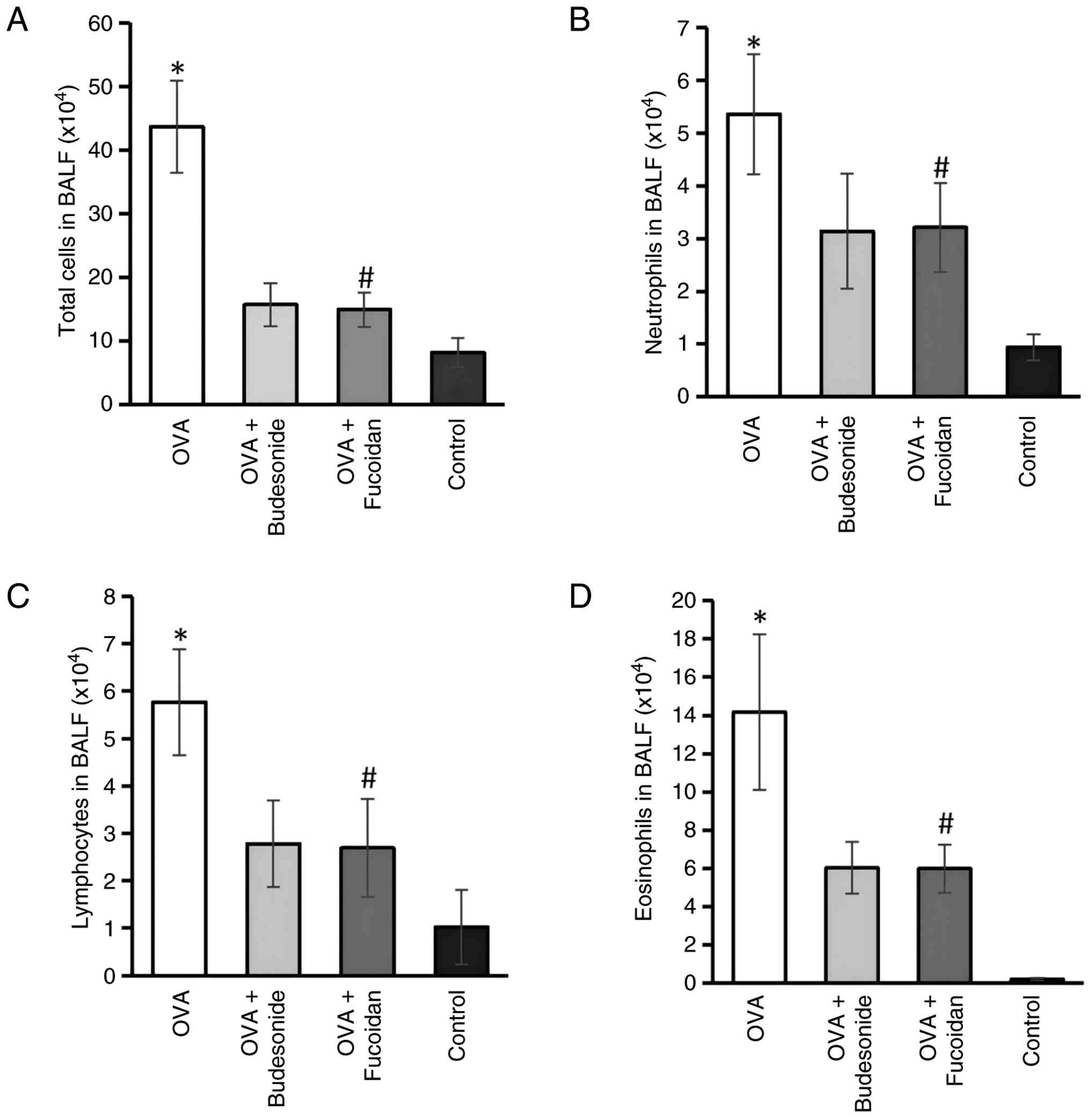
Cell count analysis of BALF (Fig. 1) revealed that the total cell number
in the OVA group was 43.67±7.26x10⁴/ml, which was
significantly higher than that in the control group
(8.18±2.28x10⁴/ml; P<0.05). Neutrophils, lymphocytes
and eosinophils were also markedly elevated in the OVA group
(5.36±1.14, 5.77±1.12 and 14.18±4.07x10⁴/ml,
respectively; P<0.05 vs. control). Following fucoidan
intervention, the total cell count decreased to
14.96±2.69x10⁴/ml, with significant reductions also
observed in each inflammatory cell subtype (neutrophils, 3.21±0.85;
lymphocytes, 2.69±1.03 and eosinophils,
5.98±1.26x10⁴/ml; all P<0.05 compared with the OVA
group). The effect of fucoidan was comparable with that of the OVA
+ budesonide group (total cells, 15.72±3.43; neutrophils,
3.14±1.09; lymphocytes, 2.78±0.92; eosinophils,
6.02±1.35x10⁴/ml; all P>0.05). These results
indicated that fucoidan can reduce the levels of inflammation and
fibrosis-related factors.
Fucoidan downregulates the levels of
remodeling-associated inflammatory factors in BALF
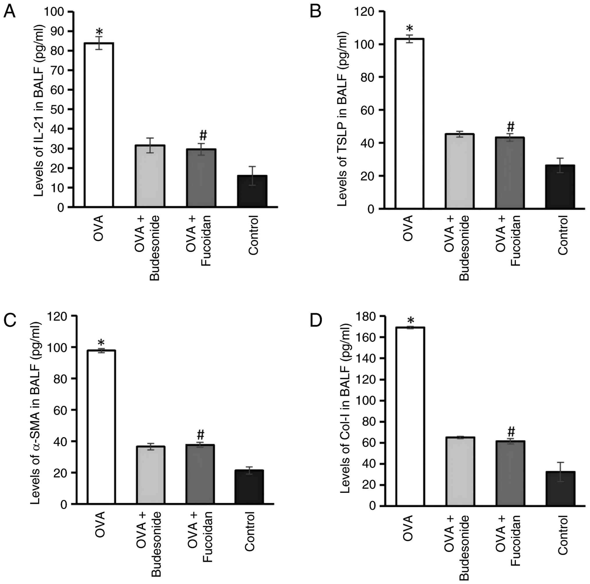
ELISA (Fig. 2)
showed that the concentrations of IL21, TSLP, αSMA and ColI in BALF
of the OVA group were 83.79±3.32, 103.15±2.42, 97.67±1.35 and
169.24±1.25 pg/ml, respectively, all significantly higher than
those in the control group (15.95±4.73, 26.31±4.32, 21.35±2.21 and
32.21±9.07 pg/ml, respectively; P<0.05). Levels of these factors
in the OVA + fucoidan group decreased to 29.47±2.94, 43.24±2.31,
37.69±1.59 and 61.32±2.56 pg/ml, respectively, which were
significantly lower than those in the OVA group (P<0.05). The
decrease was comparable with that in the OVA + budesonide group
(P>0.05). These results suggest an association between fucoidan
and reduced levels of these factors, which may contribute to the
observed attenuation of airway remodeling.
Fucoidan attenuates pathological
changes of airway remodeling in lung tissue
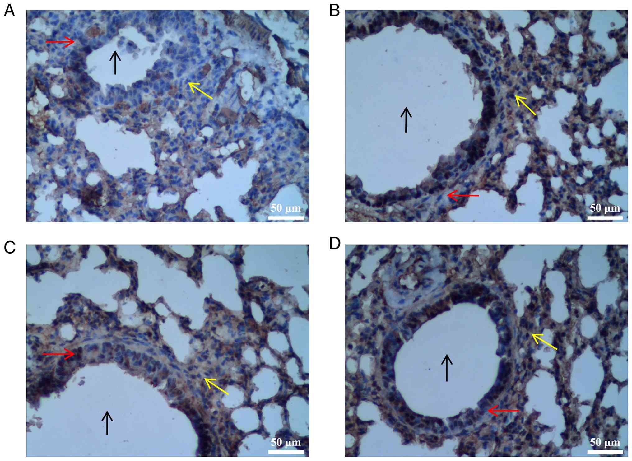
H&E staining of lung tissue (Fig. 3) revealed notable inflammatory cell
infiltration, predominantly eosinophils and lymphocytes, around
airways and blood vessels in the OVA group, along with marked
thickening of the bronchial wall and obvious luminal narrowing. OVA
+ fucoidan group showed a significant decrease in peribronchial
inflammatory cell infiltration and wall thickening, with
pathological improvement comparable with that of the OVA +
budesonide group. In control mice, the bronchial epithelium
remained intact, the airway lumen was smooth without evident folds
or narrowing, the wall showed no obvious thickening and no notable
inflammatory cell infiltration was observed around airways or
vessels.
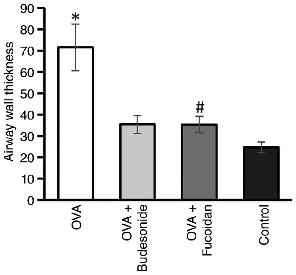
Quantitative analysis of airway wall thickness
(Fig. 4) was performed by measuring
the airway wall area (Wat) and the perimeter of the basement
membrane (Pbm, µm), with Wat/Pbm representing airway wall
thickness. The wall thickness in the OVA group was 71.56±10.87
µm²/µm, significantly higher than that in the control group
(24.68±2.51 µm²/µm; P<0.05). Wall thickness decreased to
35.37±3.78 µm²/µm in the OVA + fucoidan and 35.42±4.25 µm²/µm in
the OVA + budesonide group. Both treatment groups exhibited a
significant decrease compared with the OVA group (P<0.05), with
no statistical difference between them (P>0.05). These results
indicate that fucoidan significantly suppressed asthmainduced
structural thickening of the airway wall, with an effect similar to
that of the inhaled corticosteroid budesonide.
Fucoidan suppresses the expression of
remodelingassociated proteins in lung tissue
Representative immunohistochemical staining for
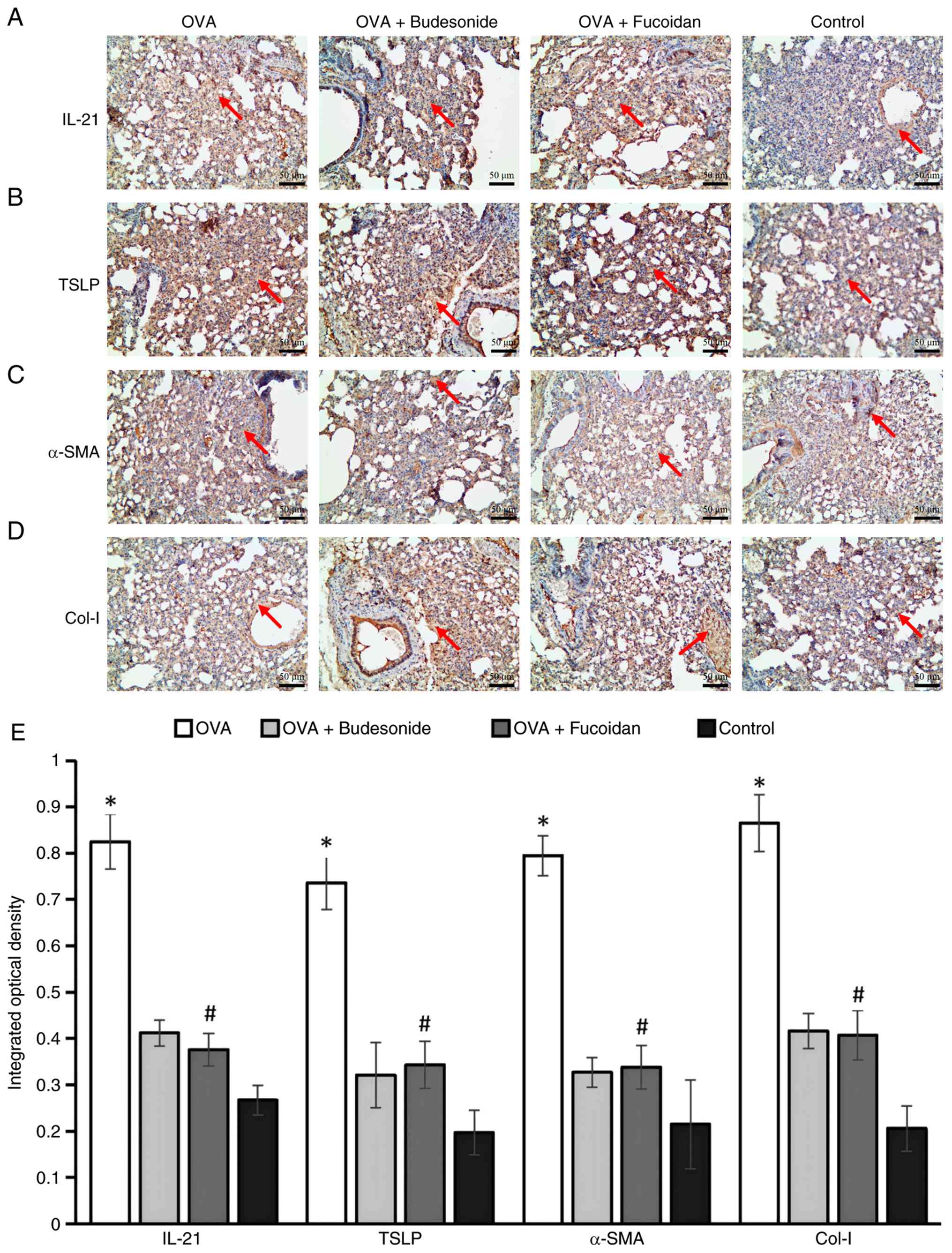
IL21, TSLP, αSMA, and ColI in lung tissue is shown in Fig. 5A-D. Compared with the control group,
the expression of all four factors was significantly enhanced in
the OVA group, with yellowishbrown positive signals primarily
distributed around alveolar epithelium, lung interstitium, bronchi
and accompanying blood vessels. Quantitative analysis was performed
using ImagePro Plus software by calculating the IOD/unit area. The
IOD values for IL21, TSLP, αSMA and ColI in the OVA group were
0.825±0.059, 0.726±0.057, 0.795±0.043 and 0.865±0.061,
respectively, all significantly higher than those in the control
group (0.267±0.032, 0.197±0.048, 0.215±0.096 and 0.206±0.049,
respectively; P<0.05). Following fucoidan intervention, the IOD
values decreased to 0.376±0.035, 0.343±0.051, 0.338±0.047 and
0.407±0.053, respectively, showing a significant decrease compared
with the OVA group (all P<0.05). The inhibitory effect of
fucoidan was comparable with that of the budesonide group
(P>0.05; Fig. 5E).
Discussion
Childhood asthma is a major global health concern.
Although the global age-standardized prevalence of childhood asthma
has declined since 1990, the absolute number of affected children
remains high, and upward trends have re-emerged in certain
high-income and industrialized regions (30,31).
Airway remodeling can begin early in life, even before clinical
diagnosis, and early structural changes are associated with
permanent loss of lung function (32). However, the disease remains poorly
controlled, imposing notable burdens and adverse impacts on
families and society (33). Airway
remodeling refers to irreversible structural alterations in the
airway resulting from persistent chronic inflammation (34). Traditionally regarded as an
end-stage outcome of prolonged airway inflammation that emerges
only in advanced, poorly controlled asthma (35), growing evidence indicates that
airway remodeling can occur early in the disease course (36,37).
Airway remodeling and inflammation may coexist, progress in
parallel and exhibit a mutually reinforcing, causal relationship
(38). Remodeling is associated
with asthma severity, difficulty of treatment and long-term
prognosis (39). Therefore,
exploring the mechanisms underlying airway remodeling and
developing novel therapeutic strategies are key for improving
asthma control and patient outcomes (40).
Increased deposition of ECM proteins, predominantly
collagen, in the reticular basement membrane region, lamina propria
or submucosa is a core feature of airway remodeling (41). This altered deposition pattern
serves as a distinguishing marker between asthmatic and healthy
airways (42). MMPs are key enzymes
in ECM metabolism (43). The
homeostasis of ECM turnover is primarily regulated by the dynamic
balance between MMPs and their tissue inhibitors (TIMPs) (44,45).
The equilibrium between MMPs and TIMPs is the central mechanism for
maintaining normal ECM renewal and structural integrity.
Hyperplasia and hypertrophy of ASM are a common manifestation of
asthmatic airway remodeling (46).
The increase in ASM mass may originate from bronchial epithelial
cells activating signaling pathways such as Wnt/β-catenin, which
drive the proliferation and migration of ASM cells (ASMCs)
(47,48).
IL-21, a pleiotropic cytokine primarily produced by
activated CD4+ T cells, plays a key role in
immune-associated and allergic disease (49). IL-21 has been demonstrated to drive
abnormal ECM deposition, leading to tissue fibrosis and remodeling,
by modulating the balance between TIMPs and MMPs (50). The present findings show that
fucoidan significantly decreased IL-21 levels in both BALF and lung
tissue of asthmatic mice, accompanied by attenuated airway
remodeling. This suggests a possible association between decreased
IL-21 levels and the anti-remodeling effect of fucoidan. However,
the present data do not establish a direct mechanistic link between
fucoidan treatment and IL-21 signaling and further mechanistic
studies are required to establish causality.
TSLP, primarily expressed and secreted by epithelial
cells, is a key initiator of allergic inflammatory responses and is
notably upregulated in the airways of patients with asthma
(51,52). Beyond driving T helper cell 2-type
inflammation, TSLP can directly act on lung fibroblasts to
upregulate α-SMA and Col-I through STAT3 signaling, thereby
promoting ASM hyperplasia and ECM deposition (10,53).
Intervention with anti-TSLP antibodies decreases airway wall
thickening in experimental mice (54). Consistently, the present study
demonstrates that fucoidan significantly reduces TSLP levels and
attenuated airway remodeling, which suggests a potential
association between TSLP modulation and the anti-remodeling effects
of fucoidan. However, the present findings do not establish a
direct mechanistic link between fucoidan and TSLP signaling and
direct experimental confirmation is needed.
In asthmatic airway remodeling, the expression of
αSMA and ColI is typically markedly upregulated. αSMA is a key
marker protein for the activation, proliferation and transition of
ASMCs toward a myofibroblast phenotype (55); its upregulation directly reflects
increased ASM mass, enhanced contractile function and phenotypic
alteration. ColI is the predominant collagen type deposited during
airway remodeling and its excessive accumulation leads to bronchial
wall thickening, stiffening and fibrosis (56,57).
Therefore, both are regarded as key indicators for assessing the
severity of airway remodeling. The present results demonstrate that
the expression of αSMA and ColI was significantly elevated in lung
tissue of the asthmatic model group, whereas fucoidan markedly
suppressed the expression of both proteins, confirming the
attenuating effect of fucoidan on asthmatic airway remodeling.
Taken together with its inhibitory effects on IL-21 and TSLP,
fucoidan may exert anti-remodeling effects through multiple
pathways, potentially involving IL-21 and TSLP. However, the causal
association remain to be established and the present findings do
not establish a direct mechanistic link between fucoidan and
IL-21/TSLP signaling.
Inhaled corticosteroids (ICSs), particularly
budesonide, are well-established as effective anti-inflammatory
drugs for asthma, controlling symptoms, improving lung function and
reducing airway inflammation and hyperresponsiveness (58,59).
Upon reaching the lung, ICSs bind to glucocorticoid receptors and
modulate the transcription of inflammatory genes, thereby
attenuating airway remodeling (60,61).
ICSs suppress the increase in ASM mass linked to remodeling
(62). In the present study, the
budesonide-treated group showed alleviated asthma symptoms,
diminished inflammatory infiltration, milder structural changes and
significantly lower levels of remodeling-associated indicators
compared with the asthma model group, corroborating the role of
ICSs in controlling asthma and mitigating airway remodeling.
However, concerns regarding the longterm side
effects of ICS remain prevalent, driving ongoing efforts to
identify alternative therapeutic agents for asthma (63). Fucoidan, a natural sulfated
polysaccharide derived from seaweed and other marine plants,
exhibits a molecular framework similar to that of heparin and
possesses a range of biological functions, including
immunomodulatory, antiinflammatory, antiviral, antioxidant,
anticoagulant and antitumor activities (60,64).
In the present OVAinduced asthmatic mouse model, oral
administration of fucoidan demonstrated effects comparable with
those of budesonide nebulization in improving asthma symptoms,
decreasing airway inflammatory cell infiltration, decreasing airway
wall thickness and downregulating the expression of IL21, TSLP,
αSMA and ColI in lung tissue. These findings suggested fucoidan may
exert antiinflammatory and antiremodeling effects similar to those
of ICSs, likely through a multitarget mechanism.
In recent years, marine-derived sulfated
polysaccharides have garnered increasing attention due to their
broad anti-inflammatory and anti-fibrotic activity (65-67).
For example, fucoidan extracted from Gracilaria
lemaneiformis mitigates allergic responses by modulating
transcription factors within T cell subsets (68). Chen et al (69) demonstrated that fucoidan from
Cladosiphon okamuranus alleviates symptoms of atopic
dermatitis. Beyond its direct anti-inflammatory effects, emerging
evidence suggests fucoidan may attenuate airway remodeling via
inhibition of epithelial-mesenchymal transition (EMT) (70). EMT is a critical early event in
asthmatic airway remodeling (71),
during which airway epithelial cells acquire a myofibroblast-like
phenotype and contribute to excessive ECM deposition and
subepithelial fibrosis (72). In
pulmonary fibrosis models (73,74),
low-molecular weight fucoidan from Laminaria japonica
inhibits TGF-β1-induced EMT through concurrent downregulation of
both TGF-β/Smad and PI3K/AKT signaling pathways (75). Similarly, fucoidan from Costaria
costata decreases TGF-β1-induced EMT in A549 cells by
inhibiting the TGF-β/Smad and PI3K/AKT/mTOR pathways (70). As these signaling cascades are
pivotal drivers of EMT in the asthmatic airway epithelium,
decreased α-SMA and Col-I expression in the present study may
partly reflect fucoidan-mediated suppression of EMT. This
hypothesis is supported by reports that fucoidan inhibits EMT
through Wnt/β-catenin and ERK pathways in other disease models
(76-78),
including renal fibrosis (via inhibition of heparanase and MMP-9),
hemangioma (via the Wnt/β-catenin pathway), and breast cancer under
hypoxia (via the HIF-1α pathway). Collectively, these findings
suggest that, in addition to its anti-inflammatory actions,
fucoidan may exert anti-remodeling effects partially through EMT
inhibition. To the best of our knowledge, however, direct evidence
in asthma is lacking and future studies incorporating in
vitro EMT assays and pathway-specific inhibitors are warranted
to confirm this mechanism.
Several limitations of the present should be
acknowledged. First, the present data demonstrate associations
rather than causation between fucoidan and the reduction of IL-21,
TSLP, α-SMA and Col-I. No loss- or gain-of-function experiments
(cytokine neutralization, gene knockout or specific pathway
inhibitors) were performed to confirm that fucoidan directly
targets these molecules or their downstream signaling cascades.
Therefore, while the present findings suggest an association
between fucoidan and the downregulation of these remodeling-related
factors, the present data do not establish a direct mechanistic
link between fucoidan and IL-21/TSLP signaling. The precise
molecular mechanisms remain to be established. Future studies
incorporating in vitro assays with recombinant cytokines or
pathway-specific inhibitors are warranted to validate causality.
Additionally, while the present study measured α-SMA and Col-I in
BALF supernatant using validated commercial ELISA kits, these
proteins are primarily tissue-bound; their presence in BALF
reflects soluble fragments released during active remodeling rather
than total tissue content. Future studies incorporating tissue
western blotting or immunohistochemical quantification may
corroborate these findings. Second, only one dose of fucoidan (50
mg/kg) was tested. Although this dose was selected based on
previous reports demonstrating efficacy in OVA-induced allergic
airway inflammation models (25-27),
dose-response associations and alternative routes (such as
inhalation) remain unexplored. Future studies incorporating
multiple doses are needed to define the optimal therapeutic
regimen. Third, lung function and airway hyperresponsiveness were
not assessed, limiting the ability to identify associations between
histological improvements with functional respiratory outcomes. The
present study did not include plethysmography or invasive pulmonary
resistance measurements due to equipment and resource constraints.
While histopathological and biochemical data indicated that
fucoidan attenuated structural remodeling, the functional value of
these changes remains to be confirmed. Future studies using forced
oscillation technique or invasive airway resistance measurements
are warranted to evaluate whether the histological benefits
translate into improved lung function. Fourth, the present study
used only female KM mice over a 35-day period; whether effects are
sex-, strain- or time-dependent is unclear. Although KM mice are an
outbred strain that may better reflect human genetic diversity than
inbred strains, their inherent variability, along with the
exclusive use of female mice, limits the generalizability of the
present findings. Future studies using male and female animals and
additional inbred strains are warranted to confirm the
reproducibility of the present results. Fifth, systematic toxicity
was not assessed. Although no mortality or notable adverse effects
were observed and body weight was monitored daily, liver and kidney
function were not evaluated. Future safety studies are warranted.
Sixth, the histological and immunohistochemical assessments were
not performed under blinded conditions. The investigators who
conducted image acquisition, scoring and IOD quantification were
aware of the group allocation, which may introduce assessment bias.
Although random field selection and a uniform threshold were
applied to minimize subjectivity, future studies should incorporate
strict blinding to ensure objective evaluation. Assessment of
asthma-like behaviors was descriptive and qualitative, based on
daily observation rather than a standardized scoring system or
objective monitoring. This may have introduced observer bias.
Future studies should incorporate validated behavioral scoring
criteria or video-based tracking to quantify symptom severity.
Finally, the present study did not examine the reversibility or
long-term sustainability of the anti-remodeling effects. Addressing
these gaps is necessary before clinical translation.
In conclusion, the present study demonstrates that
oral administration of fucoidan decreased airway inflammatory cell
infiltration and expression of IL-21, TSLP, α-SMA and Col-I and
attenuates airway wall thickening in an OVA-induced mouse model of
asthma. These effects were comparable with those of budesonide.
However, the present findings did not establish a direct
mechanistic link between fucoidan treatment and IL-21/TSLP
signaling and the underlying molecular mechanisms have not been
fully established. The limitations preclude direct clinical
extrapolation. Further mechanistic studies, dose-response
evaluation and alternative route-of-administration assessments are
necessary to determine whether fucoidan may serve as a
complementary or alternative approach to existing asthma
therapies.
Acknowledgements
Not applicable.
Funding
Funding: The present study was supported by the Qingdao
Outstanding Medical Talent Development Program [grant no. Qingwei
Kejiaozi (2024) No. 3].
Availability of data and materials
The data generated in the present study may be
requested from the corresponding author.
Authors' contributions
XW and JG wrote the manuscript and analyzed data. YQ
and JG performed experiments. YH and LH interpreted data. SZ
analyzed data. YH, SZ and LH constructed figures. RG and YQ
designed the study and confirm the authenticity of all the raw
data. All authors have read and approved the final manuscript.
Ethics approval and consent to
participate
All procedures involving animals were approved by
the Ethics Committee of the Affiliated Hospital of Qingdao
University (approval no. AHQU-MAL20150101QYL; Qingdao, Shandong,
China) and conformed to guidelines for the Care and Use of
Laboratory Animals of the Ministry of Health, China.
Patient consent for publication
Not applicable.
Competing interests
The authors declare that they have no competing
interests.
References
|
1
|
Reddel HK, Bacharier LB, Bateman ED,
Brightling CE, Brusselle GG, Buhl R, Cruz AA, Duijts L, Drazen JM,
FitzGerald JM, et al: Global initiative for asthma strategy 2021:
Executive summary and rationale for key changes. Am J Respir Crit
Care Med. 205:17–35. 2022.PubMed/NCBI View Article : Google Scholar
|
|
2
|
Camoretti-Mercado B and Lockey RF: Airway
smooth muscle pathophysiology in asthma. J Allergy Clin Immunol.
147:1983–1995. 2021.PubMed/NCBI View Article : Google Scholar
|
|
3
|
Tiotiu A, Steiropoulos P, Novakova S,
Nedeva D, Novakova P, Chong-Neto H, Fogelbach GG and Kowal K:
Airway remodeling in asthma: Mechanisms, diagnosis, treatment, and
future directions. Arch Bronconeumol. 61:31–40. 2025.PubMed/NCBI View Article : Google Scholar
|
|
4
|
Yadav V, Pandey V, Gaglani P, Srivastava
A, Soni and Subhashini : SIRT-2 inhibition by AK-7
orchestrates fibrotic cascades in airways through neuroimmune
interaction via TRPA1, TRPM8 and TGF-β signalling. Biochem
Pharmacol. 232(116689)2025.PubMed/NCBI View Article : Google Scholar
|
|
5
|
McGeachie MJ: Childhood asthma is a risk
factor for the development of chronic obstructive pulmonary
disease. Curr Opin Allergy Clin Immunol. 17:104–109.
2017.PubMed/NCBI View Article : Google Scholar
|
|
6
|
Hough KP, Curtiss ML, Blain TJ, Liu RM,
Trevor J, Deshane JS and Thannickal VJ: Airway remodeling in
asthma. Front Med (Lausanne). 7(191)2020.PubMed/NCBI View Article : Google Scholar
|
|
7
|
Holgate ST, Peters-Golden M, Panettieri RA
and Henderson WR Jr: Roles of cysteinyl leukotrienes in airway
inflammation, smooth muscle function, and remodeling. J Allergy
Clin Immunol. 111 (1 Suppl):S18–S34; discussion S34-S36.
2003.PubMed/NCBI View Article : Google Scholar
|
|
8
|
Halwani R, Al-Muhsen S and Hamid Q: Airway
remodeling in asthma. Curr Opin Pharmacol. 10:236–245.
2010.PubMed/NCBI View Article : Google Scholar
|
|
9
|
Yi JS, Cox MA and Zajac AJ:
Interleukin-21: A multifunctional regulator of immunity to
infections. Microbes Infect. 12:1111–1119. 2010.PubMed/NCBI View Article : Google Scholar
|
|
10
|
Wu J, Liu F, Zhao J, Wei Y, Lv J, Dong F,
Bi W, Wang X, Wang J, Liu W, et al: Thymic stromal lymphopoietin
promotes asthmatic airway remodelling in human lung fibroblast
cells through STAT3 signalling pathway. Cell Biochem Funct.
31:496–503. 2013.PubMed/NCBI View
Article : Google Scholar
|
|
11
|
Li Z, Cheng T, Guo Y, Gao R, Ma X, Mao X
and Han X: CD147 induces asthmatic airway remodeling and activation
of circulating fibrocytes in a mouse model of asthma. Respir Res.
25(6)2024.PubMed/NCBI View Article : Google Scholar
|
|
12
|
Dutot M, Grassin-Delyle S, Salvator H,
Brollo M, Rat P, Fagon R, Naline E and Devillier P: A
marine-sourced fucoidan solution inhibits
Toll-like-receptor-3-induced cytokine release by human bronchial
epithelial cells. Int J Biol Macromol. 130:429–436. 2019.PubMed/NCBI View Article : Google Scholar
|
|
13
|
Li Y, McGowan E, Chen S, Santos J, Yin H
and Lin Y: Immunopotentiating activity of fucoidans and relevance
to cancer immunotherapy. Mar Drugs. 21(128)2023.PubMed/NCBI View Article : Google Scholar
|
|
14
|
Asanka Sanjeewa KK, Jayawardena TU, Kim
HS, Kim SY, Shanura Fernando IP, Wang L, Abetunga DTU, Kim WS, Lee
DS and Jeon YJ: Fucoidan isolated from Padina commersonii inhibit
LPS-induced inflammation in macrophages blocking TLR/NF-ĸB signal
pathway. Carbohydr Polym. 224(115195)2019.PubMed/NCBI View Article : Google Scholar
|
|
15
|
Apostolova E, Lukova P, Baldzhieva A,
Katsarov P, Nikolova M, Iliev I, Peychev L, Trica B, Oancea F,
Delattre C and Kokova V: Immunomodulatory and anti-inflammatory
effects of fucoidan: A review. Polymers (Basel).
12(2338)2020.PubMed/NCBI View Article : Google Scholar
|
|
16
|
Ahmad T, Eapen MS, Ishaq M, Park AY,
Karpiniec SS, Stringer DN, Sohal SS, Fitton JH, Guven N, Caruso V
and Eri R: Anti-inflammatory activity of fucoidan extracts in
vitro. Mar Drugs. 19(702)2021.PubMed/NCBI View Article : Google Scholar
|
|
17
|
Yeh CW, Shih CJ, Liu TC and Chiou YL:
Effects of oligo-fucoidan on the immune response, inflammatory
status and pulmonary function in patients with asthma: A
randomized, double-blind, placebo-controlled trial. Sci Rep.
12(18150)2022.PubMed/NCBI View Article : Google Scholar
|
|
18
|
Herath KHINM, Kim HJ, Kim A, Sook CE, Lee
BY and Jee Y: the role of fucoidans isolated from the sporophylls
of undaria pinnatifida against particulate-matter-induced allergic
airway inflammation: Evidence of the attenuation of oxidative
stress and inflammatory responses. Molecules.
25(2869)2020.PubMed/NCBI View Article : Google Scholar
|
|
19
|
Bick F, Brenis Gomez CM, Lammens I, Van
Moorleghem J, De Wolf C, Dupont S, Dumoutier L, Smith NP, Villani
AC, Browaeys R, et al: IL-2 family cytokines IL-9 and IL-21
differentially regulate innate and adaptive type 2 immunity in
asthma. J Allergy Clin Immunol. 154:1129–1145. 2024.PubMed/NCBI View Article : Google Scholar
|
|
20
|
Xu BM, Zhang JH, Wang JL and Xiao JH:
TRPC3 overexpression and intervention in airway smooth muscle of
ovalbumin-induced hyperresponsiveness and remodeling. Cell Biol
Int. 42:1021–1029. 2018.PubMed/NCBI View Article : Google Scholar
|
|
21
|
Shang H, Wei H, Yue B, Xu P and Huang H:
Microsatellite analysis in two populations of Kunming mice. Lab
Anim. 43:34–40. 2009.PubMed/NCBI View Article : Google Scholar
|
|
22
|
MacArthur Clark JA and Sun D: Guidelines
for the ethical review of laboratory animal welfare people's
Republic of China national standard GB/T 35892-2018 [Issued 6
February 2018 effective from 1 September 2018]. Animal Model Exp
Med. 3:103–113. 2020.PubMed/NCBI View Article : Google Scholar
|
|
23
|
Kollias NS, Hess WJ, Johnson CL, Murphy M
and Golab G: A literature review on current practices, knowledge,
and viewpoints on pentobarbital euthanasia performed by
veterinarians and animal remains disposal in the United States. J
Am Vet Med Assoc. 261:733–738. 2023.PubMed/NCBI View Article : Google Scholar
|
|
24
|
Daubeuf F and Frossard N: Acute asthma
models to ovalbumin in the mouse. Curr Protoc Mouse Biol. 3:31–37.
2013.PubMed/NCBI View Article : Google Scholar
|
|
25
|
Maruyama H, Tamauchi H, Hashimoto M and
Nakano T: Suppression of Th2 immune responses by mekabu fucoidan
from Undaria pinnatifida sporophylls. Int Arch Allergy Immunol.
137:289–294. 2005.PubMed/NCBI View Article : Google Scholar
|
|
26
|
Olsthoorn SEM, Wang X, Tillema B,
Vanmierlo T, Kraan S, Leenen PJM and Mulder MT: Brown seaweed food
supplementation: Effects on allergy and inflammation and its
consequences. Nutrients. 13(2613)2021.PubMed/NCBI View Article : Google Scholar
|
|
27
|
Lin R, Liu X, Meng Y, Xu M and Guo J:
Effects of Laminaria japonica polysaccharides on airway
inflammation of lungs in an asthma mouse model. Multidiscip Respir
Med. 10(20)2015.PubMed/NCBI View Article : Google Scholar
|
|
28
|
Larsen K, Malmstrom J, Wildt M, Dahlqvist
C, Hansson L, Marko-Varga G, Bjermer L, Scheja A and
Westergren-Thorsson G: Functional and phenotypical comparison of
myofibroblasts derived from biopsies and bronchoalveolar lavage in
mild asthma and scleroderma. Respir Res. 7(11)2006.PubMed/NCBI View Article : Google Scholar
|
|
29
|
Yamauchi E, Shoji S, Nishihara M, Shimoda
T and Nishima S: Contribution of lung fibroblast migration in the
fibrotic process of airway remodeling in asthma. Allergol Int.
57:73–78. 2008.PubMed/NCBI View Article : Google Scholar
|
|
30
|
GBD 2021 Asthma and Allergic Diseases
Collaborators. Global, regional, and national burden of asthma and
atopic dermatitis, 1990-2021, and projections to 2050: A systematic
analysis of the Global Burden of disease study 2021. Lancet Respir
Med. 13:425–446. 2025.PubMed/NCBI View Article : Google Scholar
|
|
31
|
Xu F and Gong H: Burden of asthma among
childhood: Based on the global burden of disease study 2021.
Pediatr Pulmonol. 61(e71525)2026.PubMed/NCBI View Article : Google Scholar
|
|
32
|
Baena-Cagnani C, Rossi GA and Canonica GW:
Airway remodelling in children: When does it start? Curr Opin
Allergy Clin Immunol. 7:196–200. 2007.PubMed/NCBI View Article : Google Scholar
|
|
33
|
Subspecialty Group of Respiratory, the
Society of Pediatrics, Chinese Medical Association; Editorial
Board, Chinese Journal of Pediatrics; China Medicine Education
Association Committee on Pediatrics. Guidelines for the diagnosis
and optimal management of asthma in children (2025). Zhonghua Er Ke
Za Zhi. 63:324–337. 2025.PubMed/NCBI View Article : Google Scholar : (In Chinese).
|
|
34
|
Varricchi G, Ferri S, Pepys J, Poto R,
Spadaro G, Nappi E, Paoletti G, Virchow JC, Heffler E and Canonica
WG: Biologics and airway remodeling in severe asthma. Allergy.
77:3538–3552. 2022.PubMed/NCBI View Article : Google Scholar
|
|
35
|
Hirota N and Martin JG: Mechanisms of
airway remodeling. Chest. 144:1026–1032. 2013.PubMed/NCBI View Article : Google Scholar
|
|
36
|
Pohunek P, Warner JO, Turzikova J,
Kudrmann J and Roche WR: Markers of eosinophilic inflammation and
tissue re-modelling in children before clinically diagnosed
bronchial asthma. Pediatr Allergy Immunol. 16:43–51.
2005.PubMed/NCBI View Article : Google Scholar
|
|
37
|
Zhang WX and Li CC: Airway remodeling: A
potential therapeutic target in asthma. World J Pediatr. 7:124–128.
2011.PubMed/NCBI View Article : Google Scholar
|
|
38
|
Saglani S and Lloyd CM: Novel concepts in
airway inflammation and remodelling in asthma. Eur Respir J.
46:1796–1804. 2015.PubMed/NCBI View Article : Google Scholar
|
|
39
|
Quan J, Wen X, Su G, Zhong Y, Huang T,
Xiong Z, Huang J, Lv Y, Li S, Luo S, et al: Epithelial SIRT6
governs IL-17A pathogenicity and drives allergic airway
inflammation and remodeling. Nat Commun. 14(8525)2023.PubMed/NCBI View Article : Google Scholar
|
|
40
|
Russell RJ, Boulet LP, Brightling CE,
Pavord ID, Porsbjerg C, Dorscheid D and Sverrild A: The airway
epithelium: An orchestrator of inflammation, a key structural
barrier and a therapeutic target in severe asthma. Eur Respir J.
63(2301397)2024.PubMed/NCBI View Article : Google Scholar
|
|
41
|
Joulia R and Lloyd CM: Location, location,
location: Spatial immune-stroma crosstalk drives pathogenesis in
asthma. Immunol Rev. 330(e70013)2025.PubMed/NCBI View Article : Google Scholar
|
|
42
|
Kunchur NN, Poole JJA, Levine J, Hackett
TL, Thornhill R and Mostaco-Guidolin LB: Classification of collagen
remodeling in asthma using second-harmonic generation imaging,
supervised machine learning and texture-based analysis. Front
Bioinform. 5(1539936)2025.PubMed/NCBI View Article : Google Scholar
|
|
43
|
Li L, Jiao L, Feng D, Yuan Y, Yang X, Li
J, Jiang D, Chen H, Meng Q, Chen R, et al: Human apical-out nasal
organoids reveal an essential role of matrix metalloproteinases in
airway epithelial differentiation. Nat Commun.
15(143)2024.PubMed/NCBI View Article : Google Scholar
|
|
44
|
Chulia-Peris L, Carreres-Rey C, Gabasa M,
Alcaraz J, Carretero J and Pereda J: Matrix metalloproteinases and
their inhibitors in pulmonary fibrosis: EMMPRIN/CD147 comes into
play. Int J Mol Sci. 23(6894)2022.PubMed/NCBI View Article : Google Scholar
|
|
45
|
Janulaityte I, Januskevicius A, Rimkunas
A, Palacionyte J, Vitkauskiene A and Malakauskas K: Asthmatic
eosinophils alter the gene expression of extracellular matrix
proteins in airway smooth muscle cells and pulmonary fibroblasts.
Int J Mol Sci. 23(4086)2022.PubMed/NCBI View Article : Google Scholar
|
|
46
|
Pepe C, Foley S, Shannon J, Lemiere C,
Olivenstein R, Ernst P, Ludwig MS, Martin JG and Hamid Q:
Differences in airway remodeling between subjects with severe and
moderate asthma. J Allergy Clin Immunol. 116:544–549.
2005.PubMed/NCBI View Article : Google Scholar
|
|
47
|
Salter B, Pray C, Radford K, Martin JG and
Nair P: Regulation of human airway smooth muscle cell migration and
relevance to asthma. Respir Res. 18(156)2017.PubMed/NCBI View Article : Google Scholar
|
|
48
|
Liu Y, Li J, Chen R, Shi F and Xiong Y:
Airway epithelial cells promote in vitro airway smooth muscle cell
proliferation by activating the Wnt/β-catenin pathway. Respir
Physiol Neurobiol. 331(104368)2025.PubMed/NCBI View Article : Google Scholar
|
|
49
|
Ren HM, Lukacher AE, Rahman ZSM and Olsen
NJ: New developments implicating IL-21 in autoimmune disease. J
Autoimmun. 122(102689)2021.PubMed/NCBI View Article : Google Scholar
|
|
50
|
Xing Y, Xie S, Shi W, Zeng X, Deng W and
Tang Q: Targeting interleukin-21 inhibits stress overload-induced
cardiac remodelling via the TIMP4/MMP9 signalling pathway. Eur J
Pharmacol. 940(175482)2023.PubMed/NCBI View Article : Google Scholar
|
|
51
|
Cianferoni A and Spergel J: The importance
of TSLP in allergic disease and its role as a potential therapeutic
target. Expert Rev Clin Immunol. 10:1463–1474. 2014.PubMed/NCBI View Article : Google Scholar
|
|
52
|
Brister DL, Omer H, Whetstone CE, Ranjbar
M and Gauvreau GM: Multifactorial causes and consequences of TLSP
production, function, and release in the asthmatic airway.
Biomolecules. 14(401)2024.PubMed/NCBI View Article : Google Scholar
|
|
53
|
Cao L, Liu F, Liu Y, Liu T, Wu J, Zhao J,
Wang J, Li S, Xu J and Dong L: TSLP promotes asthmatic airway
remodeling via p38-STAT3 signaling pathway in human lung
fibroblast. Exp Lung Res. 44:288–301. 2018.PubMed/NCBI View Article : Google Scholar
|
|
54
|
Lin SC, Chou HC, Chen CM and Chiang BL:
Anti-thymic stromal lymphopoietin antibody suppresses airway
remodeling in asthma through reduction of MMP and CTGF. Pediatr
Res. 86:181–187. 2019.PubMed/NCBI View Article : Google Scholar
|
|
55
|
Holm Nielsen S, Willumsen N, Leeming DJ,
Daniels SJ, Brix S, Karsdal MA, Genovese F and Nielsen MJ:
Serological assessment of activated fibroblasts by alpha-smooth
muscle actin (α-SMA): A noninvasive biomarker of activated
fibroblasts in lung disorders. Transl Oncol. 12:368–374.
2019.PubMed/NCBI View Article : Google Scholar
|
|
56
|
Dagnachew YM, Lim HY, Wupeng L, Lim SY,
Lim SJN, Thiam CH, Tan SW, Eng JLJ, Mei D, Hazwany Mohammad Azhar
S, et al: Collagen deposition in lung parenchyma driven by
depletion of interstitial Lyve-1(+) macrophages prevents cigarette
smoke-induced emphysema and loss of airway function. Front Immunol.
15(1493395)2025.PubMed/NCBI View Article : Google Scholar
|
|
57
|
Reese CF, Gooz M, Hajdu Z and Hoffman S:
CD45+/Col I+ Fibrocytes: Major source of collagen in the fibrotic
lung, but not in passaged fibroblast cultures. Matrix Biol.
136:87–101. 2025.PubMed/NCBI View Article : Google Scholar
|
|
58
|
Pelaia G, Vatrella A, Busceti MT, Fabiano
F, Terracciano R, Matera MG and Maselli R: Molecular and cellular
mechanisms underlying the therapeutic effects of budesonide in
asthma. Pulm Pharmacol Ther. 40:15–21. 2016.PubMed/NCBI View Article : Google Scholar
|
|
59
|
Childhood Asthma Management Program
Research Group. Szefler S, Weiss S, Tonascia J, Adkinson NF, Bender
B, Cherniack R, Donithan M, Kelly HW, Reisman J, et al: Long-term
effects of budesonide or nedocromil in children with asthma. N Engl
J Med. 343:1054–1063. 2000.PubMed/NCBI View Article : Google Scholar
|
|
60
|
Abbas MF, Karim DK, Kareem HR, Kamil MM,
Al-Musawi MH, Asker MH, Ghanami M, Shahriari-Khalaji M, Sattar M,
Mirhaj M, et al: Fucoidan and its derivatives: From extraction to
cutting-edge biomedical applications. Carbohydr Polym.
357(123468)2025.PubMed/NCBI View Article : Google Scholar
|
|
61
|
Capraz F, Kunter E, Cermik H, Ilvan A and
Pocan S: The effect of inhaled budesonide and formoterol on
bronchial remodeling and HRCT features in young asthmatics. Lung.
185:89–96. 2007.PubMed/NCBI View Article : Google Scholar
|
|
62
|
Dekkers BG, Pehlic A, Mariani R, Bos IS,
Meurs H and Zaagsma J: Glucocorticosteroids and
β2-adrenoceptor agonists synergize to inhibit airway
smooth muscle remodeling. J Pharmacol Exp Ther. 342:780–787.
2012.PubMed/NCBI View Article : Google Scholar
|
|
63
|
Rayner DG, Li SA, Ferri DM, Silang I,
Fernandes A, Chu AWL, Tse S, Sumino K, Nyenhuis SM, Oppenheimer J,
et al: Values and preferences of patients and caregivers regarding
the management of moderate-to-severe asthma: A systematic review
and qualitative evidence synthesis. J Allergy Clin Immunol Pract.
14:644–655. 2026.PubMed/NCBI View Article : Google Scholar
|
|
64
|
Yang CH, Tsao CF, Ko WS and Chiou YL: The
oligo fucoidan inhibits platelet-derived growth factor-stimulated
proliferation of airway smooth muscle cells. Mar Drugs.
14(15)2016.PubMed/NCBI View Article : Google Scholar
|
|
65
|
Paulose SK and Chakraborty K:
Anti-inflammatory potential of sulfated heteropolysaccharide from
marine pharaoh cuttlefish Sepia pharaonis on LPS-activated THP-1
monocytes and RAW 264.7 macrophages. Food Res Int. 221 (Pt
1)(117183)2025.PubMed/NCBI View Article : Google Scholar
|
|
66
|
Vasuthas K, Kjesbu JS, Brambilla A,
Levitan M, Coron AE, Fonseca DM, Strand BL, Slupphaug G and Rokstad
AMA: Fucoidan alginate and sulfated alginate microbeads induce
distinct coagulation, inflammatory and fibrotic responses. Mater
Today Bio. 31(101474)2025.PubMed/NCBI View Article : Google Scholar
|
|
67
|
Wang Y, Guo X, Huang C, Shi C and Xiang X:
Biomedical potency and mechanisms of marine polysaccharides and
oligosaccharides: A review. Int J Biol Macromol. 265 (Pt
1)(131007)2024.PubMed/NCBI View Article : Google Scholar
|
|
68
|
Zhang Z, Li XM, Guo Y, Wang H, Li Z and
Lin H: Insight into the anti-allergic impacts of fucoidan from
Gracilaria lemaneiformis in mitigating allergic reactions induced
by shrimp tropomyosin via regulating Th1/Th2 cytokines and T cell
subsets. Int J Biol Macromol. 299(140228)2025.PubMed/NCBI View Article : Google Scholar
|
|
69
|
Chen BR, Hsu KT, Hsu WH, Lee BH, Li TL,
Chan YL and Wu CJ: Immunomodulation and mechanisms of fucoidan from
Cladosiphon okamuranus ameliorates atopic dermatitis symptoms. Int
J Biol Macromol. 189:537–543. 2021.PubMed/NCBI View Article : Google Scholar
|
|
70
|
Wei S, Geng L, Yu H, Wang J, Yue Y, Zhang
Q and Wu N: Isolation, characterization, and anti-idiopathic
pulmonary fibrosis activity of a fucoidan from costaria costata.
Molecules. 28(4343)2023.PubMed/NCBI View Article : Google Scholar
|
|
71
|
Zhang B, Feng X, Tian L, Xiao B, Hou L, Mo
B and Yao D: Epithelial-mesenchymal transition in asthma: Its role
and underlying regulatory mechanisms. Front Immunol.
16(1519998)2025.PubMed/NCBI View Article : Google Scholar
|
|
72
|
Liu T, Liu Y, Miller M, Cao L, Zhao J, Wu
J, Wang J, Liu L, Li S, Zou M, et al: Autophagy plays a role in
FSTL1-induced epithelial mesenchymal transition and airway
remodeling in asthma. Am J Physiol Lung Cell Mol Physiol.
313:L27–L40. 2017.PubMed/NCBI View Article : Google Scholar
|
|
73
|
Dong H, Xue T, Liu Y, He S, Yi Y, Zhang B,
Xin J, Wang Z and Li X: Low Molecular weight fucoidan inhibits
pulmonary fibrosis in vivo and in vitro via antioxidant activity.
Oxid Med Cell Longev. 2022(7038834)2022.PubMed/NCBI View Article : Google Scholar
|
|
74
|
Wang L, Zhang P, Li X, Zhang Y, Zhan Q and
Wang C: Low-molecular-weight fucoidan attenuates bleomycin-induced
pulmonary fibrosis: Possible role in inhibiting TGF-β1-induced
epithelial-mesenchymal transition through ERK pathway. Am J Transl
Res. 11:2590–2602. 2019.PubMed/NCBI
|
|
75
|
Wu N, Li Z, Wang J, Geng L, Yue Y, Deng Z,
Wang Q and Zhang Q: Low molecular weight fucoidan attenuating
pulmonary fibrosis by relieving inflammatory reaction and
progression of epithelial-mesenchymal transition. Carbohydr Polym.
273(118567)2021.PubMed/NCBI View Article : Google Scholar
|
|
76
|
Li X, Li X, Zhang Q and Zhao T: Low
molecular weight fucoidan and its fractions inhibit renal
epithelial mesenchymal transition induced by TGF-β1 or FGF-2. Int J
Biol Macromol. 105 (Pt 2):1482–1490. 2017.PubMed/NCBI View Article : Google Scholar
|
|
77
|
Zhu Z, Luo J, Li L, Wang D, Xu Q, Teng J,
Zhou J, Sun L, Yu N and Zuo D: Fucoidan suppresses proliferation
and epithelial-mesenchymal transition process via Wnt/β-catenin
signalling in hemangioma. Exp Dermatol. 33(e15027)2024.PubMed/NCBI View Article : Google Scholar
|
|
78
|
Li W, Xue D, Xue M, Zhao J, Liang H, Liu Y
and Sun T: Fucoidan inhibits epithelial-to-mesenchymal transition
via regulation of the HIF-1α pathway in mammary cancer cells under
hypoxia. Oncol Lett. 18:330–338. 2019.PubMed/NCBI View Article : Google Scholar
|