Introduction
Pterygium is a common ocular surface disorder,
characterized by a fibrovascular proliferative growth on the
cornea. Pterygial tissues may cause visual disturbance because of
corneal irregularities, corneal-surface flattening, and pupillary
occlusion. Since pterygia can be misdiagnosed with ocular surface
squamous neoplasia (OSSN) (1),
careful observation of the proliferative tissues on slit-lamp
examination is mandatory for accurate clinical diagnosis.
Histopathological studies in Japanese patients have reported that
pure pterygial tissues contained mildly proliferative multilayer
epithelial cells without atypia together with subepithelial
angiogenesis and inflammation (2-4).
Although pterygial avulsion has been reported, it is extremely rare
(5): therefore, treatment typically
involves surgery to remove pterygial tissues followed by a
pedunculated conjunctival flap, free conjunctival autograft, or
amniotic-membrane transplantation (6). Moreover, chemical agents such as
mitomycin c (MMC) and cyclosporine, an immunosuppressant, are used
during or after surgery, to reduce postoperative recurrence
(7,8). However, pterygium surgery is widely
performed in not only ophthalmic institutes but also private
clinics; therefore, a simple procedure that archives favorable
clinical outcomes without the use of specific materials or agents
is an unmet real-world need.
We reported a new technique for pterygium surgery: a
Z-shaped incision without epithelial resection (non-resection
method) in the Japanese literature approximately 10 years ago. This
technique is characterized by reduced intraoperative bleeding and
easy conjunctival-flap creation. Several ophthalmologists in
different areas of Hokkaido, Japan, perform this procedure for the
initial pterygial surgery, and our previous study reported a
single-center prospective study on this non-resection method, in
which 1 out of 10 pterygial patients showed recurrence (9). However, the major problem of that
study was the limited number of participants owing to the
single-center design and analyzing the clinical outcomes of the
non-resection method in a relatively large number of patients is
warranted. Therefore, this multicenter study aimed to clarify the
clinical outcomes of the non-resection method in patients with
pterygia. The present study differs from our previous study
(9), especially in terms of study
design, study periods and participants.
Patients and methods
Patient demographics
This multicenter retrospective observational study
was approved by the Institutional Review Board (IRB) of Hokkaido
University Hospital (IRB number: 024-0068) as the central IRB. The
study used an opt-out model in which patients were given the
opportunity to refuse participation in the study through the
website since this is a non-invasive retrospective observational
study. Therefore, the requirement for written informed consent was
waived. The study adhered to the tenets of the Declaration of
Helsinki.
Patients with pterygial eyes treated with the
non-resection method between January 1, 2018, and December 31,
2023, who were followed-up for >6 months were included. The
exclusion criteria were eyes with OSSN (10), secondary pterygia after corneal
dermoid resection (11),
pterygium-like conjunctival nevus (12), and limbal stem-cell deficiency. In
addition, patients who underwent other procedures, such as free
conjunctival flap or non-Z-shaped pedunculated conjunctival flap
with pterygium tissue resection, and eyes with insufficient
clinical data including pterygial grade were excluded.
Patients underwent slit-lamp examination at a
participating institution, and a definite diagnosis of pterygium
was made. Primary pterygium was defined as a triangle fibrous
proliferation towards the cornea with characteristic vasculatures
without a history of pterygial surgery. Recurrent pterygium was
defined as a secondary conjunctival fibrovascular proliferation
following surgery for primary pterygium. Clinical data regarding
patients' sex, age, pterygium size, and recurrence after surgery
were retrieved from the medical records of seven ophthalmic centers
[Hokkaido University Hospital (Sapporo, Japan), Health Science
University Hospital of Hokkaido (Sapporo, Japan), Abashiri-Kosei
Hospital (Abashiri, Japan), Sunagawa Hospital (Sunagawa, Japan),
Hakodate Central Hospital (Hakodate, Japan), Furano-Kyokai Hospital
(Furano, Japan) and Iwanai-Kyokai Hospital (Iwanai, Japan)] in
Hokkaido, Japan. All ophthalmologists were board-certified doctor
of ophthalmology who were members of the Japanese Ophthalmological
Society. The choice of surgical procedure depended on the
individual ophthalmologists' decision; therefore, this implies the
cohort is consecutive among selected cases receiving this
technique, but not consecutive among all pterygium surgeries,
underlying a selection bias. Recurrence was defined as
proliferation of epithelial and stromal vascular tissues after
surgery, with invasion >2 mm from the corneal limbus, which
could follow a previous study (13). A photograph of a typical recurrent
pterygium with invasion >2 mm, directly measured in the Central
Institute, was shared with all participating institutions. This
helped standardize the evaluation of recurrence across different
institutions and areas.
The preoperative size of the pterygium was divided
into four grades based on the pupillary coverage by the pterygial
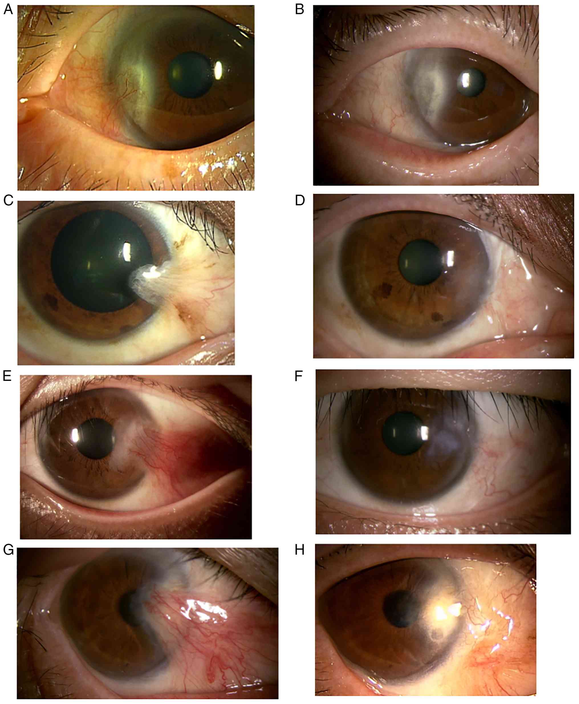
tissues according to a previous study (14). Grade 1 included eyes with pterygium
tissues located exclusively in the corneal limbus (Fig. 1A). Grade 2 showed growth beyond the
corneal limbus but not until the pupil (Fig. 1C), and Grade 3 included eyes with
tissue growth at the pupillary margin (Fig. 1E). Grade 4 included eyes with growth
beyond the pupillary margin (Fig.
1G).
Surgical procedure
A schema of this procedure is presented in a
previous paper (9). Briefly, after
administrating sufficient eye-drop anesthesia, the pterygial head
was carefully separated from the corneal surface, not to remove
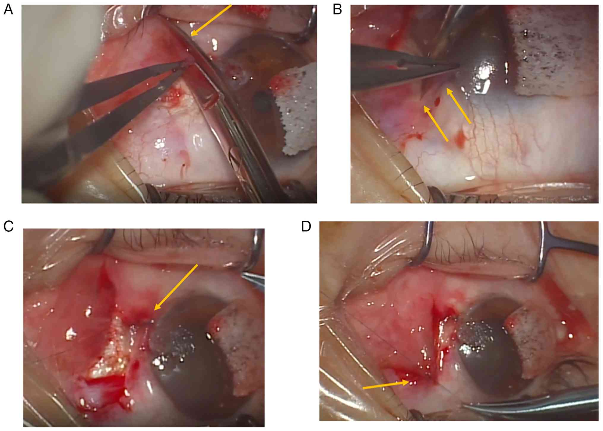
that. Next, the pterygial body was radially incised towards the
conjunctiva (Fig. 2A), and a
sub-Tenon injection of 2% lidocaine was administered. The remaining
stromal tissue on the cornea was removed by curettage and the
corneal bed was managed with Borman layer polishing using a
crescent blade or golf knife. Ocular surface hemostasis was
achieved using a built-in cataract surgery machine. The pterygial
body was incised around the fornix to facilitating manipulation and
suturing. The width of pterygial-tissue incision depended on the
size of the pterygium (Fig. 2A,
arrow). If the Tenon's capsule was thick, as in young patients, the
subepithelial Tenon's capsule was trimmed to avoid damage to the
epithelium. Cryopexy to the pterygial tissues was performed in
selected eyes, similar to that for conjunctival tumors. Next, a
sufficiently large Z- or reversed Z-shaped pedunculated
conjunctival flap was created (Fig.
2B, arrows), and sutured to the lower/upper healthy conjunctiva
(Fig. 2C, arrow). Thereafter, the
pterygial tissue was sutured to the upper/lower space (Fig. 2D, arrow). However, in patients with
conjunctival-flap overhang on the cornea, the center of the flap
was sutured to the sclera to address the corneal overhang.
Interrupted sutures were placed using 8-0 virgin silk, and tissue
glue was not required. Finally, dexamethasone was injected into the
conjunctiva and an antibiotic ointment was placed on the ocular
surface. The detailed standard operating procedure was added as
Table I. Postoperatively,
antibiotic eye drops were administered thrice daily for 1 month,
and low-dose corticosteroid eye drops 3-4 times per day were
continued for at least 3 and 6 months in eyes with primary and
recurrent pterygia, respectively. Suture removal was not required
in most cases, and postoperative inflammation resolved at
postoperative 3 months. However, sutures were removed in case of
irritation or persistence beyond postoperative 3 months.
 | Table INon-resection method of pterygial
surgery. |
Table I
Non-resection method of pterygial
surgery.
| Step | Description |
|---|
| 1 | Separation of the
pterygial head from the cornea |
| 2 | Radical incision of
the body |
| 3 | Sub-Tenon injection
of 2% lidocaine |
| 4 | Removal of the
remaining stromal tissue on the cornea |
| 5 | Polishing the corneal
bed |
| 6 | Creation of the
upper/lower conjunctival flap using a Z-shaped incision |
| 7 | Suture of the
conjunctival flap to the lower/upper conjunctiva |
| 8 | Suture of the
pterygial head to the conjunctival epithelium in the upper/lower
space |
| 9 | Dexamethasone
injection into the conjunctiva with an antibiotic ointment on the
ocular surface |
Statistical analysis
Age and pterygial size were compared between eyes
with and without recurrence using Mann-Whitney U test. To account
for within-subject correlation (clustering of both eyes from the
same patient) and adjust for center effects, multivariate logistic
regression was performed using Generalized Estimating Equations
(GEE) with an exchangeable correlation structure (Tables II and III). To address the wide variation in
follow-up duration and avoid bias from crude proportions,
Kaplan-Meier survival analysis (log-rank test) and robust Cox
proportional hazards regression were performed. Statistical
significance was set at P<0.05.
| Table IIMultivariate Cox proportional hazards
regression analysis. |
Table II
Multivariate Cox proportional hazards
regression analysis.
| A, Model A: Full
model (adjusted for patient clusters and facilities) |
|---|
| Variables | Hazard ratio | 95% CI | P-value |
|---|
| Age | 0.98 | 0.94-1.03 | 0.504 |
| Sex (male vs.
female) | 1.97 | 0.52-7.54 | 0.321 |
| Laterality (right vs.
left) | 0.79 | 0.24-2.60 | 0.695 |
| Recurrence at
operation | 2.40 | 0.46-12.37 | 0.297 |
| Grade | 4.42 | 1.33-14.67 | 0.015a |
| Cryopexy | 8.89 | 0.80-98.75 | 0.075 |
| B, Model B:
Parsimonious model (major clinical variables only) |
| Variables | Hazard ratio | 95% CI | P-value |
| Recurrence at
operation | 1.68 | 0.41-6.91 | 0.476 |
| Grade | 4.57 | 1.73-12.09 | 0.002a |
| Cryopexy | 10.94 | 2.19-54.71 | 0.004a |
| Table IIIGeneralized estimating equations
logistic regression analysis. |
Table III
Generalized estimating equations
logistic regression analysis.
| A, Model adjusted for
major clinical variables and facilities |
|---|
| Variables | Odds ratio | 95% CI | P-value |
|---|
| Recurrence at
operation | 2.10 | 0.28-15.95 | 0.470 |
| Grade | 3.85 | 1.08-13.75 | 0.038a |
| Cryopexy | 9.17 | 0.69-122.10 | 0.093 |
| B, Model including
major clinical variables only |
| Variables | Odds ratio | 95% CI | P-value |
| Recurrence at
operation | 1.52 | 0.25-9.22 | 0.650 |
| Grade | 4.36 | 1.60-11.87 | 0.004a |
| Cryopexy | 7.15 | 1.05-48.68 | 0.045a |
Results
Clinical demographics
This study included 127 eyes from 117 patients (56
men and 61 women) with pterygia. The mean age of the patients at
the time of surgery was 73 years. Pterygia were observed at nasal,
temporal, and combined sites in 124, 0, and 3 eyes, respectively.
Patients' complaints included hyperemia, visual disturbances,
proliferative tissues on the ocular surface, and conjunctival
inversion or elevation; however, diplopia was not reported by any
patient. Primary and recurrent pterygia were observed in 113 and 14
eyes, respectively. The Grades 1, 2, 3, and 4 pterygia were
observed in 3, 49, 65, and 10 eyes, respectively, and the mean size
was Garde 2.6. The follow-up duration ranged from 6 to 82 months
(mean, 31 months). In most cases, the conjunctival flap at the
corneal limbus presented a smooth surface with fine coloration
(Fig. 1B, D, F and
H); however, corneal stromal
opacity persisted in few cases (Fig.
1B and H). No complications
such as infection, persistent inflammation/hyperemia, Dellen,
corneal/sclero-conjunctival ulcers, suture-related problems,
granuloma formation or motility restriction were observed. No
patient required suture removal after surgery.
Pterygium recurrence after
surgery
Postoperative recurrence was observed in 10 eyes
(7.9%) of 10 patients (mean age, 71 years). Of these, five eyes
each were from men and women, and recurrence after surgery for
primary and recurrent pterygia was observed in seven, and three
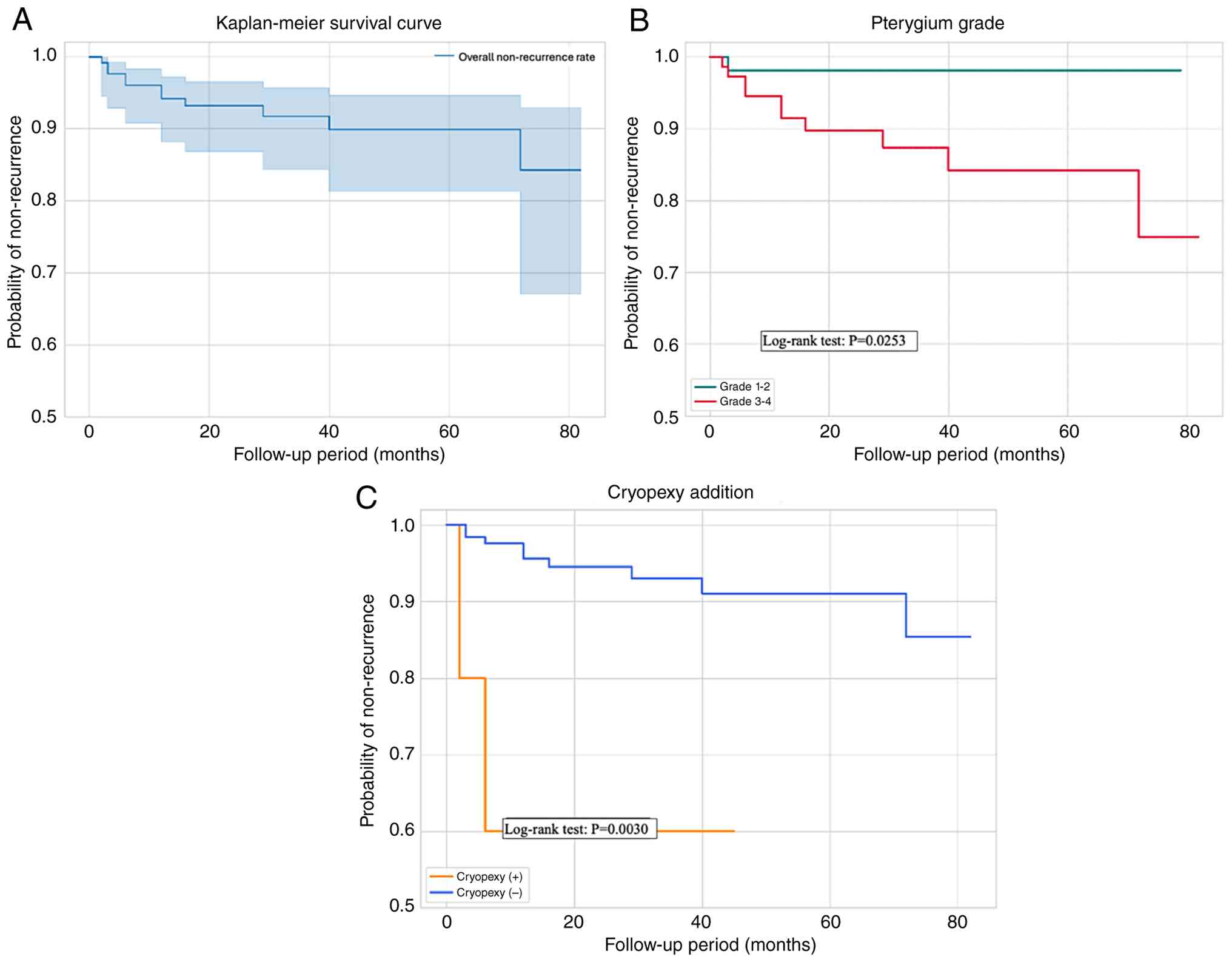
eyes, respectively. Kaplan-Meier analysis revealed no postoperative
recurrence in all enrolled patients (Fig. 3A). The mean duration from surgery to
recurrence was 20 months. The mean preoperative pterygial size was
Grade 2.6 and Grade 3.4 in eyes without and with recurrence,
respectively. The preoperative pterygium size was significantly
greater in eyes with recurrence than in those without recurrence
(U=932.50, P=0.0052, Mann-Whitney U test). By contrast, age was not
significantly different between eyes with and without recurrence
(Mann-Whitney U test). All sutured pterygial tissues shrank with a
pale coloration, and no eyes showed tumor formation during the
follow-up period. No additional surgery was required for the eyes
with pterygia after the initial surgery. In the GEE model, patient
IDs were specified as cluster, and the facility was adjusted as a
covariate. Even after accounting for clustering and facility
effects, pterygium Grade remained a significant independent risk
factor for post-operative recurrence [Odds ratio (OR), 3.85; 95%
Confidence Interval (CI), 1.08–13.75; P=0.0376 in the
facility-adjusted model; and OR, 4.36; 95% CI, 1.60-11.87; P=0.0039
in the clinical GEE model]. Recurrence occurred earlier and at a
higher rate in higher-grade eyes, and the log-rank test revealed a
statistically significant difference in recurrence between Grades
1-2 and Grades 3-4 (P=0.0253) (Fig.
3B).
Adjunctive cryopexy
There are no clear criteria for cryopexy in
pterygium surgery. In 5 eyes of 5 patients with wide pterygial
bodies or relatively young age, cryopexy was performed after
separation of the pterygial tissues from the cornea regardless of
the extent of corneal invasion by the pterygial head, because of
potential high risks of recurrence. The patients' ages ranged from
49 to 76 years (mean: 68 years). Among these, two and three eyes
had primary and recurrent pterygia, respectively. The mean grade
was 2.8. Cryopexy was performed using a conventional cryopexy probe
at -80˚C for 2-3 sec, pressing from beneath the pterygial tissues
for one cycle. Postoperative inflammation and hyperemia resolved
2-3 months after surgery. The follow-up duration of eyes that
underwent cryopexy ranged from 19 to 45 months (mean, 32.8 months).
The log-rank test revealed that addition of cryopexy was
significantly associated with recurrence (P=0.0030, Fig. 3C). Moreover, addition of cryopexy
was associated with a high hazard ratio for recurrence (hazard
ratio, 10.94; 95% CI, 2.19–54.71; P=0.0036), which likely reflects
a ‘confounding by indication’ bias, as cryopexy was selectively
performed on eyes deemed by surgeons to have a high risk of
recurrence.
Discussion
This study demonstrated that favorable clinical
outcomes of the non-resection method for pterygial surgery with low
recurrence rates, without the use of chemical agents such as MMC.
Therefore, this procedure could be performed in various ophthalmic
clinics, facilitating easy access to comprehensive pterygial
surgery for ophthalmologists globally.
In this study, the preoperative size of the
pterygium was mostly Grade 2 or 3, and 10 of 127 eyes had Grade 4
pterygia. The findings in this study suggest that this
non-resection method is appropriate for pterygial growth around the
pupil. Notably, the preoperative pterygium size was significantly
greater in eyes with recurrence than in those without recurrence
(P<0.01). Therefore, if the pterygial tissues are wide, a single
Z-shaped incision might not be sufficient to reconstruct the
corneal limbus. Particularly, if the recurrence is severe and
adhesive to the skin of the eyelid, reconstruction of the
conjunctival fornix using extensive free flaps is required.
However, this non-resection method uses pterygium tissue as a
reconstructive material; therefore, even if an additional flap or
excision is needed during surgery, it is possible to switch to
other methods. On the other hand, there would be a variability
between surgeons when this method is used. Our finding suggest that
the non-resection method is not a universal surgical procedure, but
is a useful first-line approach in pterygium surgery.
In this study, the pterygium recurrence rate after
non-resection method was 7.9%. A previous study reported that
conjunctival autograft is superior to the scleral bare procedure in
terms of postoperative recurrence rates (15). Recently, Aziza et al
(7) reported that recurrence in one
of 32 pterygial patients who underwent a scleral bare procedure
with MMC application. Noguera et al (6) reported a recurrence rate of 1% in
patients who underwent resection of pterygial tissues with
transplantation of conjunctival autograft or amniotic membrane with
MMC application. Moreover, a previous study reported that the
recurrence rate after other procedures was <10%. Therefore, the
recurrence rate after non-resection method is comparable to those
after other methods. However, the comparable rate was achieved
without the use of chemical agents such as MMC or amniotic
membranes. In fact, MMC-related scleral melt/defect is a serious
complication, with a long-term implications for the postoperative
managements of patients (16-18).
Kim et al (19) reported
that a low postoperative recurrence rate of 6% using conjunctival
mini-flap technique without the use of MMC, whereas the recurrence
rate after conjunctival autograft transplantation was 17%. The
recurrence rate in this study might be comparable to that in the
previous study (19), without any
concerns regarding serious complications owing to the use of
MMC.
In this study, the epithelium of the definitive
pterygium, which contains columnar epithelium or squamous
metaplasia without cellular atypia (3,4), was
not resected. Therefore, the pterygial epithelium could be used as
a reconstructive material, facilitating easy ocular-surface
reconstruction. Because the indication for this non-resection
method is a definitive pterygium, preoperative inspection of
proliferative tissues on slit-lamp examination is extremely
important. Moreover, cryopexy of the pterygial tissues may
contribute to the suppression of pterygial epithelial proliferation
and angiogenesis (20), which
potentially reduces recurrence in wide-edge pterygia. Conditions
such as OSSN with pterygium, pterygium-like OSSN or pterygium-like
conjunctival nevus (1,10,12),
in which resection of proliferative tissues with extensive
conjunctival reconstruction is recommended, are contraindications
for this procedure.
This study had several limitations. First, this was
a retrospective multicenter study; because choice of surgical
procedure depended on the individual ophthalmologists' decision,
the cohort included consecutive patients treated using this
technique, but did not include all consecutive pterygium surgeries.
Therefore, a selection bias could not be excluded. Furthermore,
this study design had a single-arm design, not a randomized
comparative study. A prospective head-to-head study is required to
verify the usefulness of this procedure for pterygial surgery.
Second, the sutured pterygial tissue was not histologically
confirmed. Hence, pathophysiological mechanisms underlying
shrinkage of the sutured tissues remain unknown; however, the
presumed mechanism is contact inhibition between the pterygial
epithelium and normal conjunctival epithelium (9). If the sutured tissues exhibit
postoperative growth, ophthalmologists should consider biopsy of
the tissues, for histopathological confirmation. Third, because of
the multicenter nature of the study, surgeries were conducted by
different ophthalmologists with different levels of experiences in
pterygial surgery. The differences in skills and experiences might
have affected recurrence rates. Forth, Tan's classification on
pterygial morphology classifying as atrophic, intermediate and
freshy (21), could not be
available in this multicenter analysis, because the slit-lamp
photographs were not available in several participants and
participating institutions. Fifth, the masked evaluators were not
used in analysis of clinical data, and the duration of the
pterygial surgery was unavailable in this study. Finally, visual
and refractive outcomes cannot be fully examined in this cohort;
therefore, further analyses are required to clarify the usefulness
of this method.
In conclusion, although the non-resection method
could be comparable to the previous methods for pterygial surgery
in terms of postoperative recurrence rates, it might be safely and
can be easily performed in any institute. Nevertheless, eyes with
higher grades of pterygia exhibit a tendency for recurrence.
Acknowledgements
Not applicable.
Funding
Funding: No funding was received.
Availability of data and materials
The data generated in the present study are not
publicly available to protect potentially identifiable personal
data but may be requested from the corresponding author.
Authors' contributions
SK wrote the manuscript and acquired the clinical
data. SC reviewed the manuscript and interpreted the clinical data.
MS, CY, YA, JF, SI, NK, RKi and RKa conducted critical revision and
supervised data interpretation. RKi, CY, JF, YA, RKa and NK made
substantial contributions to conception and design, and acquisition
of clinical data. SI and NK performed evaluation and interpretation
of data. MS conducted all the statistical analyses in the present
study while remaining blinded to the clinical data. SK and MS
confirm the authenticity of all the raw data. All authors have read
and approved the final version of the manuscript.
Ethics approval and consent to
participate
The present study was approved by the Institutional
Review Board of Hokkaido University (IRB no. 024-0068; Sapporo,
Japan) as the central institutional review board. The study
protocol included all the participating institutions listed in the
manuscript. Therefore, ethical approval from the other institutions
was not required. The study adopted an opt-out approach. This means
that enrolled patients were given the opportunity to refuse
participation in the study through the website. Therefore, the
requirement for written informed consent was waived.
Patient consent for publication
Not applicable.
Competing interests
The authors declare that they have no competing
interests.
References
|
1
|
Chui J, Coroneo MT, Tat LT, Crouch R,
Wakefield D and Di Girolamo N: Ophthalmic pterygium: A stem cell
disorder with premalignant features. Am J Pathol. 178:817–827.
2011.PubMed/NCBI View Article : Google Scholar
|
|
2
|
Kase S, Osaki M, Jin XH, Ohgami K, Yoshida
K, Saito W, Takahashi S, Nakanishi K, Ito H and Ohno S: Increased
expression of erythropoietin receptor in human pterygial tissues.
Int J Mol Med. 20:699–702. 2007.PubMed/NCBI
|
|
3
|
Kase S, Osaki M, Sato I, Takahashi S,
Nakanishi K, Yoshida K, Ito H and Ohno S: Immunolocalisation of
E-cadherin and beta-catenin in human pterygium. Br J Ophthalmol.
91:1209–1212. 2007.PubMed/NCBI View Article : Google Scholar
|
|
4
|
Kase S, Takahashi S, Sato I, Nakanishi K,
Yoshida K and Ohno S: Expression of p27(KIP1) and cyclin D1, and
cell proliferation in human pterygium. Br J Ophthalmol. 91:958–961.
2007.PubMed/NCBI View Article : Google Scholar
|
|
5
|
Tanaka T, Kase S and Ishida S: A case of
spontaneous avulsion of primary pterygium analyzed with anterior
segment optical coherence tomography. Am J Ophthalmol Case Rep.
26(101438)2022.PubMed/NCBI View Article : Google Scholar
|
|
6
|
Noguera SI, Nicanor KSA, Ang RET and Cruz
EM: Clinical outcomes of pterygium surgery over a ten-year period:
A review of recurrence and complication rates. BMC Ophthalmol.
25(380)2025.PubMed/NCBI View Article : Google Scholar
|
|
7
|
Aziza Y, Inatomi T, Sotozono C and
Kinoshita S: Pterygium excision with modified bare sclera technique
combined with mitomycin C. Jpn J Ophthalmol. 65:89–96.
2021.PubMed/NCBI View Article : Google Scholar
|
|
8
|
Aleksander-Ivanov Y, Cheidde L, Veiga DM,
Filho LAZ and Afshari N: Adjuvant use of topical 0.05% cyclosporine
A in primary pterygium recurrence rate: A systematic review and
Meta-analysis. Am J Ophthalmol. 278:222–238. 2025.PubMed/NCBI View Article : Google Scholar
|
|
9
|
Kase S, Chin S and Ishida S: Z-shaped
incision without epithelial resection in pterygium surgery. Int J
Ophthalmol. 17:1262–1266. 2024.PubMed/NCBI View Article : Google Scholar
|
|
10
|
Endo H, Kase S, Suzuki Y and Kase M:
Coincidence of inflamed conjunctival carcinoma in situ and primary
pterygium. Case Rep Ophthalmol. 7:208–2012. 2016.PubMed/NCBI View Article : Google Scholar
|
|
11
|
Mitamura M, Kase S, Ohguchi T and Ishida
S: A case of pterygium-like proliferation containing postoperative
limbal dermoid remnants: A clinicopathological study. BMC
Ophthalmol. 21(12)2021.PubMed/NCBI View Article : Google Scholar
|
|
12
|
Kase S, Hirooka K, Takakuwa E and Ishida
S: Pterygium-like conjunctival nevus: A clinicopathological study.
Can J Ophthalmol. 60:e498–e501. 2025.PubMed/NCBI View Article : Google Scholar
|
|
13
|
Pikkel J, Porges Y and Ophir A: Halting
pterygium recurrence by postoperative 5-fluorouracil. Cornea.
20:168–1671. 2001.PubMed/NCBI View Article : Google Scholar
|
|
14
|
Masuda A, Takahashi K, Nejima R, Minami K
and Miyata K: Pterygium excision using bulbar conjunctival
autograft with intraoperative mitomycin C for primary pterygium: A
retrospective assessment of 1832 eyes. Nippon Ganka Gakkai Zasshi.
117:743–748. 2013.PubMed/NCBI(In Japanese).
|
|
15
|
Kaufman SC, Jacobs DS, Lee WB, Deng SX,
Rosenblatt MI and Shtein RM: Options and adjuvants in surgery for
pterygium: A report by the American academy of ophthalmology.
Ophthalmology. 120:201–208. 2013.PubMed/NCBI View Article : Google Scholar
|
|
16
|
Kondo A, Mimura T, Goto M, Kamei Y, Yusuke
S, Okuma H and Matsubara M: Letter to the editor: Corneoscleral
melt 50 years after excision of pterygium. Open Ophthalmol J.
11:47–50. 2017.PubMed/NCBI View Article : Google Scholar
|
|
17
|
Bielory B: Self-retained, cryopreserved
amniotic membrane for a scleral defect caused by mitomycin C: A
case report. Am J Ophthalmol Case Rep. 36(102199)2024.PubMed/NCBI View Article : Google Scholar
|
|
18
|
Polat N: Use of an autologous lamellar
scleral graft to repair a scleral melt after mitomycin application.
Ophthalmol Ther. 3:73–76. 2014.PubMed/NCBI View Article : Google Scholar
|
|
19
|
Kim M, Chung SH, Lee JH, Lee HK and Seo
KY: Comparison of mini-flap technique and conjunctival autograft
transplantation without mitomycin C in primary and recurrent
pterygium. Ophthalmologica. 222:265–271. 2008.PubMed/NCBI View Article : Google Scholar
|
|
20
|
Fraunfelder FW: Liquid nitrogen
cryotherapy of pterygia. Ophthalmology. 120(e70)2013.PubMed/NCBI View Article : Google Scholar
|
|
21
|
Tan DT, Chee SP, Dear KB and Lim AS:
Effect of pterygium morphology on pterygium recurrence in a
controlled trial comparing conjunctival autografting with bare
sclera excision. Arch Ophthalmol. 115:1235–1240. 1997.PubMed/NCBI View Article : Google Scholar
|