Introduction
Osteoarthritis (OA) is a chronic and disabling
condition that affects the joints (1). Individuals with this condition often
experience joint discomfort, swelling and restricted movement,
which negatively affect their physical capabilities and overall
quality of life (2). Apoptosis of
chondrocytes has been identified as a key pathological feature of
OA, and mild synovial inflammation is also recognized as a
contributing factor (3,4). Currently, there is no definitive cure
for OA. In clinical settings, management typically involves
non-pharmacological approaches such as assistive tools, aerobic
activities, therapeutic exercises, lifestyle adjustments, and
pharmacological interventions such as analgesics and oral or
topical non-steroidal anti-inflammatory drugs (5,6).
Nevertheless, conclusive evidence supporting the long-term
effectiveness of these treatments for OA remains elusive. As a
result, there is an ongoing need for innovative and more effective
treatment options.
In total, ~2% of the human genome consists of
protein-coding RNAs, whereas the remainder is composed of
non-coding (nc) RNAs (7). Among
these, long nc RNAs (lncRNAs) are defined as transcripts exceeding
200 nucleotides in length. Despite their lack of translation into
functional polypeptides, lncRNAs play an essential role in
modulating the expression of protein-coding genes (8). As novel epigenetic regulators, they
influence the progression of various human diseases, including OA.
Shi et al (9) identified a
novel lncRNA, POU3F3, which modulates the microRNA (miRNA or
miR)-29a-3p/FOXO3 axis to inhibit OA progression. Chen et al
(10) reported significant
upregulation of the lncRNA HOTAIR in chondrocytes affected by OA,
which notably enhances inflammatory responses through the
PTEN/PI3K/AKT pathway. Ni et al (11) demonstrated that the lncRNA DLEU1
contributes to the progression of OA by markedly promoting
chondrocyte apoptosis. The lncRNA small nucleolar RNA host gene 6
(SNHG6), also known as U87HG, is located on human chromosome
8q13.1(12). Currently, nearly all
reports on SNHG6 are associated with cancer. It is overexpressed in
various cancers, including colorectal cancer (13), esophageal squamous carcinoma
(14) and ovarian cancer (15). Studies have demonstrated its role in
enhancing cell proliferation, metastasis and
epithelial-mesenchymal-transition in numerous cancer cell types
(13-15).
However, its role and the underlying mechanisms involved in OA
progression are poorly understood.
The present study investigated the effects of SNHG6
on apoptosis and inflammation as well as its possible a mechanism
of action in lipopolysaccharide (LPS)-induced chondrocytes. The
miR-944/post-GPI attachment to protein 1 (PGAP1) axis may be a
potential target of SNHG6 in the progression of OA.
Materials and methods
Patients and sample collection
From January 2024 to December 2025, 30 patients (17
females and 13 males; age range, 55-75 years; mean age, 64.7±6.6)
with newly diagnosed with OA were recruited at our hospital. OA
diagnosis was confirmed by analysis of both X-ray imaging and joint
fluid. The inclusion criteria of these patients were as follows: i)
Full comprehension of the study protocol and agreement to provide
synovial fluid; ii) no prior medical intervention. Patients with
other chronic inflammatory conditions who started treatment within
three months before admission were excluded from the study.
Meanwhile, 30 healthy controls (17 females and 13 males; age range,
52-73 years; mean age, 62.2±6.0) underwent systemic physiological
examination with no OA or rheumatoid arthritis were enrolled.
Synovial fluid samples (1 ml) were collected using a syringe under
local anesthesia. The ethics committee of Wenzhou People's Hospital
approved the present study protocol (approval no. KY-2023-251;
Wenzhou, China). Both healthy individuals and patients with OA
signed written informed consent. The research complied with the
principles established in the Declaration of Helsinki.
Cell culture, LPS treatment and
transfection
Primary human normal chondrocytes (cat. no. CP-H107;
https://www.procell.com.cn/p/human-chondrocyte-cells-cp-h107-68054)
were purchased from Wuhan Pricella Biotechnology Co., Ltd. at
passage 0 (P0). The use of primary human cells has obtained the
approval from the Ethical Committee of Wenzhou People's Hospital
(approval number: KY-2023-289; Wenzhou, China). They were cultured
in DMEM/F12 medium (Hyclone; Cytiva) supplemented with 1%
Penicillin-Streptomycin Solution (Beyotime Institute of
Biotechnology) and 10% fetal bovine serum (Gibco; Thermo Fisher
Scientific, Inc.). The cells were incubated at 37˚C under a
humidified atmosphere containing 5% CO2. For
experimental purposes, second-passage (P2) chondrocytes with 85%
confluency were selected and utilized. LPS was utilized to
establish the in vitro OA model, since it can efficiently
trigger chondrocyte inflammatory response, upregulate
pro-inflammatory cytokines and matrix-degrading enzymes, and
suppress the expression of cartilage-specific anabolic proteins,
which closely recapitulates the pathological alterations of
degenerated chondrocytes in OA. This model has been widely adopted
in numerous OA-related in vitro mechanistic studies due to
its high experimental stability, favorable repeatability, and
simple operation, which is particularly suitable for the initial
verification of the functional role of specific molecular axes in
chondrocyte apoptosis and inflammation. To construct an in
vitro OA cell model, normal chondrocytes were treated with LPS
(5 µg/ml; Sigma Aldrich) for 12 h (16-18).
From Genomeditech (Shanghai) Co., Ltd., mimics NC
(sense: 5'-AUGUAGUAUAGCAGAUGAUAUC-3'; anti-sense:
5'-GAUAUCAUCUGCUAUACUACAU-3')/miR-944 mimics (sense:
5'-AAAUUAUUGUACAUCGGAUGAG-3'; anti-sense:
5'-CUCAUCCGAUGUACAAUAAUUU-3') and inhibitor NC
(5'-GAUAUCAUCUGCUAUACUACAU-3')/miR-944 inhibitor
(5'-CUCAUCCGAUGUACAAUAAUUU-3'), as well as small interfering RNA
targeting SNHG6 (si-SNHG6; sense: 5'-GAUGUUGAUAACAUCACAA-3';
anti-sense: 5'-UUGUGAUGUUAUCAACAUC-3')/si-PGAP1 (sense:
5'-GGUACGAGGUGACAGUUAA-3'; anti-sense:
5'-UUAACUGUCACCUCGUACC-3')/si-NC (sense: 5'-GAGACAGUUGUACGAGUAG-3';
anti-sense: 5'-CUACUCGUACAACUGUCUC-3') and overexpression of PGAP1
(OE-PGAP1), OE-SNHG6 and OE-NC were synthesized. Cell transfection
was performed using Lipofectamine 3000 (Invitrogen; Thermo Fisher
Scientific, Inc.). The aforementioned oligonucleotides and plasmids
(50 nM) were separately co-transfected into LPS-induced
chondrocytes (1x10⁵ cells/well) for 48 h. After
transfection, chondrocytes were harvested, and the levels of gene
expression were detected via reverse transcription-quantitative
polymerase chain reaction (RT-qPCR).
RT-qPCR
Synovial fluid samples and chondrocytes were
obtained for RNA extraction using a Total RNA Extraction Kit
(Foregene Co., Ltd.). Subsequently, cDNA was synthesized using the
SeqHunt® First Strand cDNA Synthesis Kit (Seq-Hunt
Biotechnology) according to the manufacturer's instructions. PCR
analysis was performed on a Real-time PCR System (Applied
Biosystems; Thermo Fisher Scientific, Inc.) with 2X Blue Universal
SYBR qPCR Master Mix (Seq-Hunt Biotechnology) according to the
manufacturer's protocol. The thermocycling conditions were as
follows: 95˚C for 5 min, 40 cycles of denaturation at 95˚C for 10
sec, annealing at 50˚C for 1 min and extension at 72˚C for 30 sec.
The relative expression levels were quantified by the
2-ΔΔCq method (19), with GAPDH and U6 serving as the
internal controls for data normalization. All gene-specific primers
utilized in the present study are listed in Table I.
 | Table IPrimer sequences. |
Table I
Primer sequences.
| Gene name | Primer
sequence |
|---|
| lncRNASNHG6 | Forward:
5'-ATACTTCTGCTTCGTTACCT-3' |
| | Reverse:
5'-CTCATTTTCATCATTTGCT-3' |
| microRNA-944 | Forward:
5'-GCGGCGGAAATTATTGTACATC-3' |
| | Reverse:
5'-ATCCAGTGCAGGGTCCGAGG-3' |
| PGAP1 | Forward:
5'-ACAGGCTCCATCTTCCACAG-3' |
| | Reverse:
5'-ACGACATAAGCAGGAAGAGC-3' |
| GAPDH | Forward:
5'-TGCACCACCAACTGCTTAGC-3' |
| | Reverse:
5'-GGCATGCACTGTGGTCATGAG-3' |
| U6 | Forward:
5'-CTCGCTTCGGCAGCACA-3' |
| | Reverse:
5'-AACGCTTCACGAATTTGCGT-3' |
Cell viability and cell cycle
measurement
Chondrocytes were seeded in 96-well plates at a
concentration of 4x10³ cells per well and cultured
continuously for 48 h. After incubation, each well was supplemented
with 10 µl Cell Counting Kit-8 reagent (Dojindo Molecular
Technologies, Inc.) and incubated for another 2 h at 37˚C. Finally,
the absorbance values were then determined at a wavelength of 450
nm using a microplate reader (Hiwell Diatek).
To analyze the cell cycle, chondrocytes
(1x10⁵) were fixed with 75% ethanol at 4˚C for 1 h.
After fixation, the cells were centrifuged at 1,500 g for 5 min at
4˚C, then re-suspended in phosphate-buffered saline. Subsequently,
the cells were incubated with Propidium iodide/RNase A solution
(Beyotime Institute of Biotechnology) in the dark for 30 min. Data
were acquired on a flow cytometer (BD Biosciences). The
distribution of cells in the G1, S and G2 phases was quantified by
ModFit LT software (version 5.0; Verity Software House) using the
Watson pragmatic model.
Cell apoptosis measurement
Based on the manufacturer's protocol of Apoptosis
Detection Kit (Thermo Fisher Scientific, Inc.), 2x10⁵
suspended chondrocytes were stained with Annexin V-FITC (10 µl) and
PI (5 µl) at 25˚C for 15 min in the dark, followed by adding 500 µl
of binding buffer. Subsequently, the apoptotic chondrocytes were
assessed using a flow cytometer (BD Biosciences).
For further auxiliary verification and quantitative
detection of chondrocyte apoptotic levels, the One Step TUNEL
Apoptosis Assay Kit from Beyotime Institute of Biotechnology was
adopted for complementary testing. First, the cells were stabilized
in 4% paraformaldehyde for 15 min and then treated with 0.25%
Triton-X 100 for 20 min to enhance permeability. Afterwards, 50 µl
of TUNEL reaction mixture was applied, and the samples were
incubated at 37˚C for 60 min. Following phosphate-buffered saline
washes, cell nuclei were stained with DAPI (5 µg/ml) for 5 min in
the dark. Finally, the samples were analyzed using a fluorescence
microscope (Olympus Corporation).
Western blot assay
To obtain chondrocyte protein extracts, RIPA lysis
buffer (Biosharp Life Sciences) was utilized. Subsequently, the
protein samples (30 µg per lane) were subjected to separation via
10% SDS-PAGE, followed by electrotransfer onto a PVDF membrane. The
membrane was blocked with 5% non-fat milk (Biosharp Life Sciences)
for 2 h at room temperature. Thereafter, the membrane was incubated
overnight at 4˚C with primary antibodies directed against Caspase-3
(cat. no. ab32351; 1:5,000; Abcam), Caspase-9 (cat. no. ab32539;
1:5,000; Abcam), Cyclin A1 (cat. no. 13295-1-AP; 1:1,000;
Proteintech Group, Inc.), Cyclin B1 (cat. no. 55004-1-AP; 1:2,000;
Proteintech Group, Inc.), Cyclin D2 (cat. no. 10934-1-AP; 1:1,000;
Proteintech Group, Inc.) and GAPDH (cat. no. 60004-1-Ig; 1:50,000;
Proteintech Group, Inc.). Afterward, the membrane was further
incubated with HRP-conjugated secondary antibodies (cat. no. A21010
& A21020; 1:10,000; Abbkine Scientific Co., Ltd.) for 2 h at
25˚C. Protein signals were visualized using the SuperKine™ ECL Kit
(Abbkine Scientific Co., Ltd.) and quantified via ImageJ software
version 1.53 (National Institutes of Health).
Immunofluorescent test
Following routine dewaxing in xylene and gradient
rehydration in ethanol, antigen retrieval was performed on
chondrocyte specimens under standard experimental conditions. This
was followed by a blocking step using goat serum (Beijing Solarbio
Science & Technology Co., Ltd.). The samples were then
incubated overnight at 4˚C with a primary antibody specific to
PGAP1 (cat. no. 55392-1-AP; 1:50; Proteintech Group, Inc.). Next,
the sections were conjugated with an Alexa Fluor 488 secondary
antibody diluted at 1:200 (Invitrogen; Thermo Fisher Scientific,
Inc.). Following DAPI staining (1 µg/ml), fluorescence microscopy
images of the sections were acquired using an Olympus fluorescence
microscope.
ELISA
Strictly in accordance with the guidelines of
commercial ELISA kits, the concentrations of IL-1β (cat. no.
KTE6013), IL-6 (cat. no. KTE6017) and TNF-α (cat. no. KTE6032) (all
from Abbkine Scientific Co., Ltd.) were quantitatively detected and
assessed in chondrocyte samples.
Dual luciferase reporter assay
The potential targeting relationships between SNHG6
and miR-944, as well as between miR-944 and PGAP1, were predicted
using Starbase software (version 2.0; http://starbase.sysu.edu.cn). The partial sequences of
the 3'-untranslated region (UTR) of PGAP1 and SNHG6 containing the
predicted binding sites of miR-944 were synthetized by Sangon
Biotech Co., Ltd. These sequences were then inserted into the pGL3
vector to construct wild-type (Wt)-PGAP1 and Wt-SNHG6 reporter
vectors. Following the manufacturer's protocol, mutant type (Mut)
PGAP1 and SNHG6-3'-UTR sequences, which contain the miR-944 binding
sites, were generated using the Phusion Site-Directed Mutagenesis
Kit (Thermo Fisher Scientific, Inc.). Similarly, the mutated
sequences were inserted into the pGL3 vector to create Mut reporter
vectors for both PGAP1 and SNHG6. Following this, chondrocytes
(5x10³ cells/well) were co-transfected with mimics NC or
miR-944 mimics (10 nM) as well as the constructed wild-type or
mutant reporter plasmids, with the aid of Lipofectamine 3000
transfection reagent (Invitrogen; Thermo Fisher Scientific, Inc.).
At 48 h after transfection, the relative luciferase activity was
measured using the Dual-Luciferase Reporter Assay System Kit
(Promega Corporation). The activity of firefly luciferase was
normalized to that of Renilla luciferase.
Statistical analysis
SPSS 20.0 software (IBM Corp.) was employed for data
analysis, and all experimental data were expressed as the mean ±
standard deviation. Statistical differences between multiple groups
were analyzed by one-way analysis of variance followed by Tukey's
post hoc test, whereas Student's t-test (unpaired) was used for
comparisons between two independent groups. P<0.05 was
considered to indicate a statistically significant difference.
Results
Effects of SNHG6 expression on
LPS-stimulated chondrocytes
It was demonstrated that there was a notable
reduction in the expression level of SNHG6 in synovial fluid of
patients with OA, compared with the normal group (P<0.001;
Fig. 1A). SNHG6 was subsequently
silenced or overexpressed in LPS-induced chondrocytes. As shown in
Fig. 1B, SNHG6 expression levels
were significantly reduced when transfected with si-SNHG6
(P<0.0001), while was concurrently elevated upon OE-SNHG6
transfection (P<0.0001). Compared with the control group, LPS
treatment led to a significant decrease in cell viability (Fig. 1C, P<0.0001). Meanwhile,
chondrocyte viability was further inhibited following transfection
of si-SNHG6 (Fig. 1C, P<0.0001)
but was partially restored when OE-SNHG6 was transfected
(P<0.01). This finding further evidenced by the results of cell
frequency illustrated in Fig. 1D
and E. It was observed that by
contrast to the control group, G1 phase cell frequency in the LPS
group were raised. Silencing of SNHG6 further extended G1 phase,
whereas the overexpressed SNHG6 reduced G1 phase cell frequency. As
illustrated in Fig. 1F and G, the presence of LPS significantly
increased the percentage of TUNEL-positive cells (P<0.0001).
When transfected with si-SNHG6 in LPS-stimulated chondrocytes, the
percentage of TUNEL-positive cells was further increased
(P<0.01); however, the positive cells were reduced when SNHG6
was overexpressed (P<0.01). The results of flow cytometric
analysis further validated the variations in apoptotic rates across
the different groups (Fig. 1H and
I; P<0.0001). Subsequently, the
levels of proteins associated with apoptosis (Caspase-3 and
Caspase-9) and cell cycle (Cyclin A1, Cyclin B1 and Cyclin D2) were
determined. Silencing of SNHG6 significantly elevated the levels of
Caspase-3 and Caspase-9, and inhibited Cyclin A1, Cyclin B1 and
Cyclin D2 levels (Fig. 1J;
P<0.05), while high levels of SNHG6 exhibited the opposite
regulatory effects on these proteins (P<0.05). These
experimental data suggested that silencing of SNHG6 exhibited a
notably inhibiting effect on chondrocyte proliferation, and a
significantly promoting impact on apoptosis. Overexpression of
SNHG6 exerted the opposite role in LPS-stimulated chondrocytes.
Finally, the effects of SNHG6 on inflammation responses were
assessed. IL-6, TNF-α and IL-1β secretion in LPS-stimulated
chondrocytes were significantly induced by the silencing of SNHG6
(Fig. 1K; P<0.01) but suppressed
by the overexpression of SNHG6 (P<0.05).
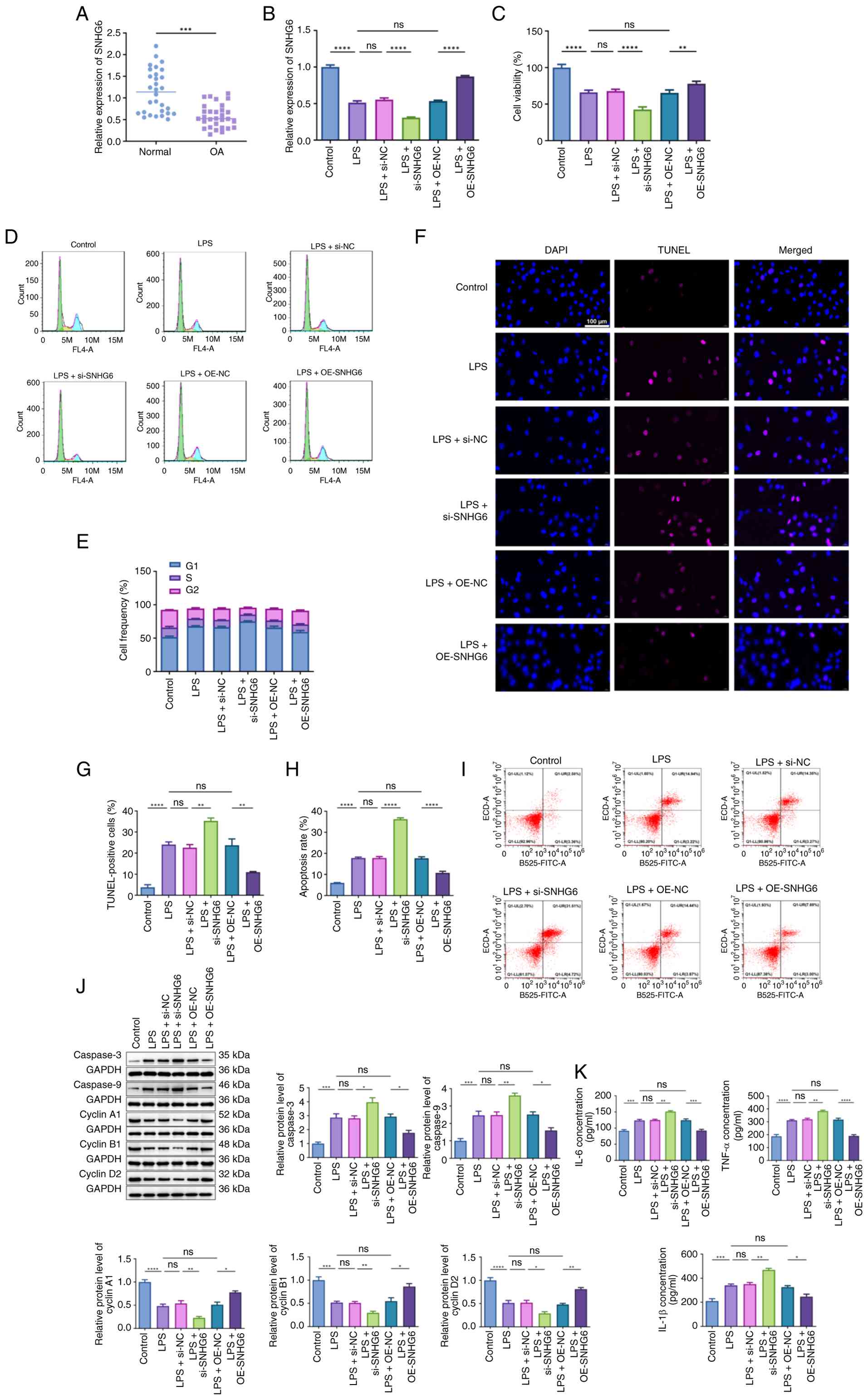 | Figure 1Effects of SNHG6 expression on
LPS-stimulated chondrocytes. (A) The expression of SNHG6 in
synovial fluid of normal individuals and patients with
osteoarthritis was detected via RT-qPCR. (B) Following transfection
of si-SNHG6 or OE-SNHG6 in LPS-induced chondrocytes, the expression
of SNHG6 was detected via RT-qPCR. (C) The viability of LPS-induced
chondrocytes that transfected with si-SNHG6 or OE-SNHG6 was
measured by Cell Counting Kit-8 assay. (D and E) Cell frequency of
LPS-induced chondrocytes was calculated via flow cytometric
analysis. (F and G) The percentages of TUNEL-positive cells were
assessed by TUNEL assay. Scale bar, 100 µm. (H and I) The apoptotic
rate of LPS-induced chondrocytes was calculated via flow cytometric
analysis. (J) The protein levels of Caspase-3, Caspase-9, Cyclin
A1, Cyclin B1 and Cyclin D2 in LPS-induced chondrocytes that
transfected with si-SNHG6 or OE-SNHG6 were determined via western
blotting. (K) The levels of IL-6, TNF-α and IL-1β in LPS-induced
chondrocytes that transfected with si-SNHG6 or OE-SNHG6 were
determined via ELISA. *P<0.05,
**P<0.01, ***P<0.001 and
****P<0.0001. SNHG6, small nucleolar RNA host gene 6;
LPS, lipopolysaccharide; RT-qPCR, reverse
transcription-quantitative PCR; si-, small interfering; OE-,
overexpression; ns, no significance; NC, negative control. |
SNHG6 binds to and negatively
regulates miR-944
Starbase software was employed to predict the
downstream targets of SNHG6 and identified potential binding sites
between SNHG6 and miR-944 (Fig.
2A). Based on dual luciferase reporter assays, no statistical
changes were observed in relative luciferase activity between the
groups of SNHG6-Mut + mimics NC and SNHG6-Mut + miR-944 mimics. By
contrast, a notable reduction in luciferase activity was observed
in SNHG6-Wt + miR-944 mimics group when compared with the SNHG6-Wt
+ mimics NC group (Fig. 2B;
P<0.05). Notably, in synovial fluid of patients with OA, a
relatively high expression of miR-944 was identified (Fig. 2C; P<0.001). Furthermore, the
silencing of SNHG6 resulted in a significant increase in miR-944
expression (Fig. 2D; P<0.0001),
while SNHG6 overexpression was found to suppress the expression of
miR-944 (P<0.0001). To further verify the regulatory
relationship between SNHG6 and miR-944, OE-SNHG6 or miR-944 mimics
was transfected into LPS-induced chondrocytes. As illustrated in
Fig. 2E and F, upregulation of miR-944 reversed the
inhibitory effect of SNHG6 overexpression on miR-944 expression
(P<0.05), while partially attenuating its promoting effect on
SNHG6 expression (P<0.01). Subsequently, functional experiments
demonstrated that upregulation of miR-944 reversed the regulatory
effects of SNHG6 overexpression on cell proliferation, including
cell viability (Fig. 2G;
P<0.001) and apoptosis (Fig. 2H
and I; P<0.001). Additionally,
miR-944 upregulation was found to significantly abolish the
suppressive effects of SNHG6 overexpression on the levels of IL-6,
TNF-α and IL-1β (Fig. 2J-L;
P<0.05).
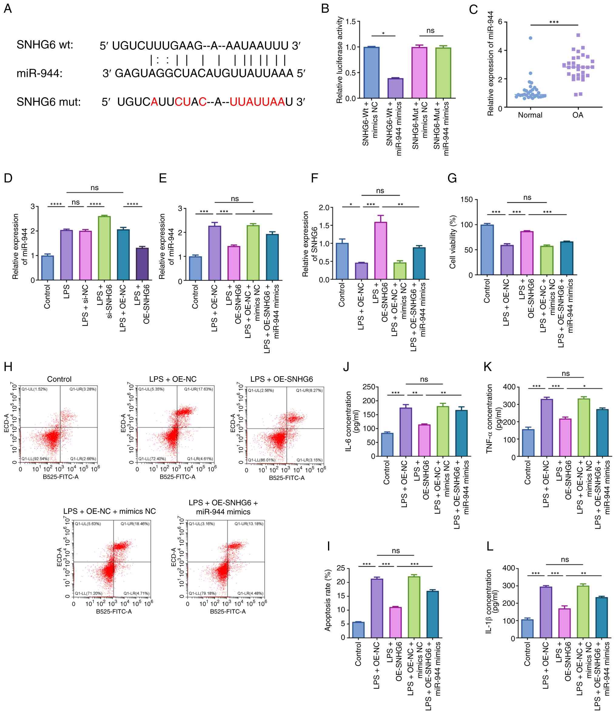 | Figure 2SNHG6 binds to and negatively
regulates miR-944. (A) StarBase showed the predicted binding site
of SNHG6 and miR-944. (B) The luciferase activities in chondrocytes
co-transfected with pGL3-SNHG6-Wt/pGL3-SNHG6-Mut and miR-944
mimics/mimics NC were determined by dual luciferase reporter assay.
(C) The expression of miR-944 in synovial fluid of normal
individuals and patients with osteoarthritis was detected via
RT-qPCR. (D) Following transfection of si-SNHG6 or OE-SNHG6 in
LPS-induced chondrocytes, the expression of miR-944 was detected
via RT-qPCR. (E and F) Following transfection of OE-SNHG6 or
miR-944 mimics in LPS-induced chondrocytes, the expression of (E)
miR-944 and (F) SNHG6 was detected via RT-qPCR. (G-L) Following
transfection of OE-SNHG6 or miR-944 mimics in LPS-induced
chondrocytes, (G) cell viability, (H and I) apoptosis and (J-L)
inflammatory cytokine levels were determined.
*P<0.05, **P<0.01,
***P<0.001 and ****P<0.0001. SNHG6,
small nucleolar RNA host gene 6; miR, microRNA; NC, negative
control; RT-qPCR, reverse transcription-quantitative PCR; si-,
small interfering; OE-, overexpression; LPS, lipopolysaccharide;
ns, no significance. |
Effects of miR-944 inhibition on
LPS-stimulated chondrocytes
Given the elevated levels of miR-944 in OA,
LPS-stimulated chondrocytes were transfected with a miR-944
inhibitor for functional validation (Fig. 3A; P<0.0001). As illustrated in
Fig. 3B and C, transfection of miR-944 inhibitor
remarkably stimulated chondrocyte viability (P<0.01). Both TUNEL
assay and flow cytometric analysis evidenced that the transfection
of miR-944 inhibitor led to a significant suppression in
chondrocyte apoptosis (Fig. 3D and
E; P<0.01). As western blotting
indicated, the levels of Caspase-3 and Caspase-9 were suppressed
(Fig. 3F; P<0.01), while Cyclin
A1, Cyclin B1 and Cyclin D2 protein levels were elevated when
miR-944 inhibitor was transfected (P<0.05). Furthermore, it was
observed that the suppression of miR-944 significantly reduced the
levels of IL-6, TNF-α and IL-1β in LPS-stimulated chondrocytes
(Fig. 3G; P<0.05).
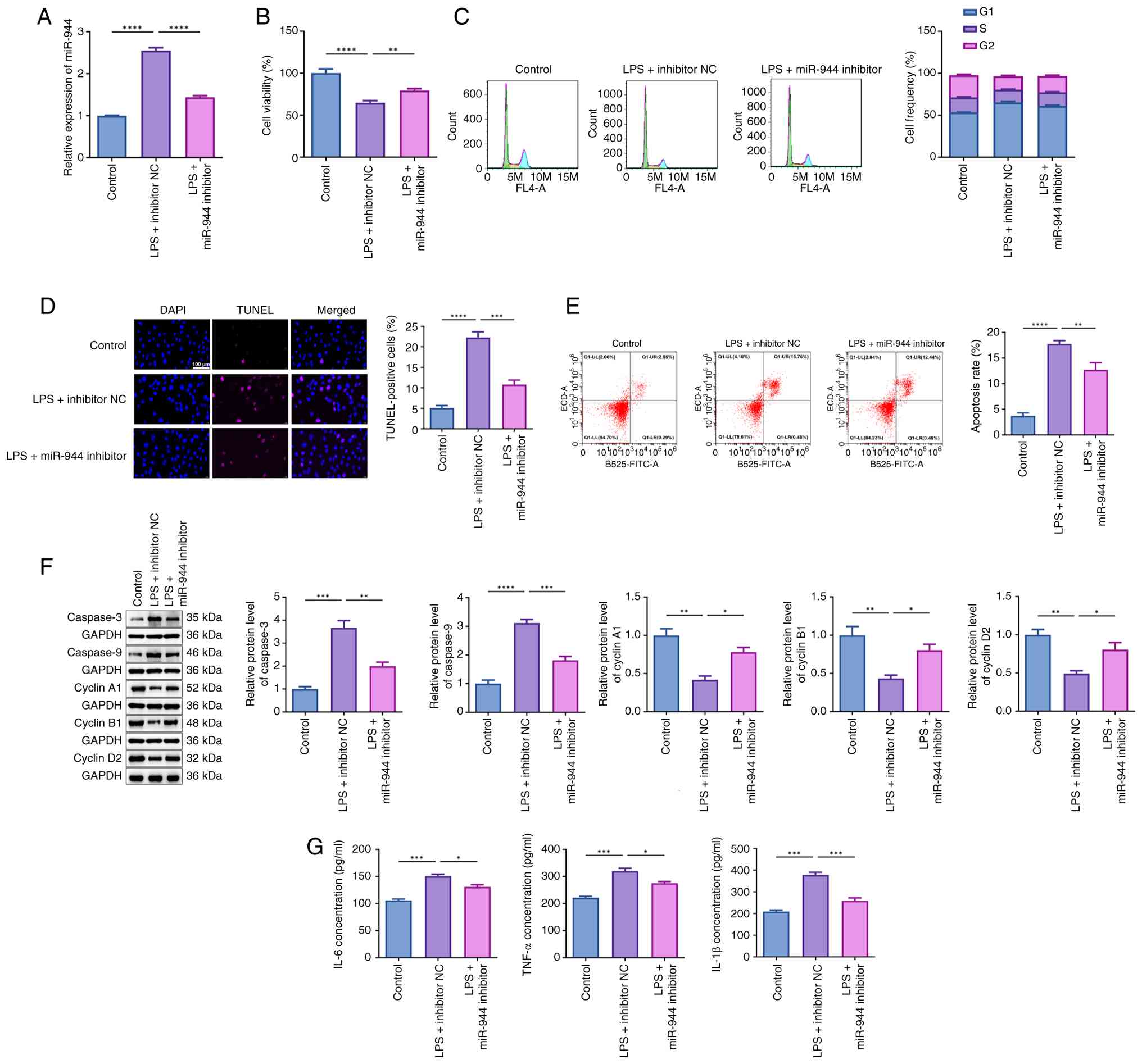 | Figure 3Effects of miR-944 inhibition on
LPS-stimulated chondrocytes. (A) Following transfection of miR-944
inhibitor or inhibitor NC in LPS-induced chondrocytes, the
expression of miR-944 was detected via reverse
transcription-quantitative PCR. (B) The viability of LPS-induced
chondrocytes that transfected with miR-944 inhibitor or inhibitor
NC was measured by Cell Counting Kit-8 assay. (C) Cell frequency of
LPS-induced chondrocytes was calculated via flow cytometric
analysis. (D) The percentages of TUNEL-positive cells were assessed
by TUNEL assay. Scale bar, 100 µm. (E) The apoptotic rate of
LPS-induced chondrocytes was calculated via flow cytometric
analysis. (F) The protein levels of Caspase-3, Caspase-9, Cyclin
A1, Cyclin B1 and Cyclin D2 in LPS-induced chondrocytes that
transfected with miR-944 inhibitor or inhibitor NC were determined
via western blotting. (G) The levels of IL-6, TNF-α and IL-1β in
LPS-induced chondrocytes that transfected with miR-944 inhibitor or
inhibitor NC were determined via ELISA. *P<0.05,
**P<0.01, ***P<0.001 and
****P<0.0001. miR, microRNA; LPS, lipopolysaccharide;
NC, negative control. |
PGAP1 is a direct downstream target of
miR-944
As the predicted results of Starbase software, a
potential binding site was identified between miR-944 and PGAP1
(Fig. 4A). Further experiments
indicated that by contrast to the PGAP1-Wt + mimics NC group, the
luciferase activity in PGAP1-Wt + miR-944 mimics group was
significantly reduced (Fig. 4B;
P<0.001), which validated the predicted results. Compared with
the normal patients, a relatively low level of PGAP1 was found in
synovial fluid of patients with OA (Fig. 4C; P<0.001). miR-944 inhibitor or
mimics (50, 100 and 200 nM) were then transfected into
chondrocytes. It was revealed that miR-944 expression was
significantly downregulated upon transfection with its inhibitor
(Fig. 4D; P<0.001), while it was
significantly upregulated after transfection with miR-944 mimics
(Fig. 4D; P<0.01). This
suggested that miR-944 inhibitor or mimics was successfully
transfected into chondrocytes. Subsequently, it was observed that
transfection of miR-944 inhibitor significantly promoted PGAP1
expression (Fig. 4E; P<0.001),
while miR-944 mimics significantly inhibited its expression
(Fig. 4E, P<0.001); both changes
occurred in a dose-dependent fashion. Further rescue experiments
demonstrated that transfection of miR-944 mimics reversed the
promoting effects of OE-PGAP1 transfection on PGAP1 expression
(Fig. 4F and G; P<0.05), whilst miR-944 inhibitor was
found to partially attenuate the inhibitory effects of si-PGAP1
transfection on PGAP1 levels (Fig.
4H and I; P<0.001).
Moreover, as shown in Fig. 4J and
K, it was found that silencing of
SNHG6 significantly inhibited PGAP1 expression (P<0.0001), while
the overexpressed SNHG6 promoted the expression levels of PGAP1
(P<0.01). Following further transfection of OE-SNHG6 or miR-944
mimics into LPS-stimulated chondrocytes, it was demonstrated that
the transfection of miR-944 mimics reversed the promoting effects
of OE-SNHG6 on PGAP1 expression (Fig.
4L; P<0.001).
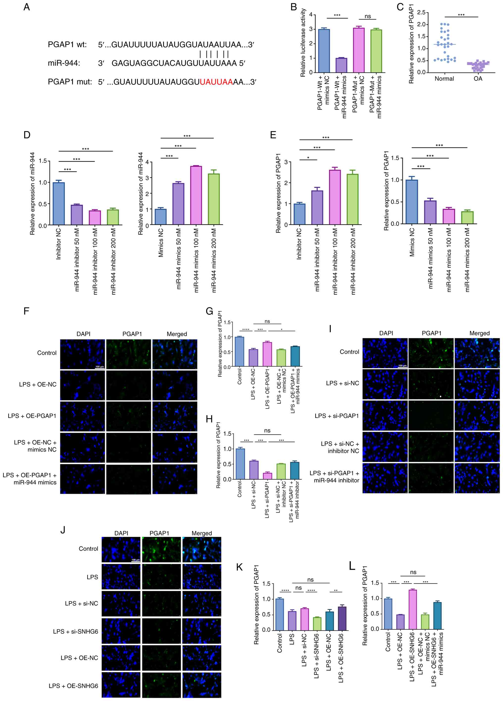 | Figure 4PGAP1 is a direct downstream target
of miR-944. (A) StarBase showed the predicted binding site of
miR-944 and PGAP1. (B) The target relationship between miR-944 and
PGAP1 was verified by dual luciferase reporter assay in
chondrocytes. (C) The expression of PGAP1 in synovial fluid of
normal individuals and patients with osteoarthritis was detected
via RT-qPCR. (D and E) Following transfection of miR-944 inhibitor
or miR-944 mimics, the expression of (D) miR-944 and (E) PGAP1 in
chondrocytes was detected by RT-qPCR. (F-I) Following transfection
of (F and G) OE-PGAP1/miR-944 mimics or (H and I) si-PGAP1/miR-944
inhibitor, the expression of PGAP1 in LPS-induced chondrocytes was
detected by immunofluorescence tests. Scale bar, 100 µm. (J and K)
Following transfection of si-SNHG6 or OE-SNHG6, the expression of
PGAP1 in LPS-induced chondrocytes was detected by
immunofluorescence tests. Scale bar, 100 µm. (L) Following
transfection of OE-SNHG6 or miR-944 mimics, the expression of PGAP1
in LPS-induced chondrocytes was detected by RT-qPCR. ns: no
significance. *P<0.05, **P<0.01,
***P<0.001 and ****P<0.0001. PGAP1,
post-GPI attachment to protein 1; miR, microRNA; RT-qPCR, reverse
transcription-quantitative PCR; si-, small interfering; OE-,
overexpression; LPS, lipopolysaccharide; NC, negative control; ns,
no significance. |
miR-944 interacts with PGAP1 to
modulate proliferation, apoptosis and inflammation in
LPS-stimulated chondrocytes
As revealed in Fig.
5A-E, overexpression of PGAP1 significantly stimulated
chondrocyte proliferation (P<0.0001), but suppressed apoptosis
(P<0.001). These results were further validated by western
blotting. It was demonstrated that the transfection of OE-PGAP1 in
LPS-stimulated chondrocytes significantly reduced Caspase-3 and
Caspase-9 levels (Fig. 5F;
P<0.01), while concurrently elevating the levels of Cyclin A1,
Cyclin B1 and Cyclin D2 (P<0.05). Notably, transfection of
miR-944 significantly reversed the regulatory effects of OE-PGAP1
transfection in LPS-stimulated chondrocytes, including
proliferation and apoptosis (Fig.
5A-F; P<0.05). Moreover, a significant reduction in
inflammation responses was observed in LPS-stimulated chondrocytes
when OE-PGAP1 was transfected (Fig.
5G; P<0.0001); however, this was partially restored upon the
transfection of miR-944 mimics (P<0.01).
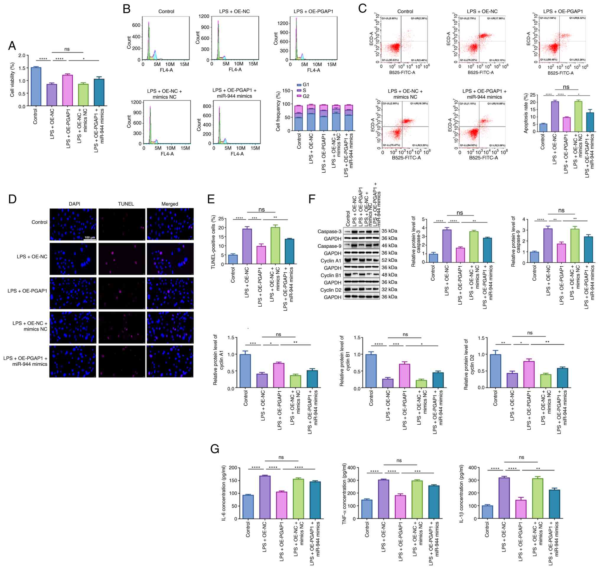 | Figure 5miR-944 interacts with PGAP1 to
modulate proliferation, apoptosis and inflammation in
LPS-stimulated chondrocytes. Following transfection of OE-PGAP1 or
miR-944 mimics, (A) the viability of LPS-induced chondrocytes was
measured by Cell Counting Kit-8 assay. (B) Cell frequency of
LPS-induced chondrocytes was calculated via flow cytometric
analysis. (C) The apoptotic rate of LPS-induced chondrocytes was
calculated via flow cytometric analysis. (D and E) The percentages
of TUNEL-positive cells were assessed by TUNEL assay. Scale bar,
100 µm. (F) The protein levels of Caspase-3, Caspase-9, Cyclin A1,
Cyclin B1 and Cyclin D2 in LPS-induced chondrocytes were determined
via western blotting. (G) The levels of IL-6, TNF-α and IL-1β in
LPS-induced chondrocytes were determined via ELISA.
*P<0.05, **P<0.01,
***P<0.001 and ****P<0.0001. miR,
microRNA; PGAP1, post-GPI attachment to protein 1; LPS,
lipopolysaccharide; OE-, overexpression; NC, negative control; ns,
no significance. |
Discussion
With the progressive intensification of global
population aging, the number of individuals affected by OA
continues to rise. Epidemiological data have confirmed that primary
OA affects ~46.3% of middle-aged and elderly individuals in China
(20). Globally, roughly 14% of
people aged 60 years and older are diagnosed with this condition,
with its incidence increasing alongside age (21). The economic impact of OA is
significant, resulting in annual losses exceeding $300 billion in
terms of income and healthcare expenses (21). In view of its high epidemiological
universality, OA has become a major factor contributing to reduced
quality of life and an increased strain on healthcare systems
(22). Although the exact
etiological origin and comprehensive pathogenic regulatory network
of OA remains unclear, recent studies increasingly highlight
inflammatory processes and chondrocyte apoptosis as critical
elements driving disease progression (23,24).
In this context, in-depth excavation and systematic clarification
of the key molecular signaling mechanisms driving the pathological
progression of OA are conducive to identifying reliable therapeutic
targets and further facilitating the targeted research and
development of novel efficient drugs for early intervention,
clinical remission and etiological blockade of OA. In the present
study, a novel molecular mechanism involved in OA progression was
identified, demonstrating that the lncRNA SNHG6/miR-944/PGAP1 axis
is linked to chondrocyte apoptosis and inflammation, thereby
mediating OA development.
Accumulating clinical and fundamental studies have
consistently documented aberrant epigenetic regulation of multiple
lncRNAs throughout the pathological progression of OA. Among these
dysregulated transcripts, the functional implications of canonical
lncRNA candidates, including HOTAIR, H19 and PVT1, in OA onset and
deteriorative progression have been systematically validated and
mechanistically elaborated in successive independent investigations
(10,25,26).
In addition to the aforementioned lncRNAs, the role of the SNHG
family of lncRNAs in the pathogenesis of OA has emerged as a
prominent topic in numerous studies. Multiple SNHG members have
been functionally annotated as critical upstream regulatory
molecules in the development of OA. Their mechanisms involve
various processes, including chondrocyte apoptosis (for example,
SNHG5) (27), inflammation
reactions (for example, SNHG1) (28), autophagy (for example, SNHG7)
(29), and the mediation of
extracellular matrix (ECM) degeneration (for example, SNHG12)
(30). These dysregulated SNHG
family members exhibit aberrant endogenous expression levels in the
synovial fluid of patients with OA, enabling differentiation
between OA patients and healthy individuals. However, as an
important member of SNHG family, the specific biological function
and potential molecular mechanism of SNHG6 in the continuous
progression of OA remain poorly elucidated and lack systematic
experimental validation. In the present study, it was observed that
compared with the normal controls, SNHG6 was significantly
downregulated in synovial fluid of patients with OA. This finding
suggested that the abnormal expression of SNHG6 may be linked to
the pathogenesis of OA. Accumulating evidence has demonstrated that
multiple members of the SNHG family of lncRNAs exert pivotal
regulatory effects on inflammatory responses and chondrocyte
apoptosis, two core pathological events that dominate the entire
progression process of OA (27,28).
On this basis, it was reasonably hypothesized that SNHG6 may also
be associated with inflammation and chondrocyte apoptosis.
Subsequently, a series of gain-of-function and loss-of-function
functional validation experiments were conducted in vitro to
systematically corroborate the aforementioned research hypothesis.
It was found that silencing of SNHG6 markedly promoted apoptosis
and inflammation. The overexpression of SNHG6 suppressed
chondrocyte apoptosis and mitigated inflammatory responses. In
addition to regulating chondrocyte apoptosis and inflammation, the
present study further revealed that SNHG6 modulated chondrocyte
proliferation via precise control of cell cycle progression, with
G1 phase arrest and dynamic changes in core cyclins (Cyclins A1, B1
and D2) showing a strong positive correlation with flow
cytometry-based cell cycle distribution data. G1 phase arrest is a
classic cytological hallmark of impaired chondrocyte proliferation
in OA, which impairs chondrocyte renewal and repair, and ultimately
accelerates articular cartilage matrix degradation (31). Flow cytometric analysis demonstrated
that LPS stimulation induced marked G1 phase accumulation of
chondrocytes and a concomitant reduction in S and G2/M phase
populations, indicative of typical G1 phase arrest in OA-like
chondrocytes. SNHG6 silencing further exacerbated this G1 phase
block, while SNHG6 overexpression effectively reversed LPS-induced
G1 phase arrest and restored normal cell cycle distribution. These
phenotypic changes were tightly aligned with western blot data for
Cyclins A1, B1 and D2; core cell cycle regulators with
non-redundant roles in driving cell cycle progression. Cyclin D2
initiates G1 phase progression and is essential for G1/S transition
via activation of CDK4/6(32);
Cyclin A1 regulates both G1/S and S/G2 transitions to maintain DNA
replication and S phase completion (33); Cyclin B1 is the master regulator of
G2/M transition, and its upregulation is a prerequisite for mitotic
entry (34). In the present study,
LPS stimulation significantly downregulated Cyclin A1, B1 and D2
expression in chondrocytes, and SNHG6 silencing further suppressed
their levels; conversely, SNHG6 overexpression significantly
upregulated these cyclins. This inverse correlation between cyclin
expression and G1 phase cell proportion is a direct reflection of
molecular cell cycle regulation: Reduced cyclin expression impairs
key cell cycle transitions (G1/S, S/G2 and G2/M), leading to G1
phase arrest and inhibited proliferation, while restored cyclin
expression relieves these molecular blocks, reduces G1 phase
accumulation, and rescues chondrocyte proliferative capacity. These
results confirmed that the SNHG6 modulated chondrocyte cell cycle
progression by fine-tuning expression of Cyclins A1, B1 and D2.
This finding deepens our mechanistic understanding of
SNHG6-mediated regulation of OA progression, suggesting that SNHG6
may act as a pathogenic factor in the progression of OA.
Notably, emerging studies have validated that SNHG
family members serve as critical miRNA sponges to mediate in the
pathogenesis of OA, such as SNHG1-miR-16-5p (35), SNHG9-miR-34a (36) and SNHG14-miR-214-3p (37). In line with these findings, it was
further hypothesized that SNHG6 could also function as a miRNA
sponge to participate in the progression of OA. Starbase online
software was utilized to screen and predict the potential
downstream target miRNAs of SNHG6, and the analytical results
identified miR-944 as a candidate downstream binding partner of
SNHG6. Meanwhile, SNHG6 was confirmed to negatively modulate the
expression levels of miR-944 in LPS-induced chondrocytes. Notably,
clinical and cellular phenotypic detection also revealed
significantly elevated expression levels of miR-944 in synovial
samples from patients with OA and LPS-induced chondrocytes.
Functional rescue experiments further verified that the inhibition
of miR-944 effectively attenuates chondrocyte apoptotic damage and
overactivates inflammatory responses. Collectively, it was
considered that SNHG6 can negatively regulate miR-944 expression to
inhibit chondrocyte apoptosis and mitigate inflammatory responses,
thereby ameliorating the progression of OA.
In fact, extensive crosstalk among lncRNAs, miRNAs
and mRNAs has been well documented throughout the pathological
progression of OA (38).
Mechanistically, lncRNA can bind to miRNA, reduce the inhibitory
effect of miRNA on mRNA, participate in regulating the progress of
inflammation, chondrocyte apoptosis and proliferation. Of note,
accumulating evidence confirms that multiple members of SNHG family
also exert this regulatory effect in the development of OA, such as
SNHG5-miR-26a-SOX2(39),
SNHG7-miR-34a-5p-SYVN1(29) and
SNHG15-miR-141-3p-BCL2L13(40). In
the present study, PGAP1 was identified as a potential target mRNA
of miR-944. PGAP1 encodes a functional protein consisting of 922
amino acid residues, which exerts the biological enzymatic activity
of glycosylphosphatidylinositol inositol-deacylase (41). It is located on the membrane of the
endoplasmic reticulum (41). In
previous studies, PGAP1 has been reported to be involved in
NEAT1-mediated miR-374b-5p network to regulate the development of
OA (42). It was therefore
hypothesized that there may be some interactions between the
SNHG6/miR-944 axis and PGAP1. In the present study, it was observed
that overexpression of SNHG6 significantly promoted PGAP1
expression, while transfection of miR-944 mimics exhibited the
opposite regulatory effect. Meanwhile, elevated miR-944 expression
was capable of abrogating the inhibitory effects of PGAP1
overexpression on chondrocyte apoptosis and inflammation.
Collectively, these findings confirmed the authors' hypothesis that
the SNHG6-miR-944-PGAP1 axis may be a novel regulatory cascade that
participates in the pathogenesis of OA.
Beyond PGAP1, miR-944 is predicted to target a
repertoire of additional functional genes and may potentially
synergize with SNHG1/5/7-mediated regulatory networks to
orchestrate chondrocyte homeostasis in physiological and
pathological conditions of OA. As a typical miRNA with pleiotropic
regulatory functions, miR-944 is not restricted to a single target
gene in its biological regulatory network; bioinformatic analysis
based on databases including Starbase, miRDB and TargetScan has
identified multiple putative target genes of miR-944 that are
implicated in chondrocyte ECM metabolism, proliferation and
inflammatory response, such as genes encoding key regulators of the
NF-κB and MAPK signaling pathways that are central to OA
progression. This multi-target regulatory characteristic is
consistent with the universal functional features of miRNAs, which
enables a single miRNA to fine-tune multiple biological processes
simultaneously. Notably, apart from SNHG6, SNHG1, SNHG5 and SNHG7,
as key members of the SNHG lncRNA family, have been well-documented
to mediate chondrocyte homeostasis in OA through distinct ceRNA
regulatory networks: SNHG1 modulates chondrocyte apoptosis and
inflammation via the miR-195/IKK-α axis (28), SNHG5 regulates chondrocyte
proliferation and apoptosis through the miR-10a-5p/H3F3B axis
(27), and SNHG7 orchestrates
chondrocyte proliferation, apoptosis and autophagy via the
miR-34a-5p/SYVN1 axis (29). These
SNHG-mediated networks independently regulate chondrocyte
biological behaviors, and miR-944 may serve as a novel molecular
node that intersects with these networks, forming synergistic
regulatory crosstalk to collectively maintain the balance of
chondrocyte survival, proliferation and inflammatory response under
physiological conditions. This synergistic effect also highlights
the complexity of the molecular regulatory network in OA.
Therefore, it is considered that individual lncRNA/miRNA/mRNA axis
does not act in isolation but are intertwined to form a
multi-layered regulatory network governing chondrocyte homeostasis.
Given the novelty of the SNHG6/miR-944/PGAP1 axis in OA reported in
the present study, the current research focuses on the regulatory
relationship between miR-944 and its validated target PGAP1. The
identification of additional target genes of miR-944 as well as the
in-depth exploration of the synergistic regulatory mechanisms
between miR-944 and SNHG1/5/7-mediated networks remain to be
elucidated in future studies. Such investigations will help to
further unravel the intricate molecular regulatory network
underlying chondrocyte dysfunction in OA and provide more
comprehensive molecular targets for the development of
disease-modifying therapies for OA.
Additionally, it is imperative to acknowledge that
the present study is subject to several other limitations. First,
the small sample size may limit the generalizability of the present
clinical findings, and larger cohorts are needed in future studies
to validate these results. Second, a single cell model cannot
capture the complex pathological microenvironment associated with
OA, including the interactions between the degradation of the
chondrocyte ECM, synovial hyperplasia, and subchondral bone
remodeling. Classic IL-1β-induced, TNF-α-induced and IL-1β + TNF-α
co-induced chondrocyte models should be established to confirm
whether SNHG6/miR-944/PGAP1 axis has a universal regulatory effect
on chondrocyte damage induced by endogenous pro-inflammatory
cytokines. Third, in vitro cell line assays inherently fall
short of recapitulating complex clinical scenarios. The current
study was restricted to preliminary cellular-level analyses,
lacking verification of biological fidelity, simulation of the
in vivo microenvironment, and assessment of translational
potential. Further exploration is also warranted to elucidate the
functional roles of these targets in animal model systems.
In conclusion, the present study constitutes a novel
and preliminary exploratory research focusing on OA pathological
mechanisms. It systematically elucidates the biological function
and intrinsic molecular regulatory mechanism of SNHG6 during OA
progression. Functional experiments validated that SNHG6 could
competitively bind to regulate the miR-944/PGAP1 signaling cascade,
thereby repressing excessive chondrocyte apoptosis and abnormal
inflammatory activation in OA. Taken together, the aforementioned
findings to some extent deepen the mechanistic understanding of the
functional role of SNHG6 in OA pathogenesis. Meanwhile, the present
study provides reliable experimental basis and novel targeted
theoretical references for the subsequent development of molecular
targeted intervention strategies and precise clinical adjuvant
treatment of OA.
Acknowledgements
Not applicable.
Funding
Funding: The present study was supported by Basic Public Welfare
Research Project of Wenzhou Science and Technology Bureau (grant
no. Y20240311).
Availability of data and materials
The data generated in the present study may be
requested from the corresponding author.
Authors' contributions
FH made substantial contributions to the conception
and design of the study. FH, ZS, JY and XZ made substantial
contributions to the acquisition, analysis and interpretation of
the data. FH drafted the manuscript. All authors critically revised
the manuscript for intellectual content and confirm the
authenticity of all the raw data. All authors read and approved the
final version of the manuscript.
Ethics approval and consent to
participate
All the individuals had been informed the study
details and provided their written consent in advance. Ethical
approval was obtained from the Ethics Committee of Wenzhou People's
Hospital (approval No. KY-2023-251; Wenzhou, China) and this
research adheres to the principles outlined in the Declaration of
Helsinki.
Patient consent for publication
Not applicable.
Competing interests
The authors declare that they have no competing
interests.
References
|
1
|
Yao Q, Wu X, Tao C, Gong W, Chen M, Qu M,
Zhong Y, He T, Chen S and Xiao G: Osteoarthritis: pathogenic
signaling pathways and therapeutic targets. Signal Transduct Target
Ther. 8(56)2023.PubMed/NCBI View Article : Google Scholar
|
|
2
|
GBD 2021 Osteoarthritis Collaborators.
Global, regional, and national burden of osteoarthritis, 1990-2020
and projections to 2050: A systematic analysis for the Global
burden of disease study 2021. Lancet Rheumatol. 5:e508–e522.
2023.PubMed/NCBI View Article : Google Scholar
|
|
3
|
Xiao SQ, Cheng M, Wang L, Cao J, Fang L,
Zhou XP, He XJ and Hu YF: The role of apoptosis in the pathogenesis
of osteoarthritis. Int Orthop. 47:1895–1919. 2023.PubMed/NCBI View Article : Google Scholar
|
|
4
|
Sanchez-Lopez E, Coras R, Torres A, Lane
NE and Guma M: Synovial inflammation in osteoarthritis progression.
Nat Rev Rheumatol. 18:258–275. 2022.PubMed/NCBI View Article : Google Scholar
|
|
5
|
Oo WM and Hunter DJ: Repurposed and
investigational disease-modifying drugs in osteoarthritis (DMOADs).
Ther Adv Musculoskelet Dis. 14(1759720X221090297)2022.PubMed/NCBI View Article : Google Scholar
|
|
6
|
Oo WM and Hunter J: Intra-articular
Therapies for Knee Osteoarthritis: Current Update. Curr Treat
Options Rheum. 9:99–119. 2023.
|
|
7
|
Winkle M, El-Daly SM, Fabbri M and Calin
GA: Noncoding RNA therapeutics - challenges and potential
solutions. Nat Rev Drug Discov. 20:629–651. 2021.PubMed/NCBI View Article : Google Scholar
|
|
8
|
Mattick JS, Amaral PP, Carninci P,
Carpenter S, Chang HY, Chen LL, Chen R, Dean C, Dinger ME,
Fitzgerald KA, et al: Long non-coding RNAs: Definitions, functions,
challenges and recommendations. Nat Rev Mol Cell Biol. 24:430–447.
2023.PubMed/NCBI View Article : Google Scholar
|
|
9
|
Shi M, Sun M, Wang C, Shen Y, Wang Y and
Yan S: Therapeutic potential of POU3F3, a novel long non-coding
RNA, alleviates the pathogenesis of osteoarthritis by regulating
the miR-29a- 3p/FOXO3 axis. Curr Gene Ther. 22:427–438.
2022.PubMed/NCBI View Article : Google Scholar
|
|
10
|
Chen X, Liu J, Sun Y, Wen J, Zhou Q, Ding
X and Zhang X: Correlation analysis of differentially expressed
long non-coding RNA HOTAIR with PTEN/PI3K/AKT pathway and
inflammation in patients with osteoarthritis and the effect of
baicalin intervention. J Orthop Surg Res. 18(34)2023.PubMed/NCBI View Article : Google Scholar
|
|
11
|
Ni C, Zhang W, Qiu S, Cheng H and Ma C:
Long non-coding RNA DLEU1 promotes progression of osteoarthritis
via miR-492/TLR8 Axis. Curr Pharm Biotechnol. 25:2166–2181.
2024.PubMed/NCBI View Article : Google Scholar
|
|
12
|
Zhang S, Qiu D, Xie X and Shen Y:
Clinicopathological and prognostic value of SNHG6 in cancers: A
systematic review and a meta-analysis. BMC Cancer.
20(343)2020.PubMed/NCBI View Article : Google Scholar
|
|
13
|
Jurkiewicz M, Szczepaniak A and Zielinska
M: Long non-coding RNAs - SNHG6 emerge as potential marker in
colorectal cancer. Biochim Biophys Acta Rev Cancer.
1879(189056)2024.PubMed/NCBI View Article : Google Scholar
|
|
14
|
Wang J, Si J, Zhao Z, Gao C, Liu T, Jia Y
and Liu L: SNHG6 facilitates the epithelial-mesenchymal transition
and metastatic potential of esophageal squamous carcinoma through
miR-26b-5p/ITGB1 axis. Sci Rep. 14(25005)2024.PubMed/NCBI View Article : Google Scholar
|
|
15
|
Su M, Huang P and Li Q: Long noncoding RNA
SNHG6 promotes the malignant phenotypes of ovarian cancer cells via
miR-543/YAP1 pathway. Heliyon. 9(e16291)2023.PubMed/NCBI View Article : Google Scholar
|
|
16
|
Zhang Y, Qiao Z, He M and Xia J: OGT
promotes the ferroptosis of chondrocytes in osteoarthritis
development via O-GlcNAcylation modification of ACSF2. Immunol Res.
73(7)2024.PubMed/NCBI View Article : Google Scholar
|
|
17
|
Wang J, Li J, Hong Z and Deng H:
Circ_0004662 promotes lipopolysaccharide-induced osteoarthritis by
targeting the miR-450b-5p/TCF4 axis. Clin Rheumatol. 45:1865–1876.
2026.PubMed/NCBI View Article : Google Scholar
|
|
18
|
Zhang H, Xiao J, Yi Y, Chen B, Lv W, Ma J,
Lin Y, Wu Y, Meng K, Cai Y, et al: Homoplantaginin ameliorates
osteoarthritis by activating Sirt3/PINK1/Parkin signaling to
promote mitophagy and attenuate inflammation in chondrocytes.
Phytomedicine. 153(157959)2026.PubMed/NCBI View Article : Google Scholar
|
|
19
|
Livak KJ and Schmittgen TD: Analysis of
relative gene expression data using real-time quantitative PCR and
the 2(-Delta Delta C(T)) Method. Methods. 25:402–408.
2001.PubMed/NCBI View Article : Google Scholar
|
|
20
|
Li Y, Xie W, Xiao W and Dou D: Progress in
osteoarthritis research by the National natural science foundation
of China. Bone Res. 10(41)2022.PubMed/NCBI View Article : Google Scholar
|
|
21
|
Kang Y, Liu C, Ji Y, Zhang H, Wang Y, Bi
W, Xu J and Guo B: The burden of knee osteoarthritis worldwide,
regionally, and nationally from 1990 to. 2019, along with an
analysis of cross-national inequalities. Arch Orthop Trauma Surg.
144:2731–2743. 2024.PubMed/NCBI View Article : Google Scholar
|
|
22
|
Hawker GA and King LK: The burden of
osteoarthritis in older adults. Clin Geriatr Med. 38:181–192.
2022.PubMed/NCBI View Article : Google Scholar
|
|
23
|
van den Bosch MHJ, Blom AB and van der
Kraan PM: Inflammation in osteoarthritis: Our view on its presence
and involvement in disease development over the years.
Osteoarthritis Cartilage. 32:355–364. 2024.PubMed/NCBI View Article : Google Scholar
|
|
24
|
Hung K and Cai HL: The role of chondrocyte
senescence in osteoarthritis pathogenesis and therapeutic
implications. Exp Gerontol. 208(112828)2025.PubMed/NCBI View Article : Google Scholar
|
|
25
|
Zhou Y, Li J, Xu F, Ji E, Wang C and Pan
Z: Long noncoding RNA H19 alleviates inflammation in osteoarthritis
through interactions between TP53, IL-38, and IL-36 receptor. Bone
Joint Res. 11:594–607. 2022.PubMed/NCBI View Article : Google Scholar
|
|
26
|
Yao N, Peng S, Wu H, Liu W, Cai D and
Huang D: Long noncoding RNA PVT1 promotes chondrocyte extracellular
matrix degradation by acting as a sponge for miR-140 in
IL-1β-stimulated chondrocytes. J Orthop Surg Res.
17(218)2022.PubMed/NCBI View Article : Google Scholar
|
|
27
|
Jiang H, Pang H, Wu P, Cao Z, Li Z and
Yang X: LncRNA SNHG5 promotes chondrocyte proliferation and
inhibits apoptosis in osteoarthritis by regulating miR-10a-5p/H3F3B
axis. Connect Tissue Res. 62:605–614. 2021.PubMed/NCBI View Article : Google Scholar
|
|
28
|
Wang Q, Deng F, Li J, Guo L and Li K: The
long non-coding RNA SNHG1 attenuates chondrocyte apoptosis and
inflammation via the miR-195/IKK-α axis. Cell Tissue Bank.
24:167–180. 2023.PubMed/NCBI View Article : Google Scholar
|
|
29
|
Tian F, Wang J, Zhang Z and Yang J: LncRNA
SNHG7/miR-34a-5p/SYVN1 axis plays a vital role in proliferation,
apoptosis and autophagy in osteoarthritis. Biol Res.
53(9)2020.PubMed/NCBI View Article : Google Scholar
|
|
30
|
Yang X, Chen H, Zheng H, Chen K, Cai P, Li
L, Li K, Du Y and He XC: LncRNA SNHG12 promotes osteoarthritis
progression through targeted down-regulation of miR-16-5p. Clin
Lab. 68:2022.PubMed/NCBI View Article : Google Scholar
|
|
31
|
Tan D, Guo J, Wang T, Yang S, Liu J, Wang
D and Li W: Chondrocyte senescence in osteoarthritis: Potential
targets and pharmacological interventions. Cartilage.
(19476035251414314)2026.PubMed/NCBI View Article : Google Scholar
|
|
32
|
Zhao J, Zhang Z, Zuo T, Yu J, Yang S, Yang
Y, Li X and Zheng J: Downregulation of miR-892b inhibits the
progression of osteoarthritis via targeting cyclin D1 and cyclin
D2. Exp Cell Res. 405(112683)2021.PubMed/NCBI View Article : Google Scholar
|
|
33
|
Radonova L, Pauerova T, Jansova D,
Danadova J, Skultety M, Kubelka M and Anger M: Cyclin A1 in oocytes
prevents chromosome segregation and anaphase entry. Sci Rep.
10(7455)2020.PubMed/NCBI View Article : Google Scholar
|
|
34
|
Li H, Qiao Y, Ding M and Liu Y: Targeting
CCNB1 repressed cartilage degradation induced by inflammation
through NF-ĸB pathway in osteoarthritis. J Histochem Cytochem.
74:235–248. 2026.PubMed/NCBI View Article : Google Scholar
|
|
35
|
Lei J, Fu Y, Zhuang Y, Zhang K and Lu D:
LncRNA SNHG1 alleviates IL-1β-induced osteoarthritis by inhibiting
miR-16-5p-mediated p38 MAPK and NF-ĸB signaling pathways. Biosci
Rep. 39(BSR20191523)2019.PubMed/NCBI View Article : Google Scholar
|
|
36
|
Zhang H, Li J, Shao W and Shen N: LncRNA
SNHG9 is downregulated in osteoarthritis and inhibits chondrocyte
apoptosis by downregulating miR-34a through methylation. BMC
Musculoskelet Disord. 21(511)2020.PubMed/NCBI View Article : Google Scholar
|
|
37
|
Wang B, Li J and Tian F: Downregulation of
lncRNA SNHG14 attenuates osteoarthritis by inhibiting FSTL-1
mediated NLRP3 and TLR4/NF-ĸB pathway through miR-124-3p. Life Sci.
270(119143)2021.PubMed/NCBI View Article : Google Scholar
|
|
38
|
Kong H, Sun ML, Zhang XA and Wang XQ:
Crosstalk among circRNA/lncRNA, miRNA, and mRNA in osteoarthritis.
Front Cell Dev Biol. 9(774370)2021.PubMed/NCBI View Article : Google Scholar
|
|
39
|
Shen H, Wang Y, Shi W, Sun G, Hong L and
Zhang Y: LncRNA SNHG5/miR-26a/SOX2 signal axis enhances
proliferation of chondrocyte in osteoarthritis. Acta Biochim
Biophys Sin (Shanghai). 50:191–198. 2018.PubMed/NCBI View Article : Google Scholar
|
|
40
|
Zhang X, Huang CR, Pan S, Pang Y, Chen YS,
Zha GC, Guo KJ and Zheng X: Long non-coding RNA SNHG15 is a
competing endogenous RNA of miR-141-3p that prevents osteoarthritis
progression by upregulating BCL2L13 expression. Int
Immunopharmacol. 83(106425)2020.PubMed/NCBI View Article : Google Scholar
|
|
41
|
Williams C, Jiang YH, Shashi V, Crimian R,
Schoch K, Harper A, McHale D, Goldstein D and Petrovski S:
Additional evidence that PGAP1 loss of function causes autosomal
recessive global developmental delay and encephalopathy. Clin
Genet. 88:597–599. 2015.PubMed/NCBI View Article : Google Scholar
|
|
42
|
Huang F, Su Z, Yang J, Zhao X and Xu Y:
Downregulation of lncRNA NEAT1 interacts with miR-374b-5p/PGAP1
axis to aggravate the development of osteoarthritis. J Orthop Surg
Res. 18(670)2023.PubMed/NCBI View Article : Google Scholar
|