Introduction
Immune-mediated necrotizing myopathy (IMNM) is a
distinct subset of idiopathic inflammatory myopathy, characterized
by proximal muscle weakness, markedly elevated serum creatine
kinase levels and histopathological features of muscle fiber
necrosis with minimal inflammatory infiltrate (1-3).
IMNM is classified into three major subgroups: Anti-signal
recognition particle (anti-SRP) antibody-positive,
anti-3-hydroxy-3-methylglutaryl-coenzyme A reductase (anti-HMGCR)
antibody-positive and seronegative IMNM (3). Among these, anti-SRP antibody-positive
IMNM is associated with severe myonecrosis and often requires
aggressive immunosuppressive therapy (2,3).
Rhabdomyolysis (RM) syndrome is a potentially fatal complication of
severe skeletal muscle injury, most commonly triggered by trauma,
exertion, drugs or infection, but increasingly recognized as a rare
yet clinically significant consequence of immune-mediated
myopathies (4,5). When RM complicates IMNM, the resulting
extensive myonecrosis portends a more complicated clinical course
with a higher risk of systemic complications such as acute kidney
injury (AKI) (6). Furthermore,
cytomegalovirus (CMV), a ubiquitous herpesvirus that may reactivate
under immunosuppression, can occasionally invade the central
nervous system (CNS) (7). The
coexistence of IMNM (which mandates aggressive immunosuppression)
and CNS CMV infection therefore creates a formidable therapeutic
dilemma, as these two conditions demand seemingly contradictory
treatment strategies. To the best of our knowledge, the
simultaneous occurrence of anti-SRP antibody-positive IMNM, severe
RM and CNS CMV infection in a single patient has rarely been
reported. A recently published case similarly described a temporal
association between anti-SRP IMNM and recent CMV infection,
highlighting the potential interplay between viral triggers and
autoimmunity (8). However, the
scenario in which all three conditions coexist, and particularly
the diagnostic challenge it poses in differentiating from
Guillain-Barré syndrome (GBS), remains poorly characterized. The
present case report therefore aims to describe the clinical
presentation, diagnostic workup and multidisciplinary management of
a 19-year-old female patient with anti-SRP antibody-positive IMNM
complicated by RM and CNS CMV infection, with the hope of raising
clinical awareness and informing timely diagnosis and treatment of
this rare and complex combination.
Case report
First admission (February-March 2024).
Patient presentation
A 19-year-old female patient was admitted to the
Affiliated Traditional Chinese Medicine Hospital, Southwest Medical
University (Luzhou, China) in February 2024 due to an abnormal
muscle enzyme profile, including increased serum creatine kinase
(CK) levels of up to 31,180 U/l (reference value, 40-200 U/l),
detected during a routine physical examination 20 days earlier.
Initial treatment at a local hospital (Yibin, China) had resulted
in no notable improvement. At 1 week before admission to the
Affiliated Traditional Chinese Medicine Hospital, Southwest Medical
University, the patient developed progressive muscle weakness and
myalgia, which eventually progressed to an inability to
ambulate.
The patient had no notable past medical history,
with no history of statin use or autoimmune disease before the
onset of the current symptoms. The patient had no notable personal
history, had received no recent vaccinations and had not suffered
any viral illnesses prior to symptom onset. There was no family
history of hereditary muscle diseases or neuromuscular
disorders.
Upon admission, vital signs were stable, and
cardiopulmonary and abdominal examinations were unremarkable.
Notably, respiratory and swallowing functions were intact, with no
dyspnea, orthopnea or dysphagia. Muscle tone in all four limbs was
normal. Muscle strength in the distal limbs was graded as 1/5 using
the Medical Research Council scale (9), while muscle strength in the proximal
limbs was nearly 0/5.
Key laboratory findings during hospitalization are
summarized in Tables I, II and III. Notably, all time points mentioned
in this report (e.g., ‘day X’) are calculated with February 2024
(the date of first hospital admission) defined as day 0. This
chronological framework remains consistent throughout the patient's
hospital course, including the intervening discharge and second
admission, to provide a unified clinical timeline. On Day 0
(admission), routine examinations, including routine blood test,
myocardial markers, kidney function, electrolytes, blood fat,
myozymogram, routine urinalysis and thyroid function, were
performed. Results revealed an elevated white blood cell count
(13.12x10⁹/l; reference range,
3.5-9.5x10⁹/l), markedly elevated CK-MB mass (>287.00
ng/ml; reference range, 0.6-6.3 ng/ml) and myoglobin (Myo;
>3,958.0 ng/ml; reference value, ≤65.8 ng/ml), as well as
elevated aspartate aminotransferase (AST, 930 U/l; reference range,
13-35 U/l), alanine aminotransferase (ALT, 590 U/l; reference
range, 7-40 U/l), lactate dehydrogenase (LDH, 2,267 U/l; reference
range, 120-250 U/l), CK-MB (817 U/l; reference range, 0-25 U/l) and
α-hydroxybutyrate dehydrogenase (1,898 U/l; reference range, 72-182
U/l), and a profound increase in CK (32,213 U/l; reference range,
40-200 U/l). Regarding RM-related complications, the following were
documented: Serum Myo was markedly elevated (>3,958.0 ng/ml;
reference value, ≤65.8 ng/ml), but routine urinalysis showed
negative results for blood and protein, indicating no gross
myoglobinuria at the time of testing, which was likely attributable
to early aggressive fluid resuscitation. Electrolyte abnormalities
included hyperphosphatemia (inorganic phosphate, 1.63 mmol/l;
reference range, 0.85-1.51 mmol/l) and mild hyponatremia (sodium,
135 mmol/l; reference range, 137-147 mmol/l). The serum creatinine
level was 37 µmol/l (reference, 41-73 µmol/l), which was below
normal, but this remained stable throughout hospitalization,
indicating no AKI and preserved renal function. The patient did not
develop compartment syndrome. As AKI did not occur, conventional
renal replacement therapy was not required, although plasma
exchange was performed for immunomodulation. On day 24,
autoantibody screening was conducted, showing positive (+++)
results for both anti-SRP and anti-SSA/Ro-52 antibodies (Table II). On day 26, serum virology
testing detected positive CMV-IgM and CMV-IgG. On day 29,
cerebrospinal fluid (CSF) analysis was performed (Table III).
 | Table IKey laboratory findings during
hospitalization. |
Table I
Key laboratory findings during
hospitalization.
| Laboratory
examinations | Results | Normal values |
|---|
| Routine blood
test | | |
|
WBCs
(x109/l) | 13.12 | 3.5-9.5 |
|
RBCs
(x1012/l) | 5.37 | 3.8-5.1 |
|
HGB,
g/l | 157 | 115-110 |
|
GR, % | 83.6 | 40-75 |
| Myocardial
marker | | |
|
CKMB-mass,
ng/ml | >287.00 | 0.6-6.3 |
|
Myo,
ng/ml | >3,958.0 | ≤65.8 |
|
hs-cTnT,
ng/ml | 0.084 | 0.000-0.0175 |
| Kidney function | | |
|
SCr,
µmol/l | 37 | 41-73 |
| Electrolytic | | |
|
Na,
mmol/l | 135 | 137-147 |
|
Cl,
mmol/l | 100 | 99-110 |
|
IP,
mmol/l | 1.63 | 0.85-1.51 |
| Blood fat | | |
|
TG,
mmol/l | 4.07 | <2.26 |
|
CHOL,
mmol/l | 6.36 | ≤5.2 |
|
HDL-C,
mmol/l | 1.12 | >1.04 |
|
LDL-C,
mmol/l | 4.08 | <3.34 |
| Myozymogram | | |
|
AST,
U/l | 930 | 13-35 |
|
ALT,
U/l | 590 | 7-40 |
|
LDH,
U/l | 2,267 | 120-250 |
|
CK, U/l | 32,213 | 40-200 |
|
CK-MB,
U/l | 817 | 0-25 |
|
α-HBDH,
U/l | 1,898 | 72-182 |
| Thyroid
function | | |
|
TSH,
µIU/ml | 5.910 | 0.25-4.20 |
|
FT3,
pmo1/l | 1.11 | 3.1-6.8 |
|
FT4,
pmo1/l | 24.40 | 12-22 |
| Routine
urinalysis | | |
|
BLD | Negative | Negative |
|
LEU | Negative | Negative |
|
PRO | Negative | Negative |
| Virus | | |
|
CMV
antibody-IgM, AU/ml | 213.30 | <8 |
|
CMV
antibody-IgG, AU/ml | >1,000.00 | <10 |
| Table IIExamination of myositis spectrum and
autoantibody spectrum of patients. |
Table II
Examination of myositis spectrum and
autoantibody spectrum of patients.
| Autoantibodies | Result | Normal result |
|---|
| Anti-JO-1
antibody | Negative | Negative |
| Anti-PL-7
antibody | Negative | Negative |
| Anti-PL-12
antibody | Negative | Negative |
| Anti-EJ
antibody | Negative | Negative |
| Anti-SRP
antibody | Positive (+++) | Negative |
| Anti-Mi-2
antibody | Negative | Negative |
| Anti-MDA5
antibody | Negative | Negative |
| Anti-TIF1γ
antibody | Negative | Negative |
| Anti-HMGCR
antibody | Negative | Negative |
| Anti-SSA/Ro-52
antibody | Positive (+++) | Negative |
| Anti-SAE1/2
antibody | Negative | Negative |
| Anti-NXP2
antibody | Negative | Negative |
| Table IIIPatient obtained CSF examination
results through lumbar puncture. |
Table III
Patient obtained CSF examination
results through lumbar puncture.
| Inspection
item | Result | Normal result |
|---|
| Germ | Negative | Negative |
| Fungus | Negative | Negative |
| Parasite | Negative | Negative |
| Virus | Cytomegalovirus
(mNGS; relative abundance 100%) | Negative |
| CSF albumin,
mg/l | 328 | 100-300 |
| CSF total protein,
mg/l | 644 | 150-450 |
| CSF pressure,
mmH2O | Not measured
(natural drip rate ~60 drops/min) | 80-180 |
| WBC
(x106/l) | 1 | 0-8 |
| HB
(x106/l) | 0 | / |
| Glucose,
mmol/l | 3.79 | 2.22-3.89 |
| Cl, mmol/l | 127 | 120-132 |
| MN, % | / | / |
| PMN, % | / | / |
| Pandy test | Extremely weak
positive (±) | Negative |
| Acid-fast stain
(acid-fast bacilli smear) | Negative (no AFB
observed) | Negative |
Histopathological examination. A right thigh
muscle biopsy was performed with the following histopathological
findings: The muscle fiber diameter ranged from 5 to 55 µm, with
marked variability in fiber size. Muscle fibers of varying
diameters were intermixed, and scattered degenerating, necrotic and
regenerating muscle fibers were observed. Internalized nuclei were
present. There was no definite lymphocytic aggregation. Mild
connective tissue hyperplasia was observed in some muscle
fascicles, along with moderate adipose tissue hyperplasia between
some muscle fascicles. Tissues were fixed in 4% paraformaldehyde
(for 24 h at 37˚) at a section thickness of 5-10 µm, and then
stained with hematoxylin (for 10 min at 37˚) and eosin (for 3 min
at 37˚). Images were acquired under a light microscope at x4 and
x20 magnification (Fig. 1).
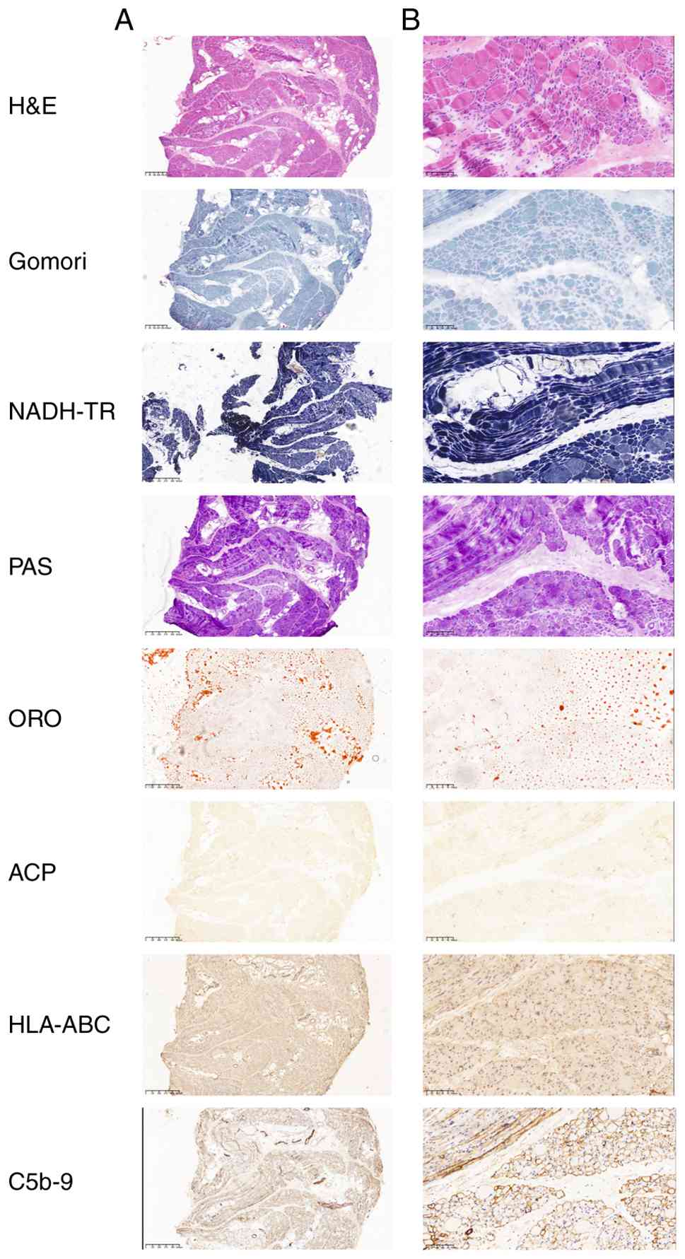 | Figure 1Histopathological and histological
staining of the patient's muscle biopsy. A number of different
staining methods were used, including H&E, Gomori, NADH-TR,
PAS, ORO, ACP, HLA-ABC and C5b-9. (A) Scale bar, 400 µm; low-power
view. (B) scale bar, 100 µm; high-power view. H&E, hematoxylin
and eosin; NADH-TR, NADH-tetrazolium reductase; PAS, periodic
acid-Schiff; ORO, Oil Red O; ACP, acid phosphatase; HLA-ABC, human
leukocyte antigen class I; C5b-9, complement membrane attack
complex. |
Special staining. Gomori staining (4%
paraformaldehyde fixation for 24 h; 2% iron alum staining for 10
min and Gomori ammonia-silver staining for 3 min) showed
histopathological changes consistent with those aforementioned.
Nicotinamide adenine dinucleotide-tetrazolium reductase staining
(fresh frozen sections, 7-µm thick; incubated in NADH-TR medium at
37˚C for 30 min in the dark) demonstrated aggregation or loss of
enzyme activity in degenerating and necrotic muscle fibers.
Periodic acid-Schiff staining (4% paraformaldehyde fixation for 24
h; Schiff reagent staining for 10-20 min in the dark at 37˚ and
hematoxylin staining for 2 min) revealed no abnormalities in
glycogen distribution within the muscle fibers. Oil Red O staining
(fresh frozen sections, 10- to 15-µm thick with no fixation; Oil
Red O working solution applied for 5-15 min and hematoxylin for 2
min) showed no abnormalities in lipid accumulation within the
muscle fibers. Acid phosphatase staining (paraffin sections, 5- to
10-µm thick; pre-cooled ACP fixation for 10 min at 2-8˚C; ACP
incubation medium applied at 37˚C for 50 min and hematoxylin for 2
min) demonstrated increased enzyme activity in degenerating and
necrotic muscle fibers, inflammatory cells and some small blood
vessels. All slides were examined under a light microscope at x4
and x20 magnification (Fig. 1).
Immunohistochemistry. Immunohistochemistry
was performed on paraffin-embedded tissue sections at a 4-µm
thickness with 4% paraformaldehyde fixation at 37˚C for 24 h.
Blocking was performed with 5% BSA (Beijing Solarbio Science &
Technology Co., Ltd.) at room temperature for 20 min, with
subsequent incubation with the following primary antibodies:
Anti-human leukocyte antigen (HLA) Class 1 ABC (rabbit monoclonal;
1:400; cat. no. EPR22172; Abcam) and Anti-C5b-9 (mouse monoclonal;
1:100; cat. no. ab55811; Abcam) at 4˚C for 24 h. Incubation was
then performed with the following secondary antibodies: Goat
Anti-Rabbit IgG H&L (HRP-conjugated; 1:500; cat. no. ab6721;
Abcam) and Goat Anti-Mouse IgG H&L (HRP-conjugated; 1:500; cat.
no. ab6789; Abcam) at 37˚C for 1 h. The results showed that HLA-ABC
expression was weakly positive, and C5b-9 deposition was observed
on the muscle fiber membranes (Fig.
1). All sections were examined under a light microscope at x4
and x20 magnification.
Pathological diagnosis. Overall, the skeletal
muscle biopsy demonstrated necrotizing myopathy with macrophagic
(histiocytic) infiltration and C5b-9 deposition, fulfilling the
histopathological criteria of the European Neuromuscular Centre
(ENMC) for IMNM.
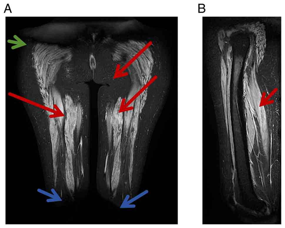
Imaging. Lower extremity T2-weighted MRI
(Fig. 2) performed on day 3
demonstrated increased T2-weighted imaging (T2WI) signal intensity
in the affected muscles, with a feather-like appearance and
heterogeneous signal intensity, consistent with RM.
Electromyography (EMG) was performed on day 2.
Electrophysiological findings were consistent with myogenic injury
involving both the upper and lower limbs.
Treatment. On day 6, treatment with 40 mg
intravenous methylprednisolone was started once a day. This was
increased to 80 mg on day 7 and the patient was discharged on day
10 with a daily dose of 60 mg oral prednisone.
Second admission (March-April 2024).
Patient presentation
The patient was re-admitted 15 days after discharge
due to worsening muscle weakness. EMG was performed, showing
evidence of peripheral neurogenic injury involving motor nerve
fibers in both the upper and lower limbs.
Diagnosis and treatment process. The patient
received methylprednisolone (40-80 mg intravenously once daily,
with dose adjustments during the treatment course). Intravenous Ig
(IVIG) therapy was administered at 20 g/day (0.4 g/kg/day based on
a body weight of 50 kg) for 6 days, in two courses on days 27-29
and 38-40. On day 27, the serum CMV-IgM level was markedly elevated
(213.30, AU/ml; normal value, <8 AU/ml; positive) and the
CMV-IgG level was >1,000.00 AU/ml (normal value, <10 AU/ml;
positive), indicating active CMV reactivation. On day 30, a lumbar
puncture was performed; CSF was sent for routine tests, and
biochemistry, microbiology and metagenomic next-generation
sequencing (mNGS) analyses. mNGS was performed by Guangzhou Jinqixi
Biotechnology Co., Ltd. DNA/RNA samples were prepared using the
Library Preparation Kit (cat. no. KS130-CAPJK-48), Reverse
Transcription Reagent (cat. no. KS630-NZL-48) and MetaCAP
Hybridization Capture Reagent (cat. no. KS129-CAPBH-6). Sample
quality and integrity were assessed using a fluorescence
quantitative analyzer and a nucleic acid protein analysis system.
Sequencing was conducted as single-end 100 bp. The sequencing
reaction was performed using the Universal Sequencing Kit
(Reversible Termination Sequencing Method; cat. no. KS107-CXR). The
final library loading concentration was 1-1.5 pmol/l. Data were
analyzed using the Pathogenic Microorganism Sequencing Data
Analysis Software (MetaCAP; Version V6.5.3; Guangzhou Jinqixi
Biotechnology Co., Ltd.). EMG performed on the same day revealed
persistent myogenic injury, with new evidence of peripheral
neurogenic involvement in the motor fibers of both upper and lower
limbs. Empirical ganciclovir (0.25 g intravenously) was initiated
on the same day as the lumbar puncture, administered from day 30 to
day 49, and resumed again on day 69. mNGS reported a relative
abundance of 100% for CMV DNA, confirming CNS CMV infection.
Plasma exchange was performed on day 31 (once daily
for 3 consecutive days, with fresh frozen plasma replacement).
Methotrexate (7.5 mg orally once weekly) was added on day 37 as a
steroid-sparing agent, after the active CNS CMV infection was
controlled.
The patient's muscle strength gradually improved and
they were discharged on day 70 with continued oral prednisone taper
starting at 50 mg/day, reduced by 5 mg every 2 weeks.
Final diagnosis, outcome and
follow-up
The final diagnosis was immune-mediated necrotizing
myopathy (SRP antibody-positive), RM and CNS cytomegalovirus
infection.
After 3 months of treatment, the patient was
discharged with upper limb muscle strength of grade 4+,
demonstrating the ability to raise both arms, clench both fists and
move against resistance. Lower limb muscle strength was grade 2+,
allowing the patient to sit independently but not to stand. At the
6-month follow-up, the condition of the patient had further
improved. Both lower limbs could be lifted off the bed and ankle
dorsiflexion could be performed. Upper limb muscle strength
remained at grade 4+, while lower limb muscle strength improved to
grade 3. Muscle tone was normal in all four extremities (Fig. 3).
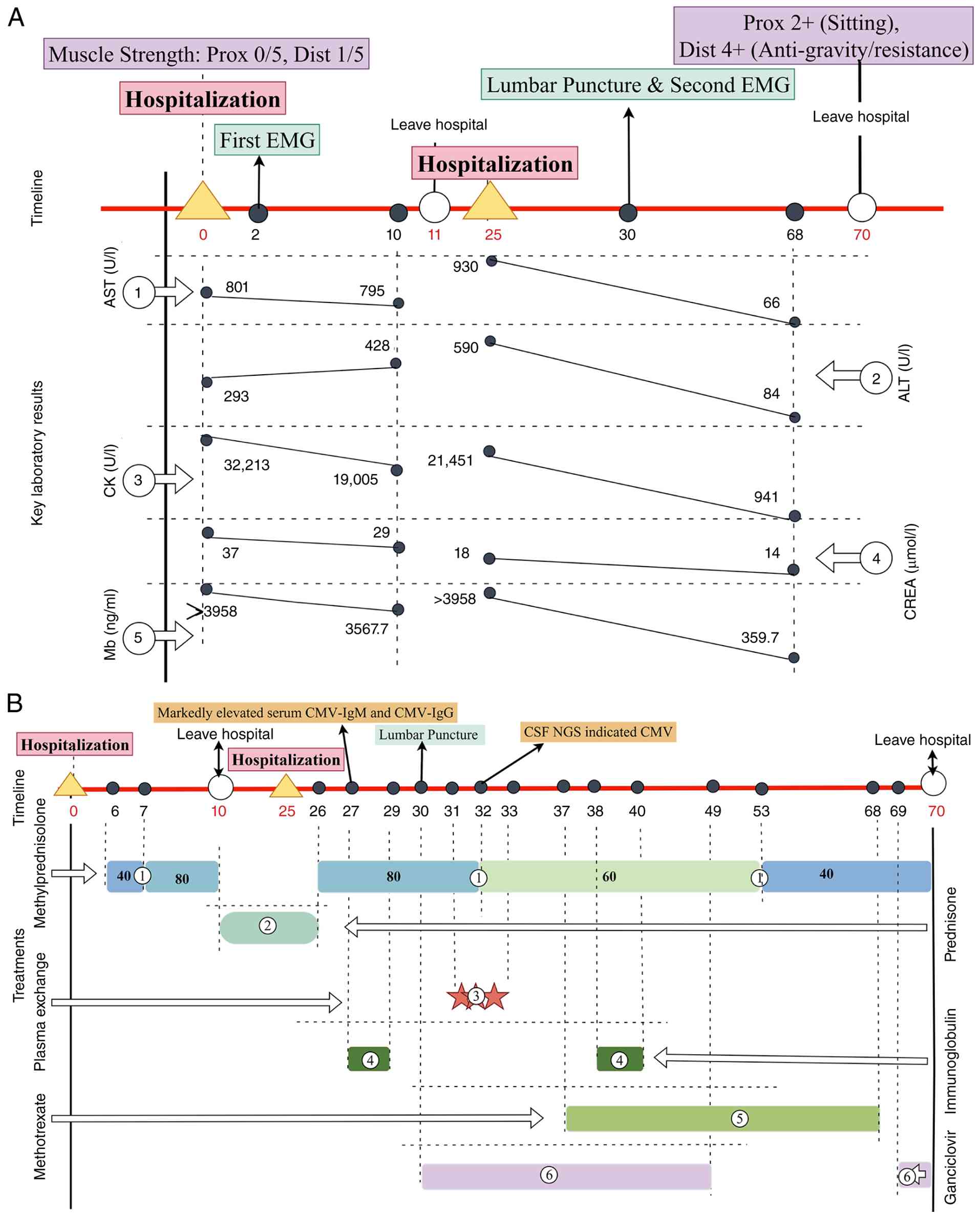 | Figure 3Timeline of the patient's muscle
strength, treatments and laboratory examinations. The red
horizontal line represents the timeline. Yellow triangles denote
hospitalization (admission) events, white circles denote discharge
events and black dots indicate specific examination or intervention
nodes. To maintain a consistent temporal framework, all numerical
labels above or below the timeline represent the number of days
relative to the first admission (the first yellow triangle, defined
as day 0). For example, the black dot labeled ‘2’ indicates an
event occurring 2 days after the first admission (day 2). (A)
Clinical course and laboratory trends. Purple boxes indicate serial
muscle strength assessments in the four limbs, graded using the
Medical Research Council scale. Red boxes mark the two hospital
admissions: February and March, 2024. Green boxes highlight key
diagnostic procedures: The first EMG assessment on day 2 and the
second EMG assessment combined with lumbar puncture on day 30.
Beneath the timeline, the trajectories of five laboratory
parameters are plotted: ① AST, ② ALT, ③ CK, ④ CREA and ⑤ Mb. (B)
Therapeutic interventions. This panel details the administration of
specific therapies. ① Methylprednisolone administered intravenously
at varying dosages (40, 60 or 80 mg daily, as indicated within the
colored boxes). ② Subsequent transition to oral prednisone
following discharge. ③ Plasma exchange performed on three
consecutive days (on days 31-33). ④ Intravenous Ig therapy
administered during two discrete courses: on days 27-29 and on days
38-40. ⑤ Methotrexate therapy initiated on day 37 and continued
until day 68. ⑥ Ganciclovir administered during two periods: from
day 30 to day 49 and from day 69. Three arrows in the upper middle
portion of the timeline indicate the following key events: Markedly
elevated serum CMV-IgM and CMV-IgG on day 27; lumbar puncture
performed on day 30; and CSF NGS suggesting CMV on day 32. EMG,
electromyography; AST, aspartate aminotransferase; ALT, alanine
aminotransferase; CK, creatine kinase; CREA, creatinine; Mb,
myoglobin; Ig, immunoglobulin; CSF, cerebrospinal fluid; NGS,
next-generation sequencing; CMV, cytomegalovirus. Created with
Figdraw (www.figdraw.com). |
Discussion
IMNM is a rare inflammatory myopathy with a subacute
or insidious onset. Although necrotizing myopathy was first
reported by Smith in 1969(1), IMNM
was not recognized as a distinct entity until 2003-2004(2). IMNM complicated by RM has rarely been
reported (10). In the present
case, EMG demonstrated both myogenic and peripheral nerve
involvement. CSF analysis revealed CMV infection and
albuminocytological dissociation. Based on the clinical
manifestations, laboratory findings, muscle pathology,
electromyographic results and CSF analysis, the patient was
diagnosed with anti-SRP antibody-positive IMNM complicated by RM
and CNS CMV infection. GBS was included in the differential
diagnosis as the patient presented with acute limb weakness,
decreased muscle tone and mild albuminocytological dissociation,
features that can mimic GBS. However, GBS was ultimately excluded
by the presence of myopathic EMG changes, normal nerve conduction
studies and a markedly elevated serum CK level (peaking at 32,213
U/l), which are inconsistent with typical GBS.
IMNM is characterized by proximal muscle weakness
and increased serum CK levels. Histopathologically, IMNM is
characterized by prominent muscle fiber necrosis and regeneration,
with little or no inflammatory lymphocytic infiltration (3). IMNM is more common in women, with the
peak age of onset between 40 and 50 years, and a higher incidence
reported during the summer months. IMNM can be classified into
three subtypes: Anti-SRP antibody-positive IMNM, anti-HMGCR
antibody-positive IMNM and seronegative IMNM. Among the patients
with anti-HMGCR antibody-positive IMNM, ~15% have a history of
statin use, whereas only 4.2% of patients with anti-HMGCR
antibody-negative IMNM have previously used statins. Simvastatin,
atorvastatin and rosuvastatin are the statins most commonly
associated with RM (4). In 2016,
the ENMC (3) proposed the
diagnostic criteria for anti-SRP antibody-associated myopathy,
which include: i) Increased serum CK levels; ii) proximal muscle
weakness; and iii) positive results for anti-SRP autoantibodies.
These criteria highlight the diagnostic importance of
myositis-specific antibodies. A muscle biopsy is not required for
the diagnosis of the first two IMNM subtypes (anti-SRP
antibody-positive and anti-HMGCR antibody-positive IMNM), whereas
patients who are negative for these serum antibodies still require
a muscle biopsy to establish the diagnosis. The present patient was
a young woman with acute-onset disease, markedly increased serum
muscle enzyme levels and prominent proximal muscle weakness.
Combined with the markedly increased serum CK level and positive
anti-SRP antibodies, the clinical presentation fulfilled the ENMC
diagnostic criteria for anti-SRP antibody-associated myopathy.
Furthermore, the biopsy findings, characterized by necrotic and
regenerating fibers, sparse lymphocytic infiltrate but macrophagic
(histiocytic) infiltration, weak HLA-ABC expression and C5b-9
deposition, fulfilled the established histopathological criteria
for IMNM. In conjunction with the clinical course and investigative
results, alternative diagnoses were systematically excluded: Toxic
myopathy was unlikely given the absence of statin or other myotoxic
drug exposure; muscular dystrophy was considered improbable due to
the acute onset, lack of family history and presence of anti-SRP
antibodies; and infectious myositis was ruled out by the absence of
viral inclusions or granulomatous changes on biopsy, with the
concurrent CMV infection localized to the CNS rather than the
skeletal muscle. RM is a clinical syndrome caused by skeletal
muscle necrosis, resulting in the release of intracellular muscle
constituents into the systemic circulation (5). The causes of RM include trauma,
strenuous exercise, drug toxicity, infection, electrolyte
disturbances, hereditary metabolic disorders and other conditions.
Patients typically present with the classic triad of myalgia,
muscle weakness and dark brown urine. In severe cases, RM can lead
to acute kidney injury and may even be life-threatening. Some
patients may also experience fatigue, fever, tachycardia, nausea,
vomiting and abdominal pain. The most common triggers of RM are
strenuous exercise, medications, trauma and infection, whereas
immune-mediated factors are relatively uncommon (4). MRI findings of RM typically include
diffuse swelling of the affected muscles with extensive involvement
and ill-defined margins. T1WI usually demonstrates decreased signal
intensity, whereas T2WI and short τ inversion recovery sequences
show increased signal intensity with patchy or flocculent
heterogeneous hyperintensity (11).
In the present case, MRI demonstrated increased T2WI signal
intensity in the affected muscles, with a feather-like appearance
and heterogeneous signal intensity, findings that are consistent
with RM. IMNM is a subtype of idiopathic inflammatory myopathy
(IIM) (3). Although IIM and RM are
distinct disease entities, they share similar clinical
manifestations, and IIM is a rare cause of RM (12). Therefore, IMNM, as a subtype of IIM,
may also be associated with the development of RM. In addition to
the aforementioned evidence, previous studies have reported
distinct pathological features in anti-SRP antibody-associated
myopathy with and without RM (13).
In patients without RM, necrotic muscle fibers are typically
angular or rounded, depending on the degree of necrosis, and are
randomly distributed throughout the muscle tissue (14). Inflammatory infiltrates are
generally minimal or absent, and when present, they are
macrophage-predominant (CD68+) with sparse perivascular
lymphocytes (14). MHC-I expression
is usually absent or only weakly expressed on non-necrotic fibers
(14). In cases with extensive
muscle fiber necrosis, such as those complicated by RM, the
pathological findings may demonstrate more prominent macrophagic
(histiocytic) infiltration associated with necrotic fibers,
increased endomysial connective tissue proliferation, sarcolemmal
MHC-I expression on affected muscle fibers and occasional C5b-9
deposition (15,16). The pathological findings from the
right thigh muscle biopsy in the present case demonstrated
macrophagic infiltration and positivity for C5b-9, supporting the
diagnosis of anti-SRP antibody-associated myopathy complicated by
RM.
As detailed in the present report, the patient
presented with severe RM yet did not develop AKI or compartment
syndrome, or require conventional renal replacement therapy. This
relatively favorable clinical course, particularly the preservation
of renal function, may be partly attributable to the early
initiation of plasma exchange, which we consider to offer dual
therapeutic benefits in patients with severe IMNM complicated by RM
(17). First, it removes
circulating pathogenic autoantibodies, including anti-SRP
antibodies, thereby reducing ongoing immune-mediated muscle fiber
injury (18). Second, it
facilitates the clearance of large amounts of circulating Myo
released from damaged muscle, potentially decreasing the risk of
cast nephropathy and acute kidney injury (19). In the present case, plasma exchange
was initiated promptly (once daily for 3 consecutive days), which
likely contributed to the removal of pathogenic antibodies and
excess Myo, thereby promoting recovery of muscle strength and
preventing renal deterioration. While plasma exchange has been
reported to improve outcomes in IMNM (18,20),
evidence for its benefit in severe RM is currently lacking
(19). Therefore, early initiation
may be a reasonable therapeutic consideration in critically ill
patients with IMNM complicated by RM, although prospective studies
are needed to confirm this hypothesis.
In addition, the following principles should be
emphasized in the clinical management of patients with IMNM
complicated by RM: i) Discontinue strenuous exercise and offending
medications, avoid trauma and other precipitating factors, and
promptly identify and treat underlying conditions such as infection
and immune-mediated disorders (21); ii) initiate aggressive fluid
resuscitation with either lactated Ringer's solution or normal
saline, guided by the patient's cardiopulmonary status. An initial
infusion rate of ~400 ml/h is recommended, with the goal of
maintaining a urine output of 1-3 ml/kg/h (18); iii) renal replacement therapy should
be considered in patients with a McMahon score ≥6 or in those who
develop AKI despite adequate fluid resuscitation (22); iv) plasma exchange may facilitate
the clearance of circulating macromolecules, including Myo,
high-sensitivity cardiac troponin T, CK-MB and LDH, thereby
potentially mitigating RM-associated AKI; however, its impact on
overall mortality remains unproven (19,23);
and v) electrolyte disturbances, particularly hyperkalemia,
hyperphosphatemia and hypocalcemia, should be identified and
corrected promptly throughout the course of RM (5). The present patient achieved a
favorable short-term outcome following treatment; however, the
long-term prognosis remains uncertain. Regarding the selection of
immunosuppressive therapy, methotrexate (7.5 mg orally once weekly)
was introduced as a steroid-sparing agent after the active CNS CMV
infection had been controlled with ganciclovir (24). Rituximab was intentionally avoided
during the acute phase due to its profound and prolonged B
cell-depleting effects, which could substantially increase the risk
of CMV reactivation or exacerbation (24). Mycophenolate mofetil and
azathioprine were not prioritized since they are mainly used as
maintenance therapies and have a relatively slow onset of action,
whereas this patient required rapid control of severe myositis and
RM. Intravenous cyclophosphamide, another treatment option for
IMNM, was also deferred due to the patient's young age and its
potential adverse effects, such as decreased fertility (24,25).
When IMNM is complicated by RM, EMG typically
reveals myogenic injury; however, a small proportion of patients
may also exhibit evidence of peripheral nerve involvement. In the
present case, EMG performed in March 2024 demonstrated peripheral
neurogenic injury involving the motor fibers of both the upper and
lower limbs, in addition to myogenic abnormalities. The peripheral
nerve involvement observed on EMG may be explained by several
mechanisms (26). First, prolonged
direct nerve compression may result in compressive neuropathy.
Second, increased intracompartmental pressure associated with
muscle injury or compartment syndrome may lead to nerve compression
and subsequent nerve injury. Third, muscle injury itself may induce
local neuroinflammation, contributing to peripheral nerve
dysfunction. In complex cases where RM is accompanied by peripheral
nerve injury, functional recovery is often prolonged, and patients
generally require a longer rehabilitation period (27). When RM is not accompanied by
peripheral nerve injury, MRI typically demonstrates the following
features: i) Diffuse involvement of the affected muscles with
heterogeneous abnormal signal intensity, ill-defined margins,
mild-to-moderate intermuscular exudation and subcutaneous edema;
ii) on T1WI, most lesions (approximately two-thirds) show
homogeneous mild hypointensity, whereas the remaining one-third
exhibit heterogeneous signal intensity; and iii) on T2WI and
fat-suppressed sequences, patchy, flocculent, feather-like or
linear areas of heterogeneous hyperintensity are observed,
accompanied by muscle swelling and a loose muscle architecture
(28). By contrast, when RM is
accompanied by peripheral nerve injury, MRI may demonstrate the
following features: i) Intramuscular hemorrhage and muscle edema in
~60% of patients; ii) more prominent high signal intensity within
the subcutaneous tissue; iii) increased muscle volume with diffuse
swelling and a loose muscle architecture; iv) muscle lesions with
predominantly rectangular or linear morphology and irregular,
sharply demarcated margins; v) variable patterns of muscle
involvement, often affecting multiple muscles within the same limb
or compartment; and vi) a higher likelihood of compartment syndrome
with increased intracompartmental pressure, resulting in secondary
peripheral nerve injury (28). In
the present patient, after systematic treatment, post-treatment
evaluation demonstrated a marked improvement in the patient's serum
CK and hemoglobin levels. Upper limb muscle strength recovered
substantially, whereas recovery of lower limb muscle strength was
slower. MRI revealed a characteristic pattern in which the central
portion of the affected muscle bundles showed no enhancement, while
the peripheral regions exhibited contrast enhancement. This imaging
pattern suggests severe irreversible necrosis in the central muscle
tissue with ongoing repair and inflammatory activity at the
periphery, which may explain the delayed recovery of lower limb
muscle strength. Nevertheless, it is important to acknowledge the
limitations of these interpretations. Although the preceding
discussion provides evidence-based explanations for the neurogenic
changes observed in this case, these conclusions are mainly based
on the clinical findings of a single patient and should therefore
be interpreted with caution. In complex clinical scenarios,
neurogenic findings may result from multiple concurrent mechanisms,
including direct nerve compression, RM-related nerve injury,
critical illness, treatment-related effects and coexisting
peripheral neuropathy (29).
Therefore, the aforementioned interpretations should be regarded as
hypothesis-generating observations based on this individual case
rather than definitive conclusions. The underlying
pathophysiological mechanisms in each patient should be evaluated
on a case-by-case basis, with treatment strategies tailored to the
individual clinical presentation.
Peripheral neuropathy, particularly GBS, can present
with clinical features similar to those observed in the current
patient, including limb weakness or paralysis and decreased muscle
tone. GBS is an acute immune-mediated polyradiculoneuropathy
characterized by multiple nerve root and peripheral nerve
involvement. Albuminocytologic dissociation in the CSF is one of
its characteristic findings (30).
In severe cases, paralysis of the respiratory and bulbar muscles
may occur, leading to life-threatening respiratory failure
(31). The diagnosis of GBS is
mainly based on the patient's clinical history,
electrophysiological findings and CSF examination, with nerve
conduction studies and EMG serving as essential diagnostic tools.
Electrophysiological findings in GBS typically include prolonged
distal motor latency and F-wave latency, temporal dispersion,
prolonged compound muscle action potential duration, reduced nerve
conduction velocity and conduction block in non-entrapment nerve
segments (32). In the present
patient, in March 2024, EMG demonstrated electrophysiological
evidence of myogenic injury in the examined muscles, characterized
by shortened motor unit potential duration, increased polyphasic
motor unit potentials and reduced amplitude. Sensory and motor
nerve conduction velocities, F-wave latencies and F-wave
persistence were all within normal limits. Although CSF analysis
revealed mildly increased protein levels, the increase was modest.
Given that systemic CMV reactivation is typically associated with
viremia, this mild protein elevation may more plausibly reflect
localized CNS CMV infection rather than non-specific systemic
inflammation. Furthermore, serum CK levels are typically normal or
only mildly increased in patients with GBS, whereas this patient
had a markedly increased CK level, peaking at 32,213 U/l, strongly
supporting a primary myopathic rather than neuropathic process.
Therefore, although the patient presented with limb weakness
resembling GBS and exhibited mild albuminocytological dissociation
in the CSF, the electrophysiological findings were not consistent
with GBS or other peripheral nervous system disorders. Based on
these combined findings, particularly the myopathic EMG changes and
the markedly elevated CK level, the patient did not meet the
diagnostic criteria for GBS. It is also essential to distinguish
clinically significant CMV CNS infection from incidental viral
detection or systemic CMV reactivation, particularly in this case,
as the patient received both immunosuppressive therapy and
ganciclovir, and the differential diagnosis with GBS relied heavily
on the interpretation of CSF albuminocytologic dissociation and EMG
findings. Several lines of evidence support that the positive CSF
mNGS result represented an active CNS infection. First, CMV DNA was
detected by mNGS in the CSF on day 32, which was after the
initiation of immunosuppressive therapy (methylprednisolone was
started on day 6 and IVIG was administered on days 27-29). This
timeline is consistent with a treatment-related opportunistic
infection, rendering incidental viral detection or systemic
reactivation unlikely. Second, although systemic CMV reactivation
typically presents with viremia, the detection of viral DNA
directly in the CSF suggests a localized pathological process
within the CNS. Importantly, the mNGS report showed a relative
abundance of 100% for CMV; however, viral load quantification (for
example, IU/ml or copies/µl) and read numbers were not available
from the assay. Third, serum CMV-IgM was markedly elevated at
213.30 AU/ml (normal value, <8 AU/ml; positive) and CMV-IgG was
>1,000.00 AU/ml (normal value, <10 AU/ml; positive),
providing serological evidence of active CMV infection. Fourth,
acid-fast staining of the CSF was negative, effectively excluding
tuberculous meningitis and other acid-fast bacillary infections as
the cause of the neurological manifestations. Fifth, the presence
of CMV in the CSF provides a plausible infectious explanation for
the observed albuminocytological dissociation and the neurogenic
findings on EMG, features that might otherwise be attributed solely
to GBS or critical illness-related neuropathy. Regarding the
pathogenesis of IMNM complicated by CNS CMV infection, two possible
mechanisms merit consideration. First, CMV infection may trigger an
autoimmune response through specific immunological mechanisms, such
as molecular mimicry and bystander activation, potentially
contributing to the development of necrotizing myopathy (8,33,34).
Alternatively, it may occur as an opportunistic infection following
immunosuppression (7). In the
present case, CMV DNA was detected in the CSF after the initiation
of immunosuppressive treatment, and serum CMV-IgM/IgG levels were
already elevated on day 27, suggesting that CMV reactivation likely
occurred as a consequence of immunosuppression and subsequently
involved the CNS, although a contributing role in triggering
autoimmunity cannot be entirely excluded.
The present case report has several limitations that
should be acknowledged. First, as a single-case report, its
findings may not be generalizable to all patients with anti-SRP
antibody-positive IMNM complicated by RM and CNS CMV infection.
Second, the follow-up period was relatively short (6 months), and
the long-term prognosis, including the risk of relapse, persistent
muscle weakness and late neurological sequelae of CNS CMV
infection, remains uncertain. Third, the CSF mNGS assay detected
CMV with a relative abundance of 100%, but viral load
quantification and read numbers were not available from the test
report, limiting the ability to precisely quantify viral burden or
monitor treatment response. Additionally, the diagnosis of CNS CMV
infection was based on a combination of CSF mNGS results,
serological evidence (positive CMV-IgM and IgG), and clinical
context, rather than being confirmed by conventional virological
methods such as CSF CMV PCR or viral culture. Further studies
involving larger cohorts and longer follow-up are needed to better
characterize the clinical course of this rare condition and to
establish evidence-based management strategies.
In conclusion, the present report describes a rare
case of anti-SRP antibody-positive IMNM complicated by RM and CNS
CMV infection. Although a single case cannot establish definitive
management recommendations, it highlights the importance of
distinguishing anti-SRP antibody-positive IMNM complicated by RM
from GBS in patients presenting with severe muscle weakness and
markedly increased muscle enzyme levels. It also underscores the
need for thorough evaluation of concomitant infections before and
during immunosuppressive therapy to facilitate timely diagnosis and
appropriate treatment.
Acknowledgements
Not applicable.
Funding
Funding: The present study was supported by the Southwest
Medical University Technology Program (grant nos. 2023QN060,
2023ZYQJ04 and 2024ZXYZX02), the National Natural Science
Foundation of China (grant no. 82205002), the China Postdoctoral
Science Foundation (grant no. 2025M783999) and the Science and
Technology Research Special Project of Sichuan Administration of
Traditional Chinese Medicine (grant no. 2024MS524).
Availability of data and materials
The mNGS data generated in the present study may be
found in the National Center for Biotechnology Information Sequence
Read Archive under BioProject accession number PRJNA1525608 or at
the following URL: https://www.ncbi.nlm.nih.gov/bioproject/PRJNA1525608.
Authors' contributions
YX, as the attending physician, was responsible for
the patient's diagnosis and treatment throughout the course,
supervised the histopathological staining procedure, reviewed the
stained slides and wrote the original draft. NZ was responsible for
data curation, visualization and analysis of mNGS data, and
assisted in writing the original draft. JL was responsible for
obtaining the medical images (MRI and CT), funding acquisition,
supervision and reviewing/editing the manuscript. QZ provided an
expert opinion on the patient's treatment and was responsible for
reviewing and editing the manuscript. QH analyzed the patient's
laboratory data, performed project administration and assisted in
reviewing and editing the manuscript. All authors have read and
approved the final version of the manuscript. YX and NZ confirm the
authenticity of all the raw data.
Ethics approval and consent to
participate
This study was approved by the Institutional Review
Board of The Affiliated Traditional Chinese Medicine Hospital,
Southwest Medical University (Luzhou, China).
Consent for publication
The patient and the patient's guardian (due to the
severe nature of the illness) provided written informed consent for
the publication of this case report, including all information and
images.
Competing interests
The authors declare that they have no competing
interests.
References
|
1
|
Smith B: Skeletal muscle necrosis
associated with cainoma. J Pathol. 97:207–210. 1969.PubMed/NCBI View Article : Google Scholar
|
|
2
|
Hoogendijk JE, Amato AA, Lecky BR, Choy
EH, Lundberg IE, Rose MR, Vencovsky J, de Visser M and Hughes RA:
119th ENMC international workshop: trial design in adult idiopathic
inflammatory myopathies, with the exception of inclusion body
myositis, 10-12 October 2003, Naarden, The Netherlands. Neuromuscul
Disord. 14:337–345. 2004.PubMed/NCBI View Article : Google Scholar
|
|
3
|
Allenbach Y, Mammen AL, Benveniste O and
Stenzel W: Immune-Mediated Necrotizing Myopathies Working Group.
224th ENMC International Workshop: Clinico-sero-pathological
classification of immune-mediated necrotizing myopathies Zandvoort,
The Netherlands, 14-16 October 2016. Neuromuscul Disord. 28:87–99.
2018.PubMed/NCBI View Article : Google Scholar
|
|
4
|
Luong MT, Danahy P and Ho GY: Spontaneous
development of immune-mediated necrotizing myopathy (IMNM) after
chronic use of statins: A case report. Cureus.
15(e42266)2023.PubMed/NCBI View Article : Google Scholar
|
|
5
|
Cabral BMI, Edding SN, Portocarrero JP and
Lerma EV: Rhabdomyolysis. Dis Mon. 66(101015)2020.PubMed/NCBI View Article : Google Scholar
|
|
6
|
Baghdasaryan EJ, Soltani H, Lada S,
Simpson C and Nathan R: Necrotizing myopathy presenting with
guillain-barré-like areflexic paraparesis, severe rhabdomyolysis,
and dialysis-requiring acute kidney injury. Cureus.
18(e115560)2026.PubMed/NCBI View Article : Google Scholar
|
|
7
|
Hsu CH, Hsuan YT, Chan YJ and Chen HP:
Clinical presentation of cytomegalovirus meningoencephalitis: A
retrospective study of 12 adult patients with a variety of
immunocompromised conditions. BMC Infect Dis.
25(575)2025.PubMed/NCBI View Article : Google Scholar
|
|
8
|
Sampaio B, Gomes F, Teixeira J, Leal M and
Rebelo M: Anti-SRP immune-mediated necrotizing myopathy and recent
cytomegalovirus infection: A case report. Int J Innov Res Med Sci.
10:22–25. 2025.
|
|
9
|
Moffat DB: Aids to the Examination of the
Peripheral Nervous System. J Anat. 151:262–263. 1987.
|
|
10
|
Pombo F, Raposo A, Martins MD, Ferreira I
and Pires L: A rare case of rhabdomyolysis. Cureus.
14(e31519)2022.
|
|
11
|
Kok SXS and Tan TJ: Clinics in diagnostic
imaging (179). Severe rhabdomyolysis complicated by myonecrosis.
Singapore Med J. 58:467–472. 2017.PubMed/NCBI View Article : Google Scholar
|
|
12
|
Joshi D, Kumar N and Rai A:
Dermatomyositis presenting with rhabdomyolysis and acute renal
failure; an uncommon manifestation. Ann Indian Acad Neurol.
12:45–47. 2009.PubMed/NCBI View Article : Google Scholar
|
|
13
|
Kao AH, Lacomis D, Lucas M, Fertig N and
Oddis CV: Anti-signal recognition particle autoantibody in patients
with and patients without idiopathic inflammatory myopathy.
Arthritis Rheum. 50:209–215. 2004.PubMed/NCBI View Article : Google Scholar
|
|
14
|
Miller T, Al-Lozi MT, Lopate G and
Pestronk A: Myopathy with antibodies to the signal recognition
particle: clinical and pathological features. J Neurol Neurosurg
Psychiatry. 73:420–428. 2002.PubMed/NCBI View Article : Google Scholar
|
|
15
|
Dimitri D, Andre C, Roucoules J, Hosseini
H, Humbel RL and Authier FJ: Myopathy associated with anti-signal
recognition peptide antibodies: clinical heterogeneity contrasts
with stereotyped histopathology. Muscle Nerve. 35:389–395.
2007.PubMed/NCBI View Article : Google Scholar
|
|
16
|
Wang L, Liu L, Hao H, Gao F, Liu X, Wang
Z, Zhang W, Lv H and Yuan Y: Myopathy with anti-signal recognition
particle antibodies: Clinical and histopathological features in
Chinese patients. Neuromuscul Disord. 24:335–341. 2014.PubMed/NCBI View Article : Google Scholar
|
|
17
|
Connolly CM and Paik JJ: Clinical pearls
and promising therapies in myositis. Expert Rev Clin Immunol.
19:797–811. 2023.PubMed/NCBI View Article : Google Scholar
|
|
18
|
Zhang H, Sun Y, Liu H, Cheng X, Ye J, Hu
Q, Jia J, Wang M, Liu T, Zhou Z, et al: Plasma exchange therapy in
refractory inflammatory myopathy with anti-signal recognition
particle antibody: A case series. Rheumatology (Oxford).
61:2625–2630. 2022.PubMed/NCBI View Article : Google Scholar
|
|
19
|
Boparai S, Lakra R, Dhaliwal L, Hansra RS,
Bhuiyan MAN, Conrad SA and Krishnan P: Therapeutic plasma exchange
in severe rhabdomyolysis: A case-control study. Cureus.
15(e39748)2023.PubMed/NCBI View Article : Google Scholar
|
|
20
|
Kruse RL, Albayda J, Vozniak SO, Lawrence
CE, Goel R, Lokhandwala PM, Ness PM, Tobian AAR, Bloch EM and Crowe
EP: Therapeutic plasma exchange for the treatment of refractory
necrotizing autoimmune myopathy. J Clin Apher. 37:253–262.
2022.PubMed/NCBI View Article : Google Scholar
|
|
21
|
Kodadek L, Carmichael Ii SP, Seshadri A,
Pathak A, Hoth J, Appelbaum R, Michetti CP and Gonzalez RP:
Rhabdomyolysis: An American association for the surgery of trauma
critical care committee clinical consensus document. Trauma Surg
Acute Care Open. 7(e000836)2022.PubMed/NCBI View Article : Google Scholar
|
|
22
|
Simpson JP, Taylor A, Sudhan N, Menon DK
and Lavinio A: Rhabdomyolysis and acute kidney injury: Creatine
kinase as a prognostic marker and validation of the McMahon Score
in a 10-year cohort: A retrospective observational evaluation. Eur
J Anaesthesiol. 33:906–912. 2016.PubMed/NCBI View Article : Google Scholar
|
|
23
|
Clark WF and Huang SS: Introduction to
therapeutic plasma exchange. Transfus Apher Sci. 58:228–229.
2019.PubMed/NCBI View Article : Google Scholar
|
|
24
|
Weeding E and Tiniakou E: Therapeutic
management of immune-mediated necrotizing myositis. Curr Treatm Opt
Rheumatol. 7:150–160. 2021.PubMed/NCBI View Article : Google Scholar
|
|
25
|
Singhal P, Ren KYM, Curtis BM, MacPherson
I and Avila-Casado C: Atypical noncrescentic antiglomerular
basement membrane disease with concurrent thin basement membrane
nephropathy. Kidney Int Rep. 3:991–996. 2018.PubMed/NCBI View Article : Google Scholar
|
|
26
|
Kim Y, Min K, Park MW, Shin HI and Kim DH:
Injury to the left sciatic and right common peroneal nerves
combined with multifocal rhabdomyolysis in a survivor of the
itaewon crowd crush: A case report. J Korean Med Sci.
38(e233)2023.PubMed/NCBI View Article : Google Scholar
|
|
27
|
Kim IS, Lee WH and Lim JY: Multiple
peripheral neuropathies adjacent to necrotizing myositis related to
rhabdomyolysis. Ann Geriatr Med Res. 21:31–34. 2017.
|
|
28
|
Kim JH, Kim YJ, Koh SH, Kim BS, Choi SY,
Cho SE, Song JH, Kim CH, Lee KH and Cho SG: Rhabdomyolysis
revisited: Detailed analysis of magnetic resonance imaging findings
and their correlation with peripheral neuropathy. Medicine
(Baltimore). 97(e11848)2018.PubMed/NCBI View Article : Google Scholar
|
|
29
|
Ejikeme C, Alyacoub R, Elkattawy S,
Shankar T and Yuridullah R: A rare complication of rhabdomyolysis:
Peripheral neuropathy. Cureus. 13(e14202)2021.PubMed/NCBI View Article : Google Scholar
|
|
30
|
Payus AO, Ibrahim A, Liew Sat Lin C and
Hui Jan T: Sensory predominant guillain-barré syndrome concomitant
with dengue infection: A case report. Case Rep Neurol. 14:281–285.
2022.PubMed/NCBI View Article : Google Scholar
|
|
31
|
Busl KM, Fried H, Muehlschlegel S,
Wartenberg KE, Rajajee V, Alexander SA, Creutzfeldt CJ, Fontaine
GV, Hocker SE, Hwang DY, et al: Guidelines for neuroprognostication
in adults with guillain-barré syndrome. Neurocrit Care. 38:564–583.
2023.PubMed/NCBI View Article : Google Scholar
|
|
32
|
Scheidl E, Canseco DD, Hadji-Naumov A and
Bereznai B: Guillain-Barré syndrome during SARS-CoV-2 pandemic: A
case report and review of recent literature. J Peripher Nerv Syst.
25:204–207. 2020.PubMed/NCBI View Article : Google Scholar
|
|
33
|
Ma X and Bu BT: Anti-SRP immune-mediated
necrotizing myopathy: A critical review of current concepts. Front
Immunol. 13(1019972)2022.PubMed/NCBI View Article : Google Scholar
|
|
34
|
Ehrenfeld M, Tincani A, Andreoli L,
Cattalini M, Greenbaum A, Kanduc D, Alijotas-Reig J, Zinserling V,
Semenova N, Amital H and Shoenfeld Y: Covid-19 and autoimmunity.
Autoimmun Rev. 19(102597)2020.PubMed/NCBI View Article : Google Scholar
|