Introduction
3-Aminopyridine-2-carboxaldehyde thiosemicarbazone
(3-AP), a metal chelator, is a potent small-molecule inhibitor of
ribonucleotide reductase (RR), an enzyme that is important for cell
division and tumor growth (1). The
RR enzyme is responsible for reducing ribonucleotides to their
corresponding deoxyribonucleotides, precursors of DNA synthesis and
repair. Human RR is composed of two heterodimeric subunits, hRRM1,
that contains the nucleotide binding site, and hRRM2, that contains
the metal binding site (1). The
hRRM2 subunit has a non-heme iron and a tyrosine-free radical,
which are required for the enzymatic reduction of ribonucleotides
(2). The RR complex contains two
ferric ions that are coordinated by four carboxylates, and two
histidine ligands that are anti-ferro-magnetically coupled by a
μ-oxo bridge, rendering it electron paramagnetic resonance
(EPR)-silent. Inhibitors of RR act by destroying the tyrosine-free
radical in the hRRM2 subunit (3).
The reaction of the di-ferric center with oxygen generates a
protein-bound tyrosyl radical that is detectable by EPR (4). Recent evidence has shown that RR
spontaneously loses the iron atoms, rendering it chelatable by
small hydrophobic chelators, such as 3-AP (4).
Inhibition of RR disrupts DNA synthesis and repair
leading to apoptotic cell death. EPR can be used to measure the
effects of 3-AP in the cell. The presence of the free tyrosyl
radical is directly proportional to the enzymatic activity of RR
(5,6). 3-AP-mediated inhibition of RR is
predicted to result in a decrease in the tyrosyl radical and an
increase in the Cu-triapine (CuT) and iron-3-AP (FeT2)
complexes (7). In addition, 3-AP
inhibits Fe uptake from transferrin and induces the transferrin
receptor at both the levels of mRNA and protein (8–10).
An increase in Fe-transferrin complexes and transferrin receptors
is also expected (9,11). Cytochrome c release occurs early in
the apoptotic cascade, and increased cyctochrome c release is
anticipated following treatment with 3-AP (12).
It has been shown that formation of the metal
chelate of 3-AP and iron is essential to its cytotoxic effect,
mediated by the formation of reactive oxygen species (ROS) that
render RR inactive (5,9). Precursors of 3-AP have been shown to
sequester large quantities of iron in humans (13). The iron-3-AP complex directly
inhibits RR and is a step that involves hydrogen peroxide (9), much as precursors to FeT2
inhibit RR (14,15). In addition, the iron-3-AP complex
is redox active, forming ROS that deplete intracellular glutathione
and cause DNA strand breaks, similar to iron-doxorubicin or the
iron-bleomycin complexes (9). Zhu
et al demonstrated that 3-AP and seven of its synthetic
derivatives decreased the activity of RR in a dose-dependent manner
in normal as well as hyroxyurea- and gemcitabine-resistant KB cell
lines, using two different forms of RR (16).
In light of recent findings, it is known that 3-AP
possesses intrinsic fluorescent properties (17). Thus, by means of fluorescence
microscopy, its uptake and intracellular distribution in living
human cancer cells can be monitored. This feature is relatively
uncommon among anticancer drugs and may help in studying resistance
mechanisms by undergoing possible phenotypic changes, as well as
the combinatorial effects of other drugs on 3-AP (17). 3-AP has anti-tumor effects, both
in vitro and in vivo, and several clinical trials are
currently being conducted to evaluate its safety and antineoplastic
activity, either as monotherapy or in combination with other
anticancer agents (18–22). It has also been shown that
inhibition of RR by 3-AP enhances radiation-mediated cytotoxicity
independent of p53 regulation by impairing repair processes that
rely on deoxyribonucleotide production. This substantially
increases the radiation sensitivity of human cancers (23). In a recent study, we validated that
the effect of 3-AP on peripheral blood mononuclear cells can be
measured by EPR spectroscopy (6).
The goal of the present study was to determine whether 3-AP affects
its targeted action in peripheral blood lymphocytes, by measuring
EPR signals formed either directly or indirectly.
Materials and methods
Sample preparation
Blood samples were evaluable from 18 patients with
locally advanced, unresectable or metastatic solid tumors who
participated in two clinical trials at the University of Wisconsin:
a phase I combination of 3-AP and doxorubicin (20), and a phase I combination of 3-AP
and irinotecan (19). Peripheral
blood lymphocytes (PBLs) were obtained as previously described
(6). Detailed information about
the dosing schedules for 3-AP, doxorubicin and irinotecan are found
in the referenced literature for the respective clinical trials.
Briefly, two 10-ml samples of blood were drawn into CPT tubes and
were immediately centrifuged to remove the PBLs. Samples were
obtained prior to administration of 3-AP and at 2, 4.5 and 22 h
after the end of 3-AP infusion. A small aliquot of cells was
removed for determining cell counts and protein concentration. The
remaining PBL samples were placed in 4-mm OD quartz EPR tubes,
frozen in liquid nitrogen and stored at −80°C until the time of
assay. Human halo-transferrin (Calbiochem; EMD Bioscienes, La
Jolla, CA, USA) was dissolved in MSH (0.225 mannitol, 0.75 M
sucrose and 20 mM HEPES buffer pH 7.5) and used as a standard
(concentration 320 μM).
EPR spectroscopy
A Varian E109 Century Series spectrometer (Varian,
Palo Alto, CA, USA) operating at X-band (9–9.5 GHz) with a 100-kHz
field modulation or a Bruker E500 ELEXSYS spectrometer with an
Oxford Instruments ESR-9 helium flow cryostat and a Bruker DM0101
cavity was employed. ESR measurements were made at 10K on frozen
intact cells. Spectra from each sample were recorded ten times and
an average was calculated.
Statistical analysis
EPR spectra recordings were taken for PBL samples
collected at baseline and at 2, 4.5 and 22 h after administration
of 3-AP. Changes in spectral parameters were evaluated by computing
the ratios at 2, 4.5 and 22 h to baseline.
The outcome variable was expressed as a ratio of the
value at the given time-point (numerator of the ratio) to the
baseline value (denominator of the ratio). The ratios were
summarized using standard descriptive statistics in terms of number
of observations, medians, ranges, means and standard deviations.
Boxplots were used to display the distributions of the ratios for
each time point. A non-parametric Wilcoxon Signed Rank test was
used to determine statistical significance. Since the a
priori hypothesis proposed that there is an increase in the
Fe-transferrin signals, heme signals and FE and Cu level values
from baseline, one-sided tests were used with the alternative
hypotheses that the ratios are >1. Exact p-values were computed,
and p-values <0.05 were defined as statistically
significant.
Results
EPR spectra of low spin (l.s.) Fe and
Cu
EPR spectra were obtained for 3-AP upon addition of
cupric or ferric ions in DMSO and PBS. This was used as a template
to recognize signals in PBLs. Typical l.s. Fe signals at
gz=2.196, gy=2.138 and gx=2.003,
and Cu signals at g11=2.191 and A11=175G were
obtained. These EPR parameters compared favorably to our previous
validation study, as well as to the EPR parameters for Fe and Cu
complexes of 2-formylpyridine monothiosemicarbazone (6,24).
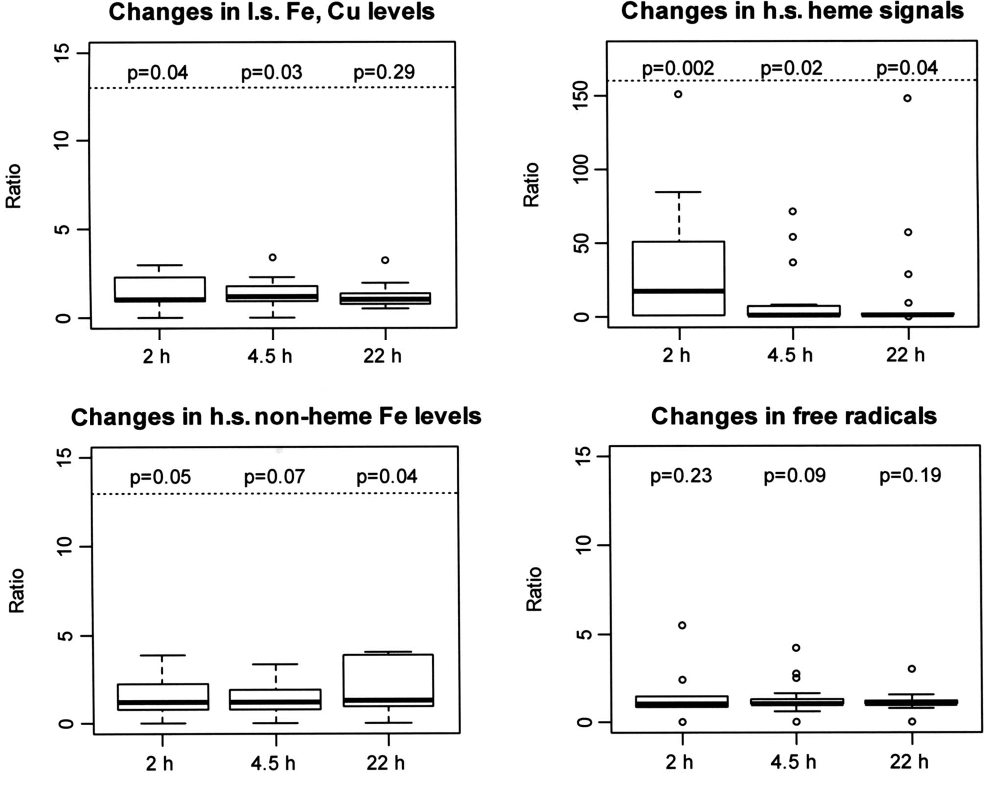
There was a significant increase in EPR signals assigned to Fe and
Cu sites at 2 and 4.5 h (p=0.04 and 0.03), reflective of 3-AP
binding to Fe or Cu, which was not observed at 22 h after 3-AP
administration (p=0.29); this suggested that 3-AP affected its
action primarily at early time-points (Fig. 1A, Table I).
 | Table I.Changes in low spin Fe, Cu levels from
baseline to 2, 4.5 and 22 h after 3-AP administration, expressed as
ratios over baseline levels. |
Table I.
Changes in low spin Fe, Cu levels from
baseline to 2, 4.5 and 22 h after 3-AP administration, expressed as
ratios over baseline levels.
| Time | No. | Median | Range | Mean | SD | p-value |
|---|
| Baseline - 2 h | 16 | 1.02 | 0.01–62.00 | 5.20 | 15.17 | 0.04a |
| Baseline - 4.5 h | 18 | 1.21 | 0.01–111.0 | 9.30 | 26.68 | 0.03a |
| Baseline - 22 h | 18 | 1.00 | 0.48–51.00 | 3.92 | 11.77 | 0.29 |
EPR spectra of peripheral blood
lymphocytes (PBLs), before and after treatment with 3-AP
EPR spectra of high spin (h.s.) heme were detected
at g=6. This represents the Fe(3+) in heme, in an oxidized state.
The heme signal was amplified significantly after the
administration of 3-AP, which was consistent with our previous
study validating the measurement of these signals using EPR
(6). There was a significant
17-fold median increase from baseline at 2 h (p=0.002), a
significant increase at 4.5 h (p=0.02) and a significant increase
at 22 h (p=0.04), indicating heightened heme Fe activity over a
prolonged period following 3-AP administration (Fig. 1B, Table II). Although commonly hypothesized,
it was difficult to establish in this case whether the heme signal
was the heme in cytochrome c and whether it was involved in
eliciting oxidative damage.
| Table II.Changes in high spin heme signals from
baseline to 2, 4.5 and 22 h after 3-AP administration (g=6),
expressed as ratios over baseline signals. |
Table II.
Changes in high spin heme signals from
baseline to 2, 4.5 and 22 h after 3-AP administration (g=6),
expressed as ratios over baseline signals.
| Time | No. | Median | Range | Mean | SD | p-value |
|---|
| Baseline - 2 h | 16 | 16.81 | 0.67–151.0 | 32.27 | 42.24 | 0.002a |
| Baseline - 4.5 h | 18 | 1.00 | 0.02–71.0 | 10.55 | 20.93 | 0.020a |
| Baseline - 22 h | 18 | 1.00 | 0.02–148.0 | 14.22 | 36.29 | 0.040a |
At g=4.3, EPR spectra for h.s. non-heme Fe were
obtained. This signal is primarily from the Fe(III) in transferrin
and, to a small extent, from Fe-phosphates and other non-heme Fe
signals. The intensity of this signal changed with a 1.22-fold
median increase at 2 h, 1.18-fold at 4.5 h and 1.31-fold at 22 h
after 3-AP administration, although not statistically significant
at 2 and 4.5 h (p=0.05 and 0.07), but significant at 22 h (p=0.04;
Fig. 1C, Table III). This Fe-transferrin signal was
the oxidized state of Fe-transferrin, possibly due to the
generation of ROS. These data are consistent with our previous
findings in that the intensity of the peak at g=4.3 increased at 2
h, with a less intense signal at 4 h. However, in our previous
study there was little or no signal at 22 h after 3-AP
administration (6). These results
suggest that effective inhibition of iron uptake occurs at later
time-points.
| Table III.Changes in high spin non-heme Fe from
baseline to 2, 4.5 and 22 h after 3-AP administration (g=4.3),
expressed as ratios over baseline signals. |
Table III.
Changes in high spin non-heme Fe from
baseline to 2, 4.5 and 22 h after 3-AP administration (g=4.3),
expressed as ratios over baseline signals.
| Time | No. | Median | Range | Mean | SD | p-value |
|---|
| Baseline - 2 h | 16 | 1.22 | 0.04–65.00 | 7.98 | 18.58 | 0.05 |
| Baseline - 4.5
h | 18 | 1.18 | 0.04–44.00 | 5.65 | 12.92 | 0.07 |
| Baseline - 22
h | 18 | 1.31 | 0.01–79.00 | 12.03 | 25.31 | 0.04a |
Other peaks were obtained at g=2.005, which were
attributable to free radicals. However, the difference in
intensities of these peaks prior to and following the
administration of 3-AP was not statistically significant (Fig. 1D, Table IV). EPR spectra for the RR tyrosyl
radical were not captured (data not shown).
| Table IV.Changes in free radicals from
baseline to 2, 4.5 and 22 h after 3-AP administration (g=2.005),
expressed as ratios over baseline values. |
Table IV.
Changes in free radicals from
baseline to 2, 4.5 and 22 h after 3-AP administration (g=2.005),
expressed as ratios over baseline values.
| Time | No. | Median | Range | Mean | SD | p-value |
|---|
| Baseline - 2 h | 16 | 1.00 | 0.01–49.00 | 4.24 | 12.00 | 0.23 |
| Baseline - 4.5
h | 18 | 1.00 | 0.02–4.18 | 1.35 | 0.94 | 0.09 |
| Baseline - 22
h | 18 | 1.00 | 0.01–120.00 | 7.69 | 28.03 | 0.19 |
Discussion
Ribonucleotide reductase (RR) plays a vital role in
DNA synthesis by catalyzing the conversion of nucleotides to
deoxynucleotides. Without a balanced supply of
deoxyribonucleotides, DNA synthesis is inhibited (1). Anticancer agents that are good
chelators of iron have been sought for the purpose of inhibiting
the RR enzyme by removing iron from RR (8). 3-AP is a potent inhibitor of RR
(1), but it is not certain whether
the inhibition is a result of removing iron from RR or whether 3-AP
forms a metal complex that inhibits RR activity, or both.
In previous studies with a precursor for 3-AP, the
preformed cupric complex of 2-formylpyridine monothiosemicarbazone
was a potent inhibitor of RR (25). 3-AP is also a tridentate chelator
that ligates Fe and other metals. Preformed Fe-3-AP is a more
potent inhibitor of RR than free 3-AP (5). It is believed that 3-AP forms a
complex with Fe(III), is reduced to Fe(II), generates ROS and
quenches RR activity (1,10). A similar scenario has been reported
for the cupric complex of 2-formylpyridine monothiosemicarbazone
(25).
In this study, EPR signals associated with iron and
copper sites in PBLs from patients were found to increase 2 h after
3-AP administration and continued to increase up to 4.5 h, with a
smaller increase observed 24 h after treatment (Fig. 1A, Table I). A significant signal observed by
EPR was that of h.s. heme iron (Fig.
1B, Table II), which was
greatly increased at 2 h after treatment and was magnified to small
extents at 4.5 and 22 h after 3-AP administration. This is
consistent with recordings from our previous study that validated
the use of EPR to capture these signals (6). One compelling hypothesis is that ROS
cause cell death and release of cytochrome c following apoptosis.
Upon release of cytochrome c, the heme is oxidized as measured by
an increase in the heme signal. Although hypothetically plausible,
the heme signal is an accumulation of all oxidized hemes, and
further investigation is necessary to attribute the increase in
heme to cytochrome c. Nonetheless, administration of 3-AP is known
to cause apoptosis (26). One of
the objectives of the present study was to measure the decrease in
the intensity of tyrosyl radical of RR by EPR. However, these
signals could not be captured. To date, the tyrosyl radical signal
from RR has only been detected in rapidly proliferating cells,
i.e., 100% cancer cells.
Another signal that was unequivocally identified was
the signal for Fe-transferrin, which was increased at 2 h after
drug administration, followed by a smaller increase at 4.5 h and a
large increase at 22 h after treatment (Fig. 1C, Table III). It appears that
Fe(III)-transferrin was not reduced. It is possible that Fe uptake
was blocked, probably through the generation of ROS from
FeT2 or CuT, which damaged transferrin or the
transferrin receptor. In support of this idea, it is known that
2-formylpyridine monothiosemicaboxylate Cu(II) inhibits cellular
iron uptake in addition to inhibiting RR (11). Assuming that ROS are generated by
either FeT2, CuT or adducts of these complexes that form
either by replacing T or by occupying the open equatorial site of
the tri-dentate cupric complex, other sites may also be damaged.
The Fe-3-AP complex has been shown to dramatically increase ROS
production in model systems (9).
Of note, the pharmacokinetic parameters of 3-AP
obtained from phase I clinical trial data demonstrate a Tmax
of 0.04±0.11 h in erythrocytes and 0.22±0.11 h in plasma (19,20).
These peak concentrations appear much earlier than the observed
pharmacodynamic effects of 3-AP at 2, 4.5 and 22 h depicted in
Fig. 1. This delayed effect could
be due to a number of factors, such as slow uptake from plasma and
a prolonged intracellular retention of 3-AP prior to formation of
metal complexes, which would suggest a slow manifestation of
cytotoxicity and potential apoptosis. Further studies are required
to determine the reasons for this delayed effect of 3-AP in
relation to its pharmacokinetics.
In conclusion, this study provides novel insight
into EPR evaluation of the effects of 3-AP in patients, identifying
an Fe-transferrin signal and potential cytochome c release from
mitochondria, in addition to detecting signals at the g=2 region.
Formation of metal complexes of 3-AP are likely essential to its
anticancer effect, whether the mechanism is related to RR
inhibition or the formation of ROS, or both (1,10).
These results provide direct evidence for the formation of metal
complexes, although not assigned, in patients receiving 3-AP
therapy and provide valuable insight into the in vivo
mechanism of the action of 3-AP.
Acknowledgements
This study was supported by the U01
CA62491, CTEP Translational Research Initiative Funds 24XS090, the
NIH GCRC Grant M01 RR03186 and the NIH-National Biomedical ESR
Center EB001980 Grant.
References
|
1.
|
Yu Y, Wong J, Lovejoy D, Kalinowski D and
Richardson D: Chelators at the cancer coalface: desferrioxamine to
Triapine and beyond. Clin Cancer Res. 12:6876–6883. 2006.
View Article : Google Scholar : PubMed/NCBI
|
|
2.
|
Nordlund P, Sjöberg B and Eklund H:
Three-dimensional structure of the free radical protein of
ribonucleotide reductase. Nature. 345:593–598. 1990. View Article : Google Scholar : PubMed/NCBI
|
|
3.
|
Qiu W, Zhou B, Darwish D, Shao J and Yen
Y: Characterization of enzymatic properties of human ribonucleotide
reductase holoenzyme reconstituted in vitro from hRRM1, hRRM2, and
p53R2 subunits. Biochem Biophys Res Commun. 340:428–434. 2006.
View Article : Google Scholar : PubMed/NCBI
|
|
4.
|
Cooper C, Lynagh G, Hoyes K, Hider R,
Cammack R and Porter J: The relationship of intracellular iron
chelation to the inhibition and regeneration of human
ribonucleotide reductase. J Biol Chem. 271:20291–20299. 1996.
View Article : Google Scholar : PubMed/NCBI
|
|
5.
|
Shao J, Zhou B, Di Bilio A, et al: A
Ferrous-Triapine complex mediates formation of reactive oxygen
species that inactivate human ribonucleotide reductase. Mol Cancer
Ther. 5:586–592. 2006. View Article : Google Scholar : PubMed/NCBI
|
|
6.
|
Kolesar J, Schelman W, Geiger P, et al:
Electron paramagnetic resonance study of peripheral blood
mononuclear cells from patients with refractory solid tumors
treated with Triapine. J Inorg Biochem. 102:693–698. 2008.
View Article : Google Scholar
|
|
7.
|
Petering DH and Antholine WE: Reviews in
Biochemical Toxicology. 9. Hodgson E, Bend JR and Philpot RM:
Elsevier; New York: 1988
|
|
8.
|
Richardson D, Sharpe P, Lovejoy D, et al:
Dipyridyl thiosemicarbazone chelators with potent and selective
antitumor activity form iron complexes with redox activity. J Med
Chem. 49:6510–6521. 2006. View Article : Google Scholar : PubMed/NCBI
|
|
9.
|
Whitnall M, Howard J, Ponka P and
Richardson D: A class of iron chelators with a wide spectrum of
potent antitumor activity that overcomes resistance to
chemotherapeutics. Proc Natl Acad Sci USA. 103:14901–14906. 2006.
View Article : Google Scholar : PubMed/NCBI
|
|
10.
|
Chaston T, Lovejoy D, Watts R and
Richardson D: Examination of the antiproliferative activity of iron
chelators: multiple cellular targets and the different mechanism of
action of Triapine compared with desferrioxamine and the potent
pyridoxal isonicotinoyl hydrazone analogue 311. Clin Cancer Res.
9:402–414. 2003.
|
|
11.
|
Narasimhan J, Antholine W, Chitambar C and
Petering D: Inhibition of iron uptake in HL60 cells by
2-formylpyridine monothiosemicarbazonato Cu(II). Arch Biochem
Biophys. 289:393–398. 1991. View Article : Google Scholar : PubMed/NCBI
|
|
12.
|
Martinou J, Desagher S and Antonsson B:
Cytochrome c release from mitochondria: all or nothing. Nat Cell
Biol. 2:E41–E43. 2000. View
Article : Google Scholar : PubMed/NCBI
|
|
13.
|
DeConti R, Toftness B, Agrawal K, et al:
Clinical and pharmacological studies wit h
5-hydroxy-2-formylpyridine thiosemicarbazone. Cancer Res.
32:1455–1462. 1972.
|
|
14.
|
Sartorelli A: Effect of chelating agents
upon the synthesis of nucleic acids and protein: inhibition of DNA
synthesis by 1-formylisoquinoline thiosemicarbazone. Biochem
Biophys Res Commun. 27:26–32. 1967. View Article : Google Scholar
|
|
15.
|
Sartorelli A, Agrawal K and Moore E:
Mechanism of inhibition of ribonucleoside diphosphate reductase by
a-(N)-heterocyclic aldehyde thiosemicarbazones. Biochem Pharmacol.
20:3119–3123. 1971. View Article : Google Scholar : PubMed/NCBI
|
|
16.
|
Zhu L, Zhou B, Chen X, Jiang H, Shao J and
Yen Y: Inhibitory mechanisms of heterocyclic carboxaldehyde
thiosemicabazones for two forms of human ribonucleotide reductase.
Biochem Pharmacol. 78:1178–1185. 2009. View Article : Google Scholar
|
|
17.
|
Kowol C, Trondl R, Arion V, Jakupec M,
Lichtscheidl I and Keppler B: Fluorescence properties and cellular
distribution of the investigational anticancer drug Triapine
(3-aminopyridine-2-carboxaldehyde thiosemicarbazone) and its
zinc(II) complex. Dalton Trans. 39:704–706. 2010. View Article : Google Scholar
|
|
18.
|
Attia S, Kolesar J, Mahoney M, et al: A
phase 2 consortium (P2C) trial of 3-aminopyridine-2-carboxaldehyde
thiosemicarbazone (3-AP) for advanced adenocarcinoma of the
pancreas. Invest New Drugs. 26:369–379. 2008. View Article : Google Scholar : PubMed/NCBI
|
|
19.
|
Choi B, Alberti D, Schelman W, et al: The
maximum tolerated dose and biologic effects of
3-aminopyridine-2-carboxaldehyde thiosemicarbazone (3-AP) in
combination with irinotecan for patients with refractory solid
tumors. Cancer Chemother Pharmacol. 66:973–980. 2010. View Article : Google Scholar : PubMed/NCBI
|
|
20.
|
Schelman W, Morgan-Meadows S, Marnocha R,
et al: A phase I study of Triapine in combination with doxorubicin
in patients with advanced solid tumors. Cancer Chemother Pharmacol.
63:1147–1156. 2009. View Article : Google Scholar : PubMed/NCBI
|
|
21.
|
Nutting C, van Herpen C, Miah A, et al:
Phase II study of 3-AP Triapine in patients with recurrent or
metastatic head and neck squamous cell carcinoma. Ann Oncol.
20:1275–1279. 2009. View Article : Google Scholar : PubMed/NCBI
|
|
22.
|
Traynor A, Lee J, Bayer G, et al: A phase
II trial of Triapine (NSC# 663249) and gemcitabine as second line
treatment of advanced non-small cell lung cancer: Eastern
Cooperative Oncology Group Study 1503. Invest New Drugs. 28:91–97.
2010.
|
|
23.
|
Kunos C, Chiu S, Pink J and Kinsella T:
Modulating radiation resistance by inhibiting ribonucleotide
reductase in cancers with virally or mutationally silenced p53
protein. Radiat Res. 172:666–676. 2009. View Article : Google Scholar : PubMed/NCBI
|
|
24.
|
Antholine W, Knight J, Whelan H and
Petering D: Studies of the reaction of 2-formylpyridine
thiosemicarbazone and its iron and copper complexes with biological
systems. Mol Pharmacol. 13:89–98. 1977.PubMed/NCBI
|
|
25.
|
Saryan L, Mailer K, Krishnamurti C,
Antholine W and Petering D: Interaction of 2-formylpyridine
thiosemicarbazonato copper (II) with Ehrlich ascites tumor cells.
Biochem Pharmacol. 30:1595–1604. 1981. View Article : Google Scholar : PubMed/NCBI
|
|
26.
|
Alvero A, Chen W, Sartorelli A, Schwartz
P, Rutherford T and Mor G: Triapine
(3-aminopyridine-2-carboxaldehyde thiosemicarbazone) induces
apoptosis in ovarian cancer cells. J Soc Gynecol Investig.
13:145–152. 2006. View Article : Google Scholar : PubMed/NCBI
|