Introduction
Gastric cancer is the second most common cause of
cancer-related deaths worldwide (1). Clinical outcomes remain poor in
patients with advanced gastric cancer, even after curative
resection. Tyrosine kinase receptors, such as epidermal growth
factor receptor (EGFR), its homolog c-erb-2 (HER2) and vascular
endothelial growth factor (VEGF), have been linked to tumor
progression or survival in gastric cancer (2–7). We
also previously reported that EGFR or HER3 expression was
correlated with tumor progression in gastric cancer (8). Several anticancer drugs designed to
inhibit signaling pathways of tyrosine kinases have been evaluated
in unresectable or metastatic gastric cancer, and trastuzumab
(anti-HER2) has been shown to be effective (9).
The main pathways stimulated by EGFR involve
phosphatidylinositol 3-kinase (PI3K)/Akt, mitogen-activated protein
kinases (MAPK) and signal transducer and activator of transcription
3 (STAT3) (10,11). The mammalian target of rapamycin
(mTOR) is one of the targets of activated Akt. Phosphorylated mTOR
(p-mTOR) stimulates initiation of translation by two targets:
ribosomal p70S6 kinase (S6K1) and eukaryotic translation initiation
factor 4E binding protein (4E-BP1). mTOR thus promotes cell growth,
proliferation and differentiation (12,13).
We previously reported that the expression of cytoplasmic mTOR is
associated with tumor progression and poorer outcomes in gastric
cancer (14). STAT3 belongs to a
family of cytoplasmic latent transcription factors that function as
both signal transducers and activators of transcription. EGFR
interacts with and activates STAT3. Phosphorylated STAT3 (p-STAT3)
translocates to the nucleus and binds to recognition sequences in
the promoter region of target genes, thereby regulating their
transcription. Thus, aberrant STAT3 signaling has been linked to
the promotion of cell cycle progression, cell survival and
malignant transformation (15,16).
STAT3 is related to tumor progression and worse survival in gastric
(17–19), colorectal (20,21),
lung (22,23) and breast cancer (24).
The present study examined whether the expression of
p-STAT3, p-mTOR and EGFR correlates with clinicopathological
features and patient outcome in gastric cancer. The expression was
evaluated immunohistochemically.
Patients and methods
Patients
The study group comprised 126 patients with primary
invasive (invasion deeper than the muscularis mucosa) gastric
adenocarcinomas who underwent gastrectomy from January 1999 through
December 2002 at the Department of Esophagogastric Surgery, Tokyo
Medical and Dental University, Japan. Each tumor was classified
according to the tumor-node-metastasis classification recommended
by the International Union against Cancer (UICC). All patients were
evaluated for recurrent disease by diagnostic imaging, including
computed tomography, ultrasonography and endoscopy, every 3–6
months. The median follow-up time was 73 months (range 2–135). Ten
patients who underwent R1 or R2 surgery received S-1 chemotherapy;
no other patient received neoadjuvant therapy. Recurrent disease
was diagnosed in 28 (24%) of 116 patients who underwent R0 surgery
and was the cause of death in these patients.
Immunohistochemical staining
Immunohistochemical staining was performed using the
streptavidin-biotin method using a Histofine SAB-PO kit (Nichirei
Co., Tokyo, Japan). Polyclonal rabbit anti-human antibodies against
p-STAT3 (Thy705) and p-mTOR (Ser2448) were purchased from Cell
Signaling Technology, Inc. (Beverly, MA, USA). Monoclonal mouse
antibodies against EGFR were from Novocastra Laboratories
(Newcastle upon Tyne, UK). All available H&E-stained slides of
the surgical specimens were reviewed. For each case, representative
paraffin blocks were selected for immunohistochemical studies.
Four-micrometer sections were cut from formalin-fixed,
paraffin-embedded tissue blocks. After deparaffinization and
rehydration, antigen retrieval treatment was performed at 121°C
(autoclave) for 5 min in 10 nmol/l sodium citrate buffer (pH 9.0),
followed by treatment with 3% hydrogen peroxide for 15 min to
quench endogenous peroxidase activity. Non-specific binding was
blocked by treating the slides with 5% EzBlock (including 5% normal
goat serum and 0.1% Tween-20) for 60 min at room temperature. The
slides were incubated with a primary antibody (dilution 1:50)
overnight at 4°C. Immunodetection was performed by the conventional
streptavidin-biotin method with a Nichirei SAB-PO kit (Nichirei
Co.). The slides were counterstained with 1% Mayer’s
hematoxylin.
Two independent observers blinded to the patients’
clinical information examined all of the slides with an optical
microscope. Positive expression was defined as >10% of the cells
with nuclear staining for p-STAT3 (17) or >10% of the cells with
cytoplasmic staining for p-mTOR, as previously described (14). Positive EGFR expression was defined
as >10% of the cells with moderate or strong membrane
staining.
Statistical analysis
The Chi-square test was used to test possible
associations of the expression of p-STAT3, p-mTOR and EGFR with
clinicopathological factors. It was also used to assess
correlations between each type of expression. Kaplan-Meier curves
were plotted to assess the effects of each type of expression on
disease-specific survival (DSS). Survival curves were compared
using the log-rank test. P-values of <0.05 were considered to
indicate statistical significance. Multivariate proportional Cox
models were used to assess the prognostic significance of all
expression levels and of several clinicopathological factors.
Statistical analysis was performed with the use of SPSS Base,
version 11.0 and SPSS Advanced Models, version 11.0 (SPSS Inc.,
Chicago, IL, USA) software.
Results
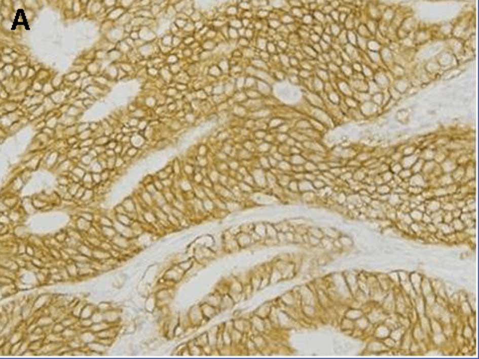
Expression of nuclear p-STAT3, cytoplasmic p-mTOR
and membranous EGFR was found in 52 (41%), 81 (64%) and 37 (29%)
tumors, respectively (Fig. 1). All
three types of expression were observed in 16 (13%) tumors, while
28 (22%) tumors showed no expression. The expression of p-STAT3
significantly correlated with that of mTOR (p=0.035). EGFR
expression correlated slightly, but not significantly with p-STAT3
and mTOR expression (p=0.060 and p=0.054).
Immunoreactivity for p-STAT3, p-mTOR and EGFR was
present in advanced cancer. Each type of expression positively
correlated with the depth of tumor invasion (T1 vs. T2–4;
p<0.001, p=0.036 and p<0.001, respectively) and lymph node
involvement (p=0.008, p=0.027 and p=0.007). Each type of expression
was also significantly associated with UICC stage (I vs. II–IV;
p<0.001, p=0.041 and p<0.001). Expression of p-STAT3 or EGFR
was significantly related to distant metastasis or recurrence
(p=0.001 and p=0.039), whereas the expression of p-mTOR was not
(p=0.21). No type of expression significantly correlated with
histological type or gender, although p-STAT3 expression tended to
be associated with older age (p=0.050). Co-expression of p-STAT3,
p-mTOR and EGFR correlated significantly with tumor depth, lymph
node involvement, UICC stage and distant metastasis or recurrence
(p<0.001, p=0.001, p<0.001 and p=0.008, respectively)
(Table I).
 | Table I.Correlation of p-STAT3, p-mTOR and
EGFR expression with clinicopathological factors. |
Table I.
Correlation of p-STAT3, p-mTOR and
EGFR expression with clinicopathological factors.
| n | Nuclear p-STAT3 | Cytoplasmic
p-mTOR | Membranous EGFR | Co-expression
values |
|---|
|
|
|
|
|---|
| Negative | Positive | p-value | Negative | Positive | p-value | Negative | Positive | p-value | 0 | 1 | 2 | 3 | p-value |
|---|
| Age | | | | | | | | | | | | | | | |
| ≥70 | 41 | 19 | 22 | 0.050 | 12 | 29 | 0.290 | 26 | 15 | 0.220 | 6 | 13 | 13 | 9 | 0.140 |
| <70 | 85 | 55 | 30 | | 33 | 52 | | 63 | 22 | | 22 | 29 | 27 | 7 | |
| Gender | | | | | | | | | | | | | | | |
| Male | 88 | 54 | 34 | 0.360 | 31 | 57 | 0.860 | 60 | 28 | 0.480 | 21 | 27 | 28 | 12 | 0.760 |
| Female | 38 | 20 | 18 | | 14 | 24 | | 29 | 9 | | 7 | 15 | 12 | 4 | |
| Tumor location | | | | | | | | | | | | | | | |
| Middle/Lower | 103 | 61 | 42 | >0.990 | 39 | 64 | 0.410 | 72 | 31 | 0.900 | 23 | 35 | 33 | 12 | 0.900 |
| Upper | 23 | 13 | 10 | | 6 | 17 | | 17 | 6 | | 5 | 7 | 7 | 4 | |
| Histopathology | | | | | | | | | | | | | | | |
| Intestinal | 48 | 32 | 16 | 0.160 | 29 | 49 | 0.660 | 35 | 13 | 0.660 | 10 | 21 | 11 | 6 | 0.210 |
| Diffuse | 78 | 42 | 36 | | 16 | 32 | | 54 | 24 | | 18 | 21 | 29 | 10 | |
| Depth of
invasion | | | | | | | | | | | | | | | |
| T1 | 49 | 39 | 10 | <0.001 | 23 | 26 | 0.036 | 45 | 4 | <0.001 | 19 | 20 | 10 | 0 | <0.001 |
| T2/3/4 | 77 | 35 | 42 | | 22 | 55 | | 44 | 33 | | 9 | 22 | 30 | 16 | |
| LN metastasis | | | | | | | | | | | | | | | |
| Negative
(N0) | 59 | 42 | 17 | 0.008 | 27 | 32 | 0.027 | 49 | 10 | 0.007 | 22 | 19 | 14 | 4 | 0.001 |
| Positive
(N1/2/3) | 67 | 32 | 35 | | 18 | 49 | | 40 | 27 | | 6 | 23 | 26 | 12 | |
| Distant metastasis
or recurrence | | | | | | | | | | | | | | | |
| Negative | 88 | 60 | 28 | 0.001 | 35 | 53 | 0.210 | 67 | 21 | 0.039 | 26 | 30 | 24 | 8 | 0.008 |
| Positive | 38 | 14 | 24 | | 10 | 28 | | 22 | 16 | | 2 | 12 | 16 | 8 | |
| Stage | | | | | | | | | | | | | | | |
| I | 63 | 47 | 16 | <0.001 | 28 | 35 | 0.041 | 54 | 9 | <0.001 | 24 | 21 | 15 | 3 | <0.001 |
| II/III/IV | 63 | 27 | 36 | | 17 | 46 | | 35 | 28 | | 4 | 21 | 25 | 13 | |
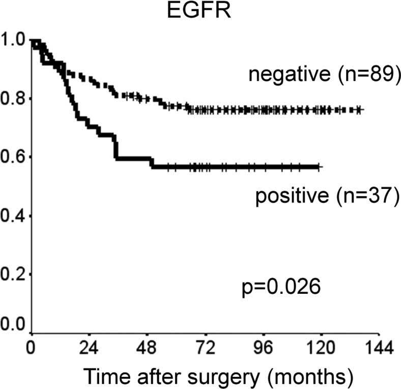
Patients with p-STAT3 or EGFR expression had
significantly shorter DSS than did those without such expression
(p=0.0018 and p=0.026). There was a slight, but insignificant
association between p-mTOR expression and shorter DSS (p=0.096)
(Fig. 2). The prognostic relevance
of the expression of each protein was assessed using a multivariate
proportional hazards model adjusted for several clinical factors
(main location of tumor, histological type, depth of tumor, lymph
node metastasis and distant metastasis) (Table II). Expression of p-STAT3 was a
marginally insignificant prognostic factor for DSS [hazard ratio
(HR)=2.0, 95% CI 0.91–4.5, p=0.082). Distant metastasis was the
only independent prognostic factor for DSS (HR=6.4, 95% CI 2.2–19,
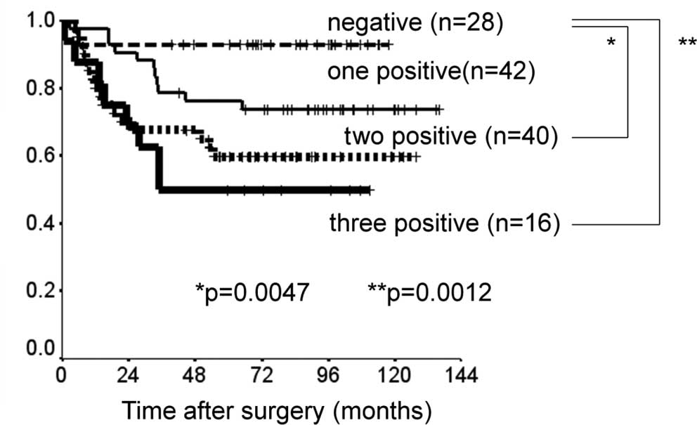
p=0.001). DSS was reduced with increased co-expression of p-STAT3,
p-mTOR and EGFR (Fig. 3). DSS at 5
years was 93% in patients with no expression of the proteins, 76%
in patients with positive expression of one type of protein, 60% in
those with positive expression of two of the proteins and 50% in
those with positive expression of all three proteins. Survival was
significantly longer in patients with no positive expression than
in those with positive expression of two or all three proteins
(none vs. all: p=0.0012; none vs. two: p=0.0047).
| Table II.Prognostic factors in a multivariate
Cox proportional hazards regression model. |
Table II.
Prognostic factors in a multivariate
Cox proportional hazards regression model.
| DSS
|
|---|
| HR | 95% CI | p-value |
|---|
| Main tumor
location | | | |
| Middle/Lower | 1.00 | | |
| Upper | 1.40 | 0.58-3.4 | 0.450 |
| Histopathology
(Lauren) | | | |
| Intestinal | 1.00 | | |
| Diffuse | 1.30 | 0.55-3.0 | 0.570 |
| Tumor depth | | | |
| T1 | 1.00 | | |
| T2–4 | 3.70 | 0.75-18 | 0.110 |
| Lymph node
metastasis | | | |
| Negative
(N0) | 1.00 | | |
| Positive
(N1–3) | 1.90 | 0.67-5.7 | 0.220 |
| Distant
metastasis | | | |
| Negative
(M0) | 1.00 | | |
| Positive
(M1) | 6.40 | 2.20-19 | 0.001 |
| EGFR | | | |
| Negative | 1.00 | | |
| Positive | 1.70 | 0.74-3.8 | 0.220 |
| p-mTOR | | | |
| Negative | 1.00 | | |
| Positive | 0.66 | 0.26-1.7 | 0.390 |
| p-STAT3 | | | |
| Negative | 1.00 | | |
| Positive | 2.00 | 0.91-4.5 | 0.082 |
Discussion
The present study showed that the co-expression of
p-STAT3, p-mTOR and EGFR, members of the EGFR signaling pathway,
was significantly associated with tumor progression and less
favorable survival in gastric cancer. The expression levels of
these proteins appeared to be related to clinical outcomes, but
were not independent prognostic factors. In addition, the
co-expression of these proteins was associated with poorer DDS.
STAT3 translocates to the nucleus, where it
activates gene transcription and modulates cell proliferation,
apoptosis and differentiation. Therefore, nuclear expression of
p-STAT3, the activated form of STAT3, may be more intimately
related to tumor progression than its cytoplasmic expression.
Immunohistochemically, STAT3 is detected much more frequently in
gastric cancer than in surrounding normal tissue (18). In the present study, p-STAT3
expression was rarely detected in normal gastric mucosa. Nuclear
expression of p-STAT3 was significantly associated with tumor
progression and clinical outcomes in gastric cancer, whereas
cytoplasmic expression of p-STAT3 was not (data not shown). Several
studies showed that immunohistochemical staining for p-STAT3 or
STAT3 was related to poor survival in gastric cancer, without
specifying the site of staining. Park et al reported that
p-STAT3 was expressed predominantly in the nucleus in colon cancer.
However, when they analyzed nuclear and cytoplasmic expression
together, p-STAT3 was found to be associated with clinical stage
(21). In breast cancer, STAT3 was
detected mainly in the cytoplasm and its expression was related to
the overall 5-year survival rate (24). This discrepancy in the location of
staining may be attributed to differences in the type of cancer or
in the antibodies used for immunostaining. Lee et al found
that immunohistochemical staining for nuclear p-STAT3 was an
independent prognostic factor in gastric cancer. The prognostic
value appeared to be enhanced when p-STAT3 and matrix
metalloproteinase-10 (MMP-10) were combined (19). STAT3 activation is implicated in
the modulation of MMP-10 activity. Several molecular targets
involving the STAT3 signaling pathway may have a substantial effect
on patient outcome in gastric cancer.
EGFR expression has been considered a prognostic
factor for patients with gastric cancer (2,3). In
the present study, EGFR expression was significantly associated
with tumor progression and poor survival in gastric cancer, but was
not an independent prognostic factor. EGFR consists of an
extracellular ligand-binding domain, a transmembrane domain and an
intracytoplasmic tyrosine kinase domain. EGFR expression is
detected mainly in the membrane of cancer cells, but has also been
detected in the nucleus in several studies (25,26).
The EGFR antibodies used in the present study were rarely detected
in the cytoplasm or nucleus. EGFR interacts with STAT3 in the
nucleus, leading to transcriptional activation of inducible nitric
oxide synthases (26). Several
inhibitors of EGFR have been used to treat unresectable or
recurrent gastric cancer, but provided no benefit in such patients
or in those with colorectal or lung cancer (27). Inhibition of EGFR alone may be
inadequate and synchronous inhibition of downstream substrates,
such as mTOR and STAT3, may be essential for improving outcomes in
gastric cancer.
We previously reported that the cytoplasmic
expression of p-mTOR positively correlated with tumor progression
and survival in gastric cancer. However, few studies have
demonstrated the impact of mTOR expression on survival in patients
with cancer (28). Akt, which is
an upstream regulator of mTOR, was not associated with outcomes in
patients with several types of cancer in previous studies,
including ours (14,29,30).
Therefore, activation of Akt/mTOR alone may not have an impact on
tumor growth or patient survival. Ma et al demonstrated that
mTOR activates STAT3/p63/Jagged signal cascade in vitro
(31). In the present study,
p-mTOR expression correlated significantly with p-STAT3 expression.
Activation of STAT3, by not only EGFR/STAT3 but also mTOR/STAT3
signaling, may lead to poor survival in gastric cancer.
In conclusion, co-expression of p-STAT3, p-mTOR and
EGFR may play an important role in tumor progression and clinical
outcomes in patients with gastric cancer.
References
|
1.
|
Parkin DM, Bray F, Ferlay J and Pisani P:
Global cancer statistics, 2002. CA Cancer J Clin. 55:74–108. 2005.
View Article : Google Scholar
|
|
2.
|
Yonemura Y, Sugiyama K, Fushida S, Kamata
T, Ohoyama S, Kimura H, Yamaguchi A and Miyazaki I: Tissue status
of epidermal growth factor and its receptor as an indicator of poor
prognosis in patients with gastric cancer. Anal Cell Pathol.
3:343–350. 1991.PubMed/NCBI
|
|
3.
|
Hirono Y, Tsugawa K, Fushida S, Ninomiya
I, Yonemura Y, Miyazaki I, Endou Y, Tanaka M and Sasaki T:
Amplification of epidermal growth factor receptor gene and its
relationship to survival in human gastric cancer. Oncology.
52:182–188. 1995. View Article : Google Scholar : PubMed/NCBI
|
|
4.
|
Allgayer H, Babic R, Gruetzner KU,
Tarabichi A, Schildberg FW and Heiss MM: c-erbB-2 is of independent
prognostic relevance in gastric cancer and is associated with the
expression of tumor-associated protease systems. J Clin Oncol.
18:2201–2209. 2000.PubMed/NCBI
|
|
5.
|
García I, Vizoso F, Martín A, Sanz L,
Abdel-Lah O, Raigoso P and García-Muñiz JL: Clinical significance
of the epidermal growth factor receptor and HER2 receptor in
resectable gastric cancer. Ann Surg Oncol. 10:234–241.
2003.PubMed/NCBI
|
|
6.
|
Hirashima Y, Yamada Y, Matsubara J,
Takahari D, Okita N, Takashima A, Kato K, Hamaguchi T, Shirao K,
Shimada Y, Taniguchi H and Shimoda T: Impact of vascular
endothelial growth factor receptor 1, 2, and 3 expression on the
outcome of patients with gastric cancer. Cancer Sci. 100:310–315.
2009. View Article : Google Scholar : PubMed/NCBI
|
|
7.
|
Jüttner S, Wissmann C, Jöns T, Vieth M,
Hertel J, Gretschel S, Schlag PM, Kemmner W and Höcker M: Vascular
endothelial growth factor-D and its receptor VEGFR-3: two novel
independent prognostic markers in gastric adenocarcinoma. J Clin
Oncol. 24:228–240. 2006.PubMed/NCBI
|
|
8.
|
Hayashi M, Inokuchi M, Takagi Y, Yamada H,
Kojima K, Kumagai J, Kawano T and Sugihara K: High expression of
HER3 is associated with a decreased survival in gastric cancer.
Clin Cancer Res. 14:7843–7849. 2008. View Article : Google Scholar : PubMed/NCBI
|
|
9.
|
Bang YJ, van Cutsem E, Feyereislova A,
Chung HC, Shen L, Sawaki A, Lordick F, Ohtsu A, Omuro Y, Satoh T,
Aprile G, Kulikov E, Hill J, Lehle M, Rüschoff J and Kang YK; for
the ToGA Trial Investigators: Trastuzumab in combination with
chemotherapy versus chemotherapy alone for treatment of
HER2-positive advanced gastric or gastro-oesophageal junction
cancer (ToGA): a phase 3, open-label, randomised controlled trial.
Lancet. 376:687–697. 2010. View Article : Google Scholar
|
|
10.
|
Grandis JR, Drenning SD, Chakraborty A,
Zhou MY, Zeng Q, Pitt AS and Tweardy DJ: Requirement of Stat3 but
not Stat1 activation for epidermal growth factor receptor-mediated
cell growth in vitro. J Clin Invest. 102:1385–1392. 1998.
View Article : Google Scholar : PubMed/NCBI
|
|
11.
|
Coffer PJ and Kruijer W: EGF receptor
deletions define a region specifically mediating STAT transcription
factor activation. Biochem Biophys Res Commun. 210:74–81. 1995.
View Article : Google Scholar : PubMed/NCBI
|
|
12.
|
Schmelzle T and Hall MN: TOR, a central
controller of cell growth. Cell. 103:253–262. 2000. View Article : Google Scholar : PubMed/NCBI
|
|
13.
|
Petroulakis E, Mamane Y, Le Bacquer O,
Shahbazian D and Sonenberg N: mTOR signaling: implications for
cancer and anticancer therapy. Br J Cancer. 94:195–199. 2006.
View Article : Google Scholar : PubMed/NCBI
|
|
14.
|
Murayama T, Inokuchi M, Takagi Y, Yamada
H, Kojima K, Kumagai J, Kawano T and Sugihara K: Relation between
outcomes and localisation of p-mTOR expression in gastric cancer.
Br J Cancer. 100:782–788. 2009. View Article : Google Scholar : PubMed/NCBI
|
|
15.
|
Bowman T, Garcia R, Turkson J and Jove R:
STATs in oncogenesis. Oncogene. 15:2474–2488. 2000. View Article : Google Scholar
|
|
16.
|
Levy DE and Lee CK: What does Stat3 do? J
Clin Invest. 109:1143–1148. 2002. View Article : Google Scholar : PubMed/NCBI
|
|
17.
|
Yakata Y, Nakayama T, Yoshizaki A, Kusaba
T, Inoue K and Sekine I: Expression of p-STAT3 in human gastric
carcinoma: significant correlation in tumour invasion and
prognosis. Int J Oncol. 30:437–442. 2007.PubMed/NCBI
|
|
18.
|
Kim DY, Cha ST, Ahn DH, Kang HY, Kwon CI,
Ko KH, Hwang SG, Park PW, Rim KS and Hong SP: STAT3 expression in
gastric cancer indicates a poor prognosis. J Gastroenterol Hepatol.
24:646–651. 2009. View Article : Google Scholar : PubMed/NCBI
|
|
19.
|
Lee J, Kang WK, Park JO, Park SH, Park YS,
Lim HY, Kim J, Kong J, Choi MG, Sohn TS, Noh JH, Bae JM, Kim S, Lim
do H, Kim KM and Park CK: Expression of activated signal transducer
and activator of transcription 3 predicts poor clinical outcome in
gastric adenocarcinoma. APMIS. 117:598–606. 2009. View Article : Google Scholar : PubMed/NCBI
|
|
20.
|
Kusaba T, Nakayama T, Yamazumi K, Yakata
Y, Yoshizaki A, Inoue K, Nagayasu T and Sekine I: Expression of
p-STAT3 in human colorectal adenocarcinoma and adenoma; correlation
with clinicopathological factors. J Clin Pathol. 58:833–838. 2005.
View Article : Google Scholar : PubMed/NCBI
|
|
21.
|
Park JK, Hong R, Kim KJ, Lee TB and Lim
SC: Significance of p-STAT3 expression in human colorectal
adenocarcinoma. Oncol Rep. 20:597–604. 2008.PubMed/NCBI
|
|
22.
|
Kim HS, Park YH, Lee J, Ahn JS, Kim J,
Shim YM, Kim JH, Park K, Han J and Ahn MJ: Clinical impact of
phosphorylated signal transducer and activator of transcription 3,
epidermal growth factor receptor, p53, and vascular endothelial
growth factor receptor 1 expression in resected adenocarcinoma of
lung by using tissue microarray. Cancer. 116:676–685. 2010.
View Article : Google Scholar
|
|
23.
|
Haura EB, Zheng Z, Song L, Cantor A and
Bepler G: Activated epidermal growth factor receptor-Stat-3
signaling promotes tumor survival in vivo in non-small cell lung
cancer. Clin Cancer Res. 11:8288–8294. 2005. View Article : Google Scholar : PubMed/NCBI
|
|
24.
|
Sheen-Chen SM, Huang CC, Tang RP, Chou FF
and Eng HL: Prognostic value of signal transducers and activators
of transcription 3 in breast cancer. Cancer Epidemiol Biomarkers
Prev. 17:2286–2290. 2008. View Article : Google Scholar : PubMed/NCBI
|
|
25.
|
Lin SY, Makino K, Xia W, Matin A, Wen Y,
Kwong KY, Bourguignon L and Hung MC: Nuclear localization of EGF
receptor and its potential new role as a transcription factor. Nat
Cell Biol. 3:802–808. 2001. View Article : Google Scholar : PubMed/NCBI
|
|
26.
|
Lo HW, Hsu SC, Ali-Seyed M, Gunduz M, Xia
W, Wei Y, Bartholomeusz G, Shih JY and Hung MC: Nuclear interaction
of EGFR and STAT3 in the activation of the iNOS/NO pathway. Cancer
Cell. 7:575–589. 2005. View Article : Google Scholar : PubMed/NCBI
|
|
27.
|
Pinto C, Di Fabio F, Barone C, Siena S,
Falcone A, Cascinu S, Rojas Llimpe FL, Stella G, Schinzari G,
Artale S, Mutri V, Giaquinta S, Giannetta L, Bardelli A and Martoni
AA: Phase II study of cetuximab in combination with cisplatin and
docetaxel in patients with untreated advanced gastric or
gastro-oesophageal junction adenocarcinoma (DOCETUX study). Br J
Cancer. 101:1261–1268. 2009. View Article : Google Scholar
|
|
28.
|
Zhou X, Tan M, Stone Hawthorne V, Klos KS,
Lan KH, Yang Y, Yang W, Smith TL, Shi D and Yu D: Activation of the
Akt/mammalian target of rapamycin/4E-BP1 pathway by ErbB2
overexpression predicts tumor progression in breast cancers. Clin
Cancer Res. 10:6779–6788. 2004. View Article : Google Scholar : PubMed/NCBI
|
|
29.
|
Nam SY, Lee HS, Jung GA, Choi J, Cho SJ,
Kim MK, Kim WH and Lee BL: Akt/PKB activation in gastric carcinomas
correlates with clinicopathologic variables and prognosis. APMIS.
111:1105–1113. 2003. View Article : Google Scholar : PubMed/NCBI
|
|
30.
|
Chadha KS, Khoury T, Yu J, Black JD, Gibbs
JF, Kuvshinoff BW, Tan D, Brattain MG and Javle MM: Activated Akt
and Erk expression and survival after surgery in pancreatic
carcinoma. Ann Surg Oncol. 13:933–939. 2006. View Article : Google Scholar : PubMed/NCBI
|
|
31.
|
Ma J, Meng Y, Kwiatkowski DJ, et al:
Mammalian target of rapamycin regulates murine and human cell
differentiation through STAT3/p63/Jagged/Notch cascade. J Clin
Invest. 120:103–114. 2010. View
Article : Google Scholar : PubMed/NCBI
|