Introduction
Hepatocellular carcinoma (HCC) is one of the most
common and aggressive human malignant neoplasias worldwide. Despite
considerable diagnostic and therapeutic advances in recent years,
the overall outcome of HCC patients remains dismal (1). To improve the effectiveness of
diagnosis and therapy, and thus reduce mortality, it is crucial to
develop novel biomarkers for early detection, for monitoring tumor
progression and for predicting the prognosis of HCC.
The Janus family of tyrosine kinases (JAK) and the
signal transducer and activator of transcription (STAT) family have
been shown to play important roles in diverse signal transduction
pathways which are involved in many biological processes, including
cell proliferation, differentiation, survival and apoptosis, as
well as cancer development and progression (2–4).
Studies have also demonstrated a strong association between the
expression of STAT proteins and the progression of various human
malignances (5,6). Among the STAT family members, STAT3
is the most commonly activated in human epithelial cancers
(7–12). STAT3 is a latent cytoplasmic
transcription factor which is tyrosine-phosphorylated by tyrosine
kinase signals. Notably, STAT3 is constitutively activated, and
contributes to oncogenesis and progression by directly or
indirectly regulating the expression of genes required for cell
proliferation, survival and angiogenesis (7). By contrast, suppressors of cytokine
signaling (SOCS) have been implicated as negative regulators of
cytokine signaling, including the JAK-STAT pathway (13). In particular, SOCS3 is regarded to
be an endogenous inhibitor of STAT3 by suppression of its
activation (14,15). Although several studies have
implicated their roles in a diverse group of human carcinomas, the
relationship between STAT3 and SOCS3 in HCC remains poorly
understood.
Herein, we determined the expression of pSTAT3
protein and STAT3 mRNA in 138 HCC and 110 adjacent non-tumorous
hepatic tissue specimens on tissue microarray, and explored the
correlation of pSTAT3 and SOCS3 expression in HCC with
clinicopathologic features. We further demonstrated the
relationship between pSTAT3 and SOCS3 expression in HCC. Therefore,
the detection of pSTAT3 and SOCS3 expression may be helpful as a
combined prognostic indicator in HCC.
Materials and methods
Patients and specimens
The patient population consisted of 138 consecutive
HCC patients who underwent partial hepa-tectomy at the First
Affiliated Hospital of Anhui Medical University (Hefei, Anhui,
China) between 2004 and 2007. HCC patients who had undergone
chemotherapy or radiation therapy before surgery were excluded, as
were patients with rheumatic disease, acute infection, HIV or other
types of cancer. The pathological tumor stage was defined according
to the sixth edition of the tumor-node-metastasis (TNM)
classification of the International Union against Cancer. Tumor
differentiation was defined according to the Edmondson grading
system (16). Complete follow-up
data were obtained from all HCC patients. Primary study end points
were post-operative overall survival (OS) and post-operative
relapse-free survival (RFS). OS and RFS were defined as the time
from the date of surgery to the date of death from HCC or to the
date of local recurrence or detection of distant metastasis,
respectively. All tissue diagnoses were confirmed by permanent
histology. Institutional ethics committee approval for the project
was granted before the study was commenced and was in compliance
with the Helsinki Declaration. Written informed consent was
obtained from all patients.
Tissue microarray (TMA) construction
Paraffin-embedded tumor and adjacent non-tumorous
tissue specimens were obtained from the archive of the Department
of Pathology, The First Affiliated Hospital of Anhui Medical
University, China. We reviewed all the H&E-stained sections
from each paraffin-embedded, formalin-fixed block to identify
target areas. Three to five representative 1-mm cores were obtained
from each case and inserted in a grid pattern into a recipient
paraffin block by using a tissue arrayer (Hengtai Instruments Inc.,
Liaoning, China).
Immunohistochemical analysis
We performed immunohistochemical analyses of pSTAT3,
SOCS3, Ki67 and vascular endothelial growth factor (VEGF) protein
expression in TMA sections (3-μm) using a Two-Step histostaining
kit (Maixin, Fuzhou, China) with monoclonal antibodies against
human pSTAT3 (1:100; Santa Cruz Biotechnology, Inc., Santa Cruz,
CA, USA), SOCS3 (1:200; Santa Cruz Biotechnology), Ki67 (working
solution, Maixin) and VEGF (working solution, Maixin). The assay
was performed as described previously (17). In brief, sections were
deparaffinized in xylene, rehydrated in a graded series of ethanol
solutions and heated in a microwave oven in 0.01 M sodium citrate
buffer (pH 6.0) for 10 min for antigen retrieval. The sections were
then immersed in 3% hydrogen peroxide in methanol for 10 min to
block endogenous peroxidase activity. After rinsing with
phosphate-buffered saline (PBS), the sections were incubated with
primary antibodies in a moist chamber for 1 h at ambient
temperature, and then incubated in horseradish
peroxidase-conjugated secondary antibody for 20 min. To visualize
positive signals, the sections were incubated with
3,3′-diaminobenzidine solution for 5 min and then counterstained
with hematoxylin solution, dehydrated and mounted. All experiments
included separate known positive and negative controls. For the
negative control samples, the primary antibody was replaced by PBS
only.
In situ hybridization
For detecting the expression of STAT3 mRNA in HCC
and adjacent non-tumorous tissues, an in situ hybridization
assay was performed as previously described (17,18).
Briefly, 3-μm TMA sections were deparaffinized, rehydrated and then
digested and refixed in 4% paraformaldehyde. The sections were then
replaced with hybridization solution and incubated with the
biotinylated oligonucleotide probe complementary to STAT3 mRNA
(5′CCTTGGATTGAGAGTCAAGATTGGGCATAT3′) (Boshide, Wuhan, China) and
scrambled sequence (negative control) at 40°C for 20 h. After
washing, the sections were incubated with a mouse anti-digoxin
antibody followed by binding to the streptavidin-biotin-peroxidase
complex. Next, the sections were stained with 3,3′-diaminobenzidine
solution and counterstained with a hematoxylin solution.
Scoring of stained sections
The stained sections were reviewed and scored for
expression of pSTAT3, SOCS3, Ki67 and VEGF protein and STAT3 mRNA
using an Olympus microscope (Olympus America, Inc., Melville, NY,
USA) independently by two experienced pathologists who had no
knowledge of the patient identities or clinical status. Positive
staining for pSTAT3 expression was defined as >25% nuclear
staining with more than moderate intensity (8). Cytoplasmic staining in >30% of
cells was considered positive for SOCS3 (19), and cytoplasmic and/or membranous
staining in >50% of cells was considered positive for VEGF
(8). The Ki67 labeling index was
scored by counting 500 cells and determining the percentage of
cells that stained positively for Ki67: score +, 0–25%; score ++,
26–50%; score +++, 51–75%; score ++++, >75% (20). For the expression of STAT3 mRNA,
the core tissue was scored as positive when >10% of the
cytoplasm was stained.
Statistical analysis
All statistical analyses were performed using SPSS
software system for Windows (version 13.0; SPSS, Chicago, IL, USA).
Differences between groups were compared using the Pearson’s
Chi-square test for qualitative variables and the Student’s t-test
for continuous variables. Kaplan-Meier curves were constructed to
determine patient RFS and OS rates. The statistical differences in
survival among subgroups were compared using the log-rank test.
P<0.05 was considered statistically significant.
Results
Expression of pSTAT3 and SOCS3 in HCC and
adjacent non-tumorous tissue specimens
To determine the expression of pSTAT3 and SOCS3 in
hepatic tissues, immunohistochemical staining was performed on 138
HCC and 110 corresponding adjacent non-tumor tissues (at least 2 cm
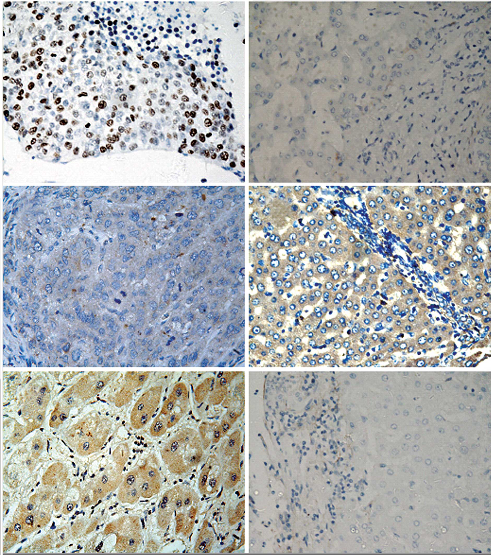
from the carcinoma margin). pSTAT3 protein was mainly expressed in
the nucleus of tumor cells or non-tumorous hepatic cells, whereas
immunoreactive SOCS3 protein was predominantly located in the
cytoplasm of cells (Fig. 1). As
shown in Table I, pSTAT3 protein
expression was detected in 75 (54.3%) of the HCC tissues and 35
(35.8%) of the non-tumor tissue (P<0.001), while the expression
of SOCS3 protein was significantly decreased in the HCC compared to
that in the non-tumor tissue (46.4 and 72.7%, P<0.001).
 | Table I.Expression of pSTAT3 and SOCS3 in the
HCC and adjacent non-tumor tissue specimens. |
Table I.
Expression of pSTAT3 and SOCS3 in the
HCC and adjacent non-tumor tissue specimens.
| | Positive expression,
n (%)
|
|---|
| Group | No. | pSTAT3 protein | SOCS3 protein | STAT3 mRNA |
|---|
| HCC | 138 | 75 (54.3)a | 64 (46.4)a | 70 (50.7)a |
| Non-tumorous | 110 | 35 (31.8) | 80 (72.7) | 33 (30.0) |
To further confirm the expression of pSTAT3 found in
the immunohistochemical analysis, digoxigenin-labeled antisense
oligonucleotide probe was utilized to detect STAT3 mRNA in the same
cohort of hepatic tissue specimens. In situ hybridization
staining revealed that the expression of STAT3 mRNA was located in
the cytoplasm of cells (Fig. 1),
and STAT3 mRNA expression was also significantly higher in the HCC
than in the non-tumorous hepatic tissues (50.7 and 30.0%,
respectively; P<0.001). Furthermore, Spearman’s correlation
analyses revealed that the expression of STAT3 mRNA was
significantly correlated with the expression of pSTAT3 protein
(correlation coefficient, rs=0.763; P<0.001).
Correlation between the expression of
pSTAT3 and SOCS3 protein
To detect the relationship between pSTAT3 and SOCS3,
we performed Spearman’s correlation analyses of protein expression
in the HCC tissue specimens. As shown in Table II, SOCS3 protein expression was
deficient in 74 of 138 HCC cases, in which the nuclear accumulation
of pSTAT3 protein was detected in 61 (82.4%). Among the 64 cases
which expressed SOCS3 protein, only 14 (21.9%) HCC cases exhibited
stronger positivity for pSTAT3. Correlation analysis revealed that
reduced SOCS3 protein expression was significantly correlated with
the high expression of pSTAT3 protein (rs=−0.606,
P<0.001).
| Table II.Correlation between the expression of
pSTAT3 and SOCS3 in HCC tissues. |
Table II.
Correlation between the expression of
pSTAT3 and SOCS3 in HCC tissues.
| pSTAT3
|
|---|
| Negative, no.
(%) | Positive, no.
(%) |
|---|
| SOCS3 | Negative, n (%) | 13 (17.6) | 61 (82.4)a |
| Positive, n (%) | 50 (78.1) | 14 (21.9) |
Association of the expression of pSTAT3
and SOCS3 with clinicopathological features of HCC
Next, we demonstrated the association of pSTAT3 and
SOCS3 protein with the clinicopathological features of the HCC
patients. As shown in Table III,
pSTAT3 expression was associated with tumor size (P=0.001) and
higher clinical stage (P=0.002). In addition, pSTAT3 expression was
also correlated with the expression of proliferation-associated
antigen Ki67 and the pro-angiogenic factor VEGF (P=0.000 and 0.001,
respectively). Furthermore, a trend was found for the decreased
expression of SOCS3 and large tumor size and high clinical stage,
although these did not reach significance (P=0.137 and 0.251,
respectively). However, SOCS3 expression was conversely correlated
with the expression of Ki67 and VEGF (P=0.034 and 0.001,
respectively).
| Table III.Association of pSTAT3 and SOCS3
expression with clinicopathological parameters of the HCC
patients. |
Table III.
Association of pSTAT3 and SOCS3
expression with clinicopathological parameters of the HCC
patients.
| Parameter | No. | pSTAT3 expression, n
(%) | P-value | SOCS3-positive
expression, n (%) | P-value |
|---|
| Age (years) | | | 0.133 | | 0.967 |
| ≤55 | 86 | 51 (59.3) | | 40 (46.5) | |
| >55 | 52 | 24 (46.2) | | 24 (46.2) | |
| Gender | | | 0.252 | | 0.222 |
| Male | 115 | 60 (52.2) | | 56 (48.7) | |
| Female | 23 | 15 (65.2) | | 8 (34.8) | |
| Cirrhosis | | | 0.533 | | 0.986 |
| Yes | 125 | 69 (55.2) | | 58 (46.4) | |
| No | 13 | 6 (46.2) | | 6 (46.2) | |
| HBsAg | | | 0.888 | | 0.513 |
| Yes | 111 | 60 (54.1) | | 53 (47.7) | |
| No | 27 | 15 (55.6) | | 11 (40.7) | |
| Tumor size
(cm) | | | 0.001 | | 0.137 |
| <5 | 41 | 13 (31.7) | | 23 (56.1) | |
| ≥5 | 97 | 62 (63.9) | | 41 (42.3) | |
| Grade | | | 0.417 | | 0.299 |
| I | 5 | 4 (80.0) | | 4 (80.0) | |
| II | 128 | 69 (53.9) | | 58 (45.3) | |
| III | 5 | 2 (40.0) | | 2 (40.0) | |
| Stage | | | 0.002 | | 0.251 |
| I–II | 106 | 50 (47.2) | | 52 (49.1) | |
| III–IV | 32 | 25 (78.1) | | 12 (37.5) | |
| Ki67 | | | 0.000 | | 0.034 |
| <50% | 60 | 22 (36.7) | | 34 (56.7) | |
| ≥50% | 78 | 53 (67.9) | | 30 (38.5) | |
| VEGF | | | 0.001 | | 0.001 |
| Negative | 53 | 12 (22.6) | | 37 (69.8) | |
| Positive | 85 | 63 (74.1) | | 27 (31.8) | |
Association of the expression of pSTAT3
and SOCS3 with patient survival
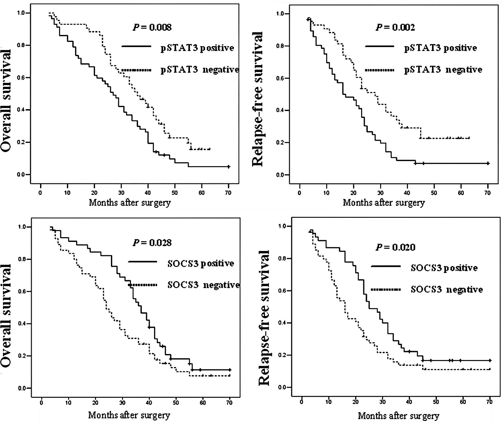
To determine whether the expression of pSTAT3 and
SOCS3 is associated with OS and RFS of HCC patients, we performed
Kaplan-Meier analyses. As shown in Fig. 2, patients whose primary tumors
expressed pSTAT3 protein had a significantly poorer OS and RFS
after curative resection compared to those without pSTAT3
expression (P=0.034 and 0.001, respectively). In addition, a
significant association of SOCS3 protein expression with patient OS
and RFS was found, which showed that patients with SOCS3-positive
expression had increased OS and RFS rates when compared with
patients without SOCS3 expression (P=0.028 and 0.020,
respectively).
Discussion
In the present study, the expression of pSTAT3
protein and STAT3 mRNA was significantly increased, while the
expression of SOCS3 was decreased in HCC compared to adjacent
non-tumor tissues. Furthermore, the expression of pSTAT3 and SOCS3
was conversely correlated in the HCC tissue specimens. In addition,
high expression of pSTAT3 and low expression of SOCS3 protein were
associated with disease progression and poor patient outcome. Our
data offer clinical evidence that pSTAT3 and SOCS3 play fundamental
roles in HCC development and progression. The data also suggest
that pSTAT3 and SOCS3 may serve as potential biomarkers for
predicting the prognosis of HCC patients.
The STAT family comprises seven members: STAT1–4,
STAT5a, STAT5b and STAT6 (6).
Since the STAT family regulates the expression of multiple genes
involved in both physiological and pathological conditions, it
seems to be one of the most promising biomarkers for predicting
disease progression and the prognosis of patients with various
cancer types. Among the STAT family members, activated STAT3 plays
a vital role in a diverse group of human cancers. Yang et al
(9) detected pSTAT3 expression in
69 HCC and corresponding adjacent non-tumor parenchyma by
immunohistochemistry, and found that 49.3% of HCC and 5.8% of
non-tumorous tissues were positive for pSTAT3 nuclear staining.
This observation differed quantitatively, but not qualitatively
with our observation that pSTAT3-positive expression was observed
in 54.3% of HCC and 31.8% of non-tumor tissues. This discrepancy
may be attributed to the different population of patients,
subjective interpretation by pathologists or assay achieving.
To avoid the limitations of immunohistochemistry, we
also performed in situ hybridization for a more precise
detection of STAT3 mRNA expression. The result of STAT3 mRNA
expression detected from in situ hybridization was
consistent with pSTAT3 protein expression. In other types of
carcinomas, Deng et al (10) reported that the expression of both
pSTAT3 protein and STAT3 mRNA was significantly higher in gastric
cancer than in normal gastric tissues. They further demonstrated
that the level of pSTAT3 immunohistochemical expression was
positively associated with the status of lymph node metastasis and
poor patient survival. Constitutive activation of STAT3 has also
been implicated in lung cancer development. Kim et al
(11) detected pSTAT3 and VEGFR1
expression by immmunohistochemistry and found that pSTAT3 was
expressed with higher frequency (51.2%) in 162 lung
adenocarcinomas. Their data further revealed that the coexpression
of pSTAT3 and VEGFR1 was significantly correlated with increased
lymph node involvement and a trend towards a decreased OS.
Similarly, in urothelial carcinoma, Huang et al (12) demonstrated that the expression of
pSTAT3 protein was positively associated with tumor invasiveness
and high histological grade. In our HCC patient population,
increased pSTAT3 expression was observed, in accord with previous
studies, and our data further demonstrated that pSTAT3 expression
correlated with higher tumor stage and decreased patient survival.
Moreover, pSTAT3 expression has been implicated in tumor cell
proliferation, and tumoral VEGF production and angiogenesis
(8,21,22).
Thus, we also observed a correlation between pSTAT3 expression and
tumor expression of Ki67 and VEGF.
SOCS are a family of proteins that regulate negative
feedback to the signaling cascade of JAK/STAT activating cytokine
(23,24). There are eight members of the SOCS
family: the cytokine inducible Src homology 2 domain-containing
protein and SOCS1–7 (24). In the
present study, decreased expression of SOCS3 was found to correlate
with poor patient survival and expression of Ki67, VEGF and pSTAT3,
suggesting a tumor suppressor role of SOCS3 in HCC. Indeed, the
function of SOCS3 has been examined in vitro and in
vivo as a negative regulator of STATs. Forced expression of
SOCS3 was found to result in growth inhibition of human lung
adenocarcinoma cells (25,26). Conditional knockouts of SOCS3
displayed sustained interleukin (IL)-6-mediated activation of
STAT3, suggesting SOCS3 as a crucial inhibitor of STAT3 in
vivo (27). However,
contradictory reports exist concerning its role as a protector of
tumor cells or tumor suppressor in studies using clinical
specimens. Huang et al found that SOCS3 expression was
higher in non-invasive urothelial carcinoma than in invasive
urothelial carcinoma (12).
Further study of breast carcinoma (28) showed that the expression of SOCS3
was significantly decreased in breast carcinoma cell lines and
clinical specimens, compared to normal and adjacent non-tumor
breast tissue. Their data further revealed that deficient
expression of SOCS3 was significantly associated with lymph node
metastasis, blood vessel invasion, expression of VEGF and Ki-67 and
reduced disease-free survival, concordant with our study in HCC.
Moreover, Nakagawa et al (29) demonstrated that decreased
expression of SOCS3 mRNA was correlated with tumor lymph node
metastasis in breast carcinoma. However, Yang et al
(21) detected the expression of
SOCS3 in 87 HCC patients and observed that 67.8% of HCC lesions
showed moderate to very strong SOCS3 staining, and increased
expression of SOCS3 was positively associated with tumor vascular
invasion and poor patient OS. In addition, it has been reported
that forced expression of SOCS3 protects chronic myelogenous
leukaemia and cutaneous T-cell lymphoma against growth inhibition
by IFN-α (30,31). These contradictions in the
literature reflect the complex role of SOCS3 in different types of
malignancies and require further investigation.
In general, our study suggests that altered
expression of pSTAT3 and SOCS3 may play important roles in HCC
development and progression. However, further studies are required
to verify the usefulness of pSTAT3 and SOCS3 as a biomarker in
different ethnic populations.
Acknowledgements
This study was supported, in part, by
a grant from the Anhui Medical University.
References
|
1.
|
He J, Gu D, Wu X, et al: Major causes of
death among men and women in China. N Engl J Med. 353:1124–1134.
2005. View Article : Google Scholar : PubMed/NCBI
|
|
2.
|
Hirano T, Ishihara K and Hibi M: Roles of
STAT3 in mediating the cell growth, differentiation and survival
signals relayed through the IL-6 family of cytokine receptors.
Oncogene. 19:2548–2556. 2000. View Article : Google Scholar : PubMed/NCBI
|
|
3.
|
Valentino L and Pierre J: JAK/STAT signal
transduction: regulators and implication in hematological
malignancies. Biochem Pharmacol. 71:713–721. 2006. View Article : Google Scholar : PubMed/NCBI
|
|
4.
|
Buettner R, Mora LB and Jove R: Activated
STAT signaling in human tumors provides novel molecular targets for
therapeutic intervention. Clin Cancer Res. 8:945–954.
2002.PubMed/NCBI
|
|
5.
|
Bowman T, Garcia R, Turkson J and Jove R:
STATs in oncogenesis. Oncogene. 19:2474–2488. 2000. View Article : Google Scholar : PubMed/NCBI
|
|
6.
|
Yu H and Jove R: The STATs of cancer – new
molecular targets come of age. Nat Rev Cancer. 4:97–105. 2004.
|
|
7.
|
Kanda N, Seno H, Konda Y, et al: STAT3 is
constitutively activated and supports cell survival in association
with survivin expression in gastric cancer cells. Oncogene.
23:4921–4929. 2004. View Article : Google Scholar : PubMed/NCBI
|
|
8.
|
Choi JH, Ahn MJ, Park CK, Han HX, Kwon SJ,
Lee YY and Kim IS: Phospho-Stat3 expression and correlation with
VEGF, p53, and Bcl-2 in gastric carcinoma using tissue microarray.
APMIS. 114:619–625. 2006. View Article : Google Scholar : PubMed/NCBI
|
|
9.
|
Yang SF, Wang SN, Wu CF, Yeh YT, Chai CY,
Chunag SC, Sheen MC and Lee KT: Altered p-STAT3 (tyr705) expression
is associated with histological grading and intratumour microvessel
density in hepatocellular carcinoma. J Clin Pathol. 60:642–648.
2007. View Article : Google Scholar : PubMed/NCBI
|
|
10.
|
Deng JY, Sun D, Liu XY, Pan Y and Liang H:
STAT-3 correlates with lymph node metastasis and cell survival in
gastric cancer. World J Gastroenterol. 16:5380–5387. 2010.
View Article : Google Scholar : PubMed/NCBI
|
|
11.
|
Kim HS, Park YH, Lee J, Ahn JS, Kim J,
Shim YM, Kim JH, Park K, Han J and Ahn MJ: Clinical impact of
phosphorylated signal transducer and activator of transcription 3,
epidermal growth factor receptor, p53, and vascular endothelial
growth factor receptor 1 expression in resected adenocarcinoma of
lung by using tissue microarray. Cancer. 116:676–685. 2010.
View Article : Google Scholar
|
|
12.
|
Huang WT, Yang SF, Wu CC, Chen WT, Huang
YC, Su YC and Chai CY: Expression of signal transducer and
activator of transcription 3 and suppressor of cytokine signaling 3
in urothelial carcinoma. Kaohsiung J Med Sci. 25:640–646. 2009.
View Article : Google Scholar : PubMed/NCBI
|
|
13.
|
Kile BT and Alexander WS: The suppressors
of cytokine signalling (SOCS). Cell Mol Life Sci. 58:1627–1635.
2001. View Article : Google Scholar : PubMed/NCBI
|
|
14.
|
Bai L, Yu Z, Qian G, Qian P, Jiang J, Wang
G and Bai C: SOCS3 was induced by hypoxia and suppressed STAT3
phosphorylation in pulmonary arterial smooth muscle cells. Respir
Physiol Neurobiol. 152:83–91. 2006. View Article : Google Scholar : PubMed/NCBI
|
|
15.
|
O’Shea JJ, Gadina M and Schreiber RD:
Cytokine signaling in 2002: new surprises in the Jak/Stat pathway.
Cell. 109:S121–S131. 2002.PubMed/NCBI
|
|
16.
|
Edmondson HA and Steiner PE: Primary
carcinoma of the liver: a study of 100 cases among 48, 900
necropsies. Cancer. 7:462–503. 1954. View Article : Google Scholar : PubMed/NCBI
|
|
17.
|
Wu ZS, Wu Q, Yang JH, Wang HQ, Ding XD,
Yang F and Xu XC: Prognostic significance of MMP-9 and TIMP-1 serum
and tissue expression in breast cancer. Int J Cancer.
122:2050–2056. 2008. View Article : Google Scholar : PubMed/NCBI
|
|
18.
|
Wu ZS, Wu Q, Wang CQ, Wang XN, Wang Y,
Zhao JJ, Mao SS, Zhang GH, Zhang N and Xu XC: MiR-339-5p inhibits
breast cancer cell migration and invasion in vitro and may be a
potential biomarker for breast cancer prognosis. BMC Cancer.
10:5422010. View Article : Google Scholar : PubMed/NCBI
|
|
19.
|
Li Y, de Haar C, Chen M, Deuring J,
Gerrits MM, Smits R, Xia B, Kuipers EJ and van der Woude CJ:
Disease-related expression of the IL6/STAT3/SOCS3 signalling
pathway in ulcerative colitis and ulcerative colitis-related
carcinogenesis. Gut. 59:227–235. 2010. View Article : Google Scholar : PubMed/NCBI
|
|
20.
|
Brown DC and Gatter KC: Ki67 protein: the
immaculate deception? Histopathology. 40:2–11. 2002. View Article : Google Scholar
|
|
21.
|
Yang SF, Yeh YT, Wang SN, Hung SC, Chen
WT, Huang CH and Chai CY: SOCS-3 is associated with vascular
invasion and overall survival in hepatocellular carcinoma.
Pathology. 40:558–563. 2008. View Article : Google Scholar : PubMed/NCBI
|
|
22.
|
Niu G, Wright KL, Huang M, et al:
Constitutive Stat3 activity up-regulates VEGF expression and tumor
angiogenesis. Oncogene. 21:2000–2008. 2002. View Article : Google Scholar : PubMed/NCBI
|
|
23.
|
Alexander WS and Hilton DJ: The role of
suppressors of cytokine signaling (SOCS) proteins in regulation of
the immune response. Annu Rev Immunol. 22:503–529. 2004. View Article : Google Scholar : PubMed/NCBI
|
|
24.
|
Yoshimura A, Nishinakamura H, Matsumura Y
and Hanada T: Negative regulation of cytokine signaling and immune
responses by SOCS proteins. Arthritis Res Ther. 7:100–110. 2005.
View Article : Google Scholar : PubMed/NCBI
|
|
25.
|
He B, You L, Uematsu K, Zang K, Xu Z, Lee
AY, Costello JF, McCormick F and Jablons DM: SOCS-3 is frequently
silenced by hypermethylation and suppresses cell growth in human
lung cancer. Proc Natl Acad Sci USA. 100:14133–14138. 2003.
View Article : Google Scholar : PubMed/NCBI
|
|
26.
|
Yu ZB, Bai L, Qian P, Xiao YB, Wang GS,
Qian GS, Bai CX and Min JX: Restoration of SOCS3 suppresses human
lung adenocarcinoma cell growth by downregulating activation of
Erk1/2, Akt apart from STAT3. Cell Biol Int. 33:995–1001. 2009.
View Article : Google Scholar : PubMed/NCBI
|
|
27.
|
Croker BA, Krebs DL, Zhang JG, et al:
SOCS3 negatively regulates IL-6 signaling in vivo. Nat Immunol.
4:540–545. 2003. View
Article : Google Scholar : PubMed/NCBI
|
|
28.
|
Ying M, Li D, Yang L, Wang M, Wang N, Chen
Y, He M and Wang Y: Loss of SOCS3 expression is associated with an
increased risk of recurrent disease in breast carcinoma. J Cancer
Res Clin Oncol. 136:1617–1626. 2010. View Article : Google Scholar : PubMed/NCBI
|
|
29.
|
Nakagawa T, Iida S, Osanai T, Uetake H,
Aruga T, Toriya Y, Takagi Y, Kawachi H and Sugihara K: Decreased
expression of SOCS-3 mRNA in breast cancer with lymph node
metastasis. Oncol Rep. 19:33–39. 2008.PubMed/NCBI
|
|
30.
|
Sakai I, Takeuchi K, Yamauchi H, Narumi H
and Fujita S: Constitutive expression of SOCS3 confers resistance
to IFN-alpha in chronic myelogenous leukemia cells. Blood.
100:2926–2931. 2002. View Article : Google Scholar : PubMed/NCBI
|
|
31.
|
Brender C, Lovato P, Sommer VH, Woetmann
A, Mathiesen AM, Geisler C, Wasik M and Ødum N: Constitutive SOCS-3
expression protects T-cell lymphoma against growth inhibition by
IFNalpha. Leukemia. 19:209–213. 2005. View Article : Google Scholar : PubMed/NCBI
|