Introduction
Dentigerous cyst, also known as follicular cyst, is
an odontogenic cyst caused by fluid accumulation between the
reduced enamel epithelium and the enamel surface of a formed tooth.
It is thought to be a developmental abnormality derived from the
reduced enamel epithelium of the tooth forming organ (1).
A dentigerous cyst is most frequently found in
individuals in the age group between 20 and 40 years (2). Most typical dentigerous cysts are
those associated with the third molar teeth of the mandible,
followed by maxillary third molars, maxillary canines and premolars
of both the maxillary and mandibular bones (3). They are occasionally associated with
supernumerary teeth (3,4). Stafne first described dentigerous
cysts associated with supernumerary teeth and found an incidence of
5.5% among 200 supernumerary teeth (8). Most supernumerary teeth are noted in
the anterior maxillary region. The most common supernumerary tooth
which appears in the maxillary midline has also been named a
mesiodens due to its position in the center of the maxilla
(6).
In the present study, we review the literature
spanning the past 22 years concerning dentigerous cysts associated
with supernumerary teeth in the anterior maxilla, and present four
additional cases with emphasis on the clinicopathological
characteristics of this type of dentigerous cyst. A search of
Medline from 1988 to 2010 was conducted, using the key words
‘dentigerous cyst’, ‘mesiodens’ and ‘supernumerary tooth’.
Case reports
Case 1
A 55-year-old Chinese female attended our clinic
with a chief complaint of painless swelling in the palatine for a
duration of 6 months. There was no history of allergic symptoms and
systemic illness. On intraoral examination, a soft, fluctuant,
painless swelling was palpable in the anterior portion of the hard
palatine. There was no history of trauma or no dental treatment
around the lesion. The maxillary incisors were sensitive to
percussion, but responded positively to vitality tests. The
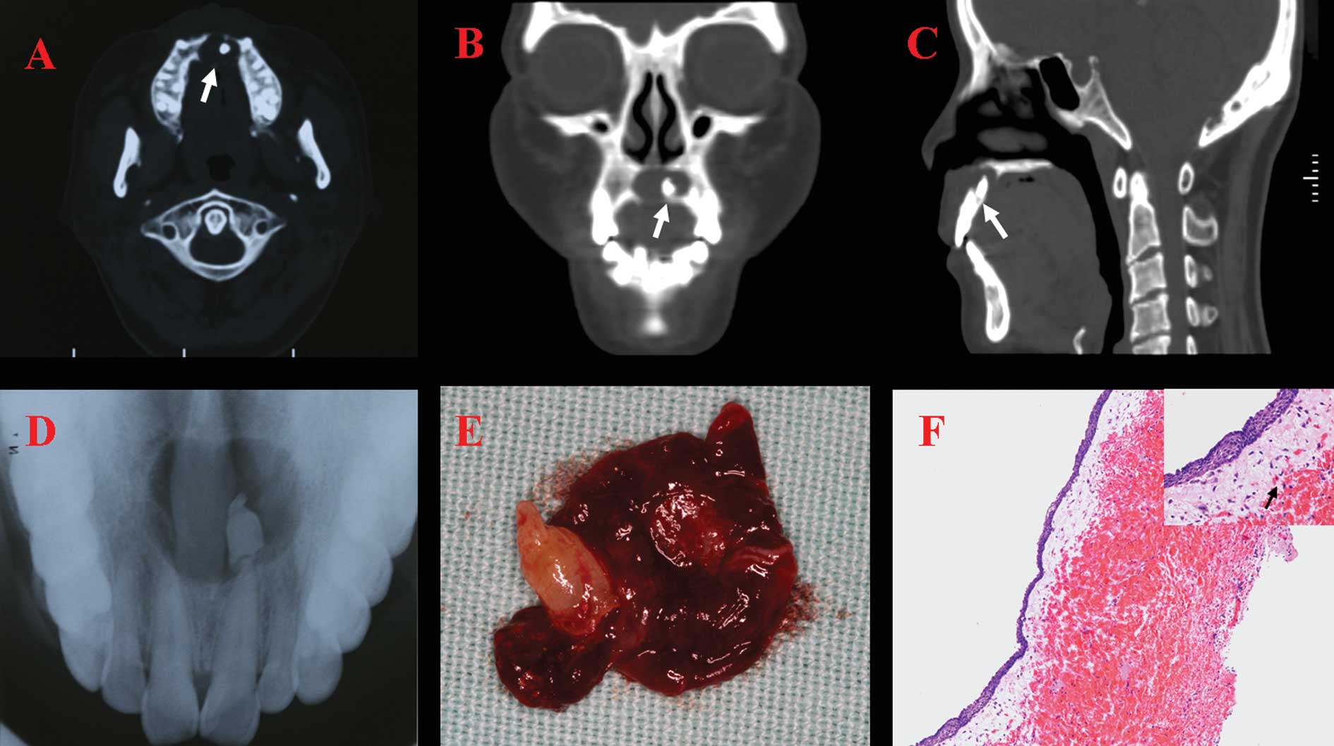
patient’s neurological exam was normal. The upper occlusal
radiograph of the lesion revealed an impacted supernumerary tooth
with a short root apical to the maxillary left central incisor
which was associated with a well-defined radiolucent area in the
anterior region of the maxilla. It also showed that the
pericementum of the maxillary central incisors was continuous and
intact, and the apices of the left tooth existed in the cyst.
Displacement of the roots of the maxillary central incisors was
evident (Fig. 1D). Computed
tomography (CT) scan revealed soft tissue density measuring 1.9×1.4
cm in the midline, containing the impacted supernumerary tooth
(Fig. 1A–C). A clinical diagnosis
of an infected dentigerous cyst associated with a supernumerary
tooth (a mesiodens) was made.
After the acute stage subsided, routine blood
investigations were normal. With the patient under local
anesthesia, a full-thickness mucoperiosteal flap was reflected at
the right mucopalatine fold from the maxillary right second
premolar tooth to the maxillary left second premolar. A cyst
measuring 2 cm in diameter was enucleated. The cyst consisted of a
smooth outer surface and a well-formed wall that was 1-mm thick,
surrounding the impacted supernumerary tooth (Fig. E). During
surgery, there was no connection between the supernumerary tooth
and the apices of the maxillary left center incisor. Histological
examination of the specimen confirmed the initial diagnosis
(Fig. 1F). Postoperative
antibiotics were prescribed for 5 days, and the stitches were
removed after 1 week. One month later, the patient presented no
complications after receiving cyst enucleation.
Case 2
A 46-year-old Chinese male attended our clinic with
a chief complaint of painless swelling in the palatine for a
duration of one year. There was no history of trauma or no dental
treatment around the lesion. Intraoral examination revealed a
solitary and well-defined swelling in the right premaxillary
region. There was no history of trauma, and vitality of the
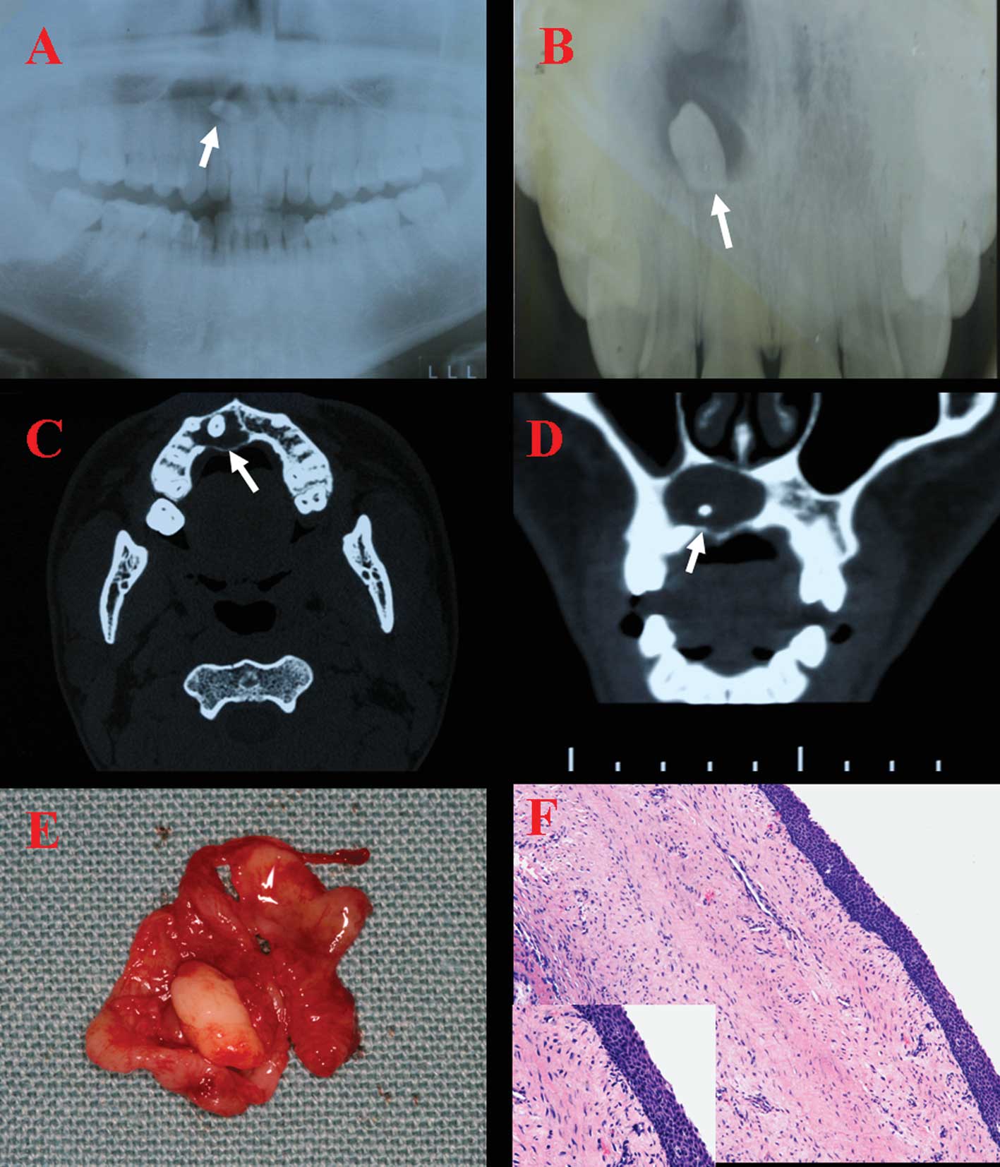
associated teeth was intact. The radiological examination included
panoramic and occlusal radiographs that revealed one supernumerary
tooth involved in a solitary, well-defined radiolucency, extending
from the midline to the right maxillary lateral region, apically to
the right upper central incisor (Fig.
2A and B). CT revealed a supernumerary tooth surrounded by a
soft tissue mass measuring 1.6×2.04 cm in the right premaxillary
region, with the palatal cortical expanded. There was no bone
destruction (Fig. 2C and D). A
tentative diagnosis of dentigerous cyst associated with a
supernumerary tooth was made.
Routine blood investigations were normal, and the
cyst was enucleated along with the supernumerary tooth under local
anesthesia. The macroscopic findings revealed cystic lining
attachments to the supernumerary tooth (Fig. 2E). Routine histological examination
of the enucleated specimen confirmed the initial diagnosis
(Fig. 2F). After surgery,
antibiotics were prescribed for 5 days, and the stitches were
removed after 1 week. The postoperative period of the patient was
uneventful.
Case 3
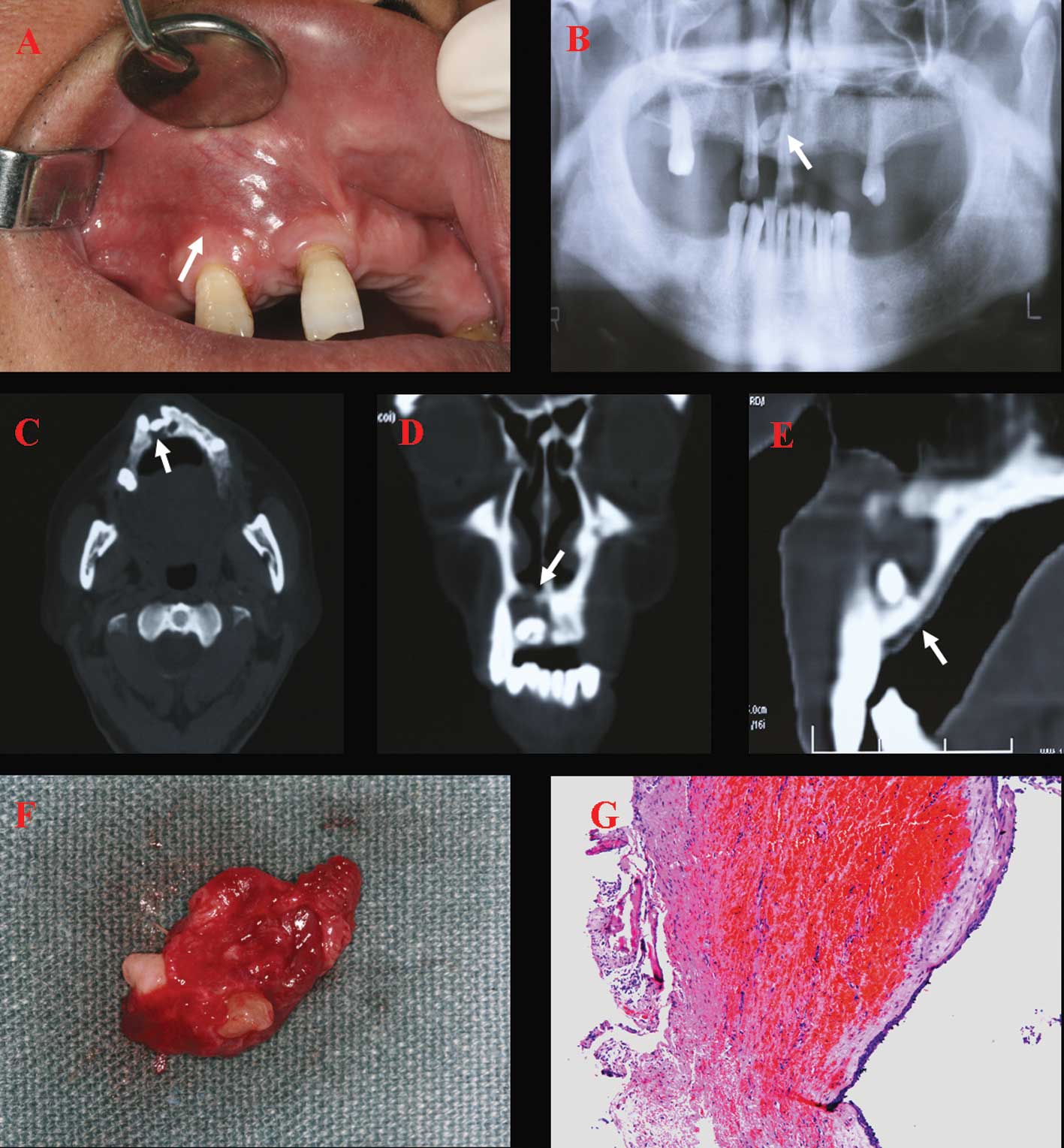
A 53-year-old Chinese male attended our clinic with
a chief complaint of a painless slow-growing mass on the upper lip
for a duration of one year. The right upper lateral incisor had
been extracted several years earlier. Intraoral examination
revealed a soft, fluctuant, labial cyst-like swelling measuring
2.5×1.5 cm on the right upper lip (Fig. 3A). The maxillary right canine and
maxillary right central incisor were vital and not sensitive to
percussion. A panoramic radiograph revealed a relatively large and
well-defined radiolucency between the maxillary right canine and
the right central incisor, enveloping an unerupted horizontal
supernumerary tooth (Fig. 3B). CT
showed a supernumerary tooth surrounded by a soft tissue mass
measuring 2.4 cm horizontally, 1.5 cm vertically and 1.3 cm
sagittally, with the labial cortical bone expanded and heavily
eroded (Fig. 3C–E). A tentative
diagnosis of infected dentigerous cyst associated with a
supernumerary tooth was made.
The patient’s general health and development were
normal except for this lesion. Under local anesthesia, the cyst was
wholly enucleated together with the unerupted super-numerary tooth.
The extracted specimen consisted of a brown cyst measuring
2.5×0.5×1.0 cm with a small monoradicular malformed supernumerary
tooth (Fig. 3F). Routine
histological examination of the enucleated specimen confirmed the
initial diagnosis (Fig. 2F). After
surgery, antibiotics were prescribed for 5 days, and the stitches
were removed after 1 week. The postoperative period of the patient
was uneventful.
Case 4
A 23-year-old Chinese male attended our clinic with
a chief complaint of painless swelling in the palatine for a
duration of one year. There was no history of allergic symptoms and
systemic illness. On intraoral examination, a soft, painless
swelling was palpable in the anterior portion of the hard palatine.
There was no history of trauma or no dental treatment around the
lesion. Maxillary anterior teeth were vital and not sensitive to
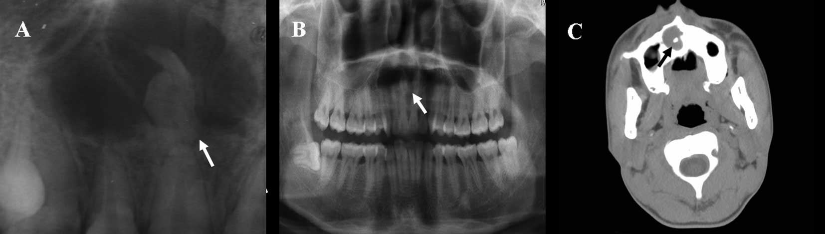
percussion. The radiological examination including panoramic and
occlusal radiographs revealed an impacted inverted supernumerary
tooth with a short root apical to the maxillary right lateral
incisor which was associated with a well-defined radiolucent area
extending from the maxillary right first molar to the maxillary
right cental incisor. None of the apices of these teeth existed in
the cyst (Fig. 4A and B). CT
demonstrated that the radiolucent lesion measuring 1.74×1.5 cm in
the right premaxillary region involved an inverted-shaped
tooth-like calcified structure (Fig.
4C).
Routine blood investigations were abnormal with a
hemoglobin level <68 g/l. Surgery could not be performed because
of severe anemia.
Discussion
Supernumerary teeth are commonly located in the
anterior maxillary region and can often cause developmental and
eruption disturbances of adjacent permanent teeth, leading to
crowding, displacement, diastema and, in some cases, radicular
resorption and dentigerous cyst formation (6–8). The
four cases described the dentigerous cyst formation involving a
supernumerary tooth in the anterior maxilla. In addition,
displacement of the adjacent central incisors was only observed in
case 1 and none of the 4 patients had root resorption.
Dentigerous cysts are the second most common
odontogenic cysts of the jaws after periapical or radicular cysts,
while a dentigerous cyst associated with a supernumerary tooth is a
rare entity. A review of the literature since 1988 disclosed 16
reported cases of dentigerous cysts associated with premaxillay
supernumerary teeth (1,3,10–15).
Our four cases bring the total number of documented cases to 20
(Table I). The reported prevalence
of dentigerous cysts associated with supernumerary teeth ranges
between 1 and 9.9% in the literature (6,7,9,11,16).
Most reports revealed a peak incidence of dentigerous cysts in the
second and third decades of life. The age range for reported cases
varies widely, from 9 to 71 years of age. The mean age of the 16
previously reported cases, not including our four cases, was 30.5
years. The mean age of the present four cases was 44.3 years. Among
the 20 cases summarized in Table
I, the incidence is significantly higher in men (n=14) compared
to women (n=6). This finding is similar to previous studies, which
found that dentigerous cysts appear to have a distinct predilection
for the male gender (4,17).
 | Table I.Documented cases in the literature of
dentigerous cysts associated with supernumerary teeth. |
Table I.
Documented cases in the literature of
dentigerous cysts associated with supernumerary teeth.
|
Authors/Reference | Patient age
(years) | Gender | Site (premaxilla
region) | Symptoms | Treatment |
|---|
| Lustmann and Bodner
(3) | 9 | Female | Right central
incisor | - | Enucleation |
| Lustmann and Bodner
(3) | 12 | Male | Left cental and
lateral incisor | - | Marsupialization |
| Lustmann and Bodner
(3) | 37 | Male | Entire
premaxilla | Swelling, pain | Enucleation |
| Lustmann and Bodner
(3) | 38 | Male | Midline to the left,
maxillary second premolar | Swelling | Enucleation |
| Lustmann and Bodner
(3) | 68 | Female | Midline to the left
maxillary premolar | Swelling, pain | Enucleation |
| Lustmann and Bodner
(3) | 71 | Female | Left premaxillary
region | Asymptomatic | Enucleation |
| Awang and Siar
(6) | 34 | Male | Midline to the upper
left first premolar | Swelling | Enucleation |
| Awang and Siar
(6) | 24 | Female | From the upper right
canine to the upper left central incisor | Swelling | Enucleation |
| Scolozzi, et
al (21) | 42 | Male | Premaxilla | Swelling | Enucleation and
autogenous cancellous bone graft from the iliac crest |
| Dinkar, et al
(15) | 14 | Female | Maxillary anterior
region | Pain, swelling | Enucleation |
| Khan, et al
(20) | 24 | Male | Incisor region | Swelling | Enucleation |
| Gulses, et al
(13) | 10 | Male | Right central
incisor | Asymptomatic | Enucleation |
| Kumar, et al
(10) | 14 | Male | Central incisor
region | Swelling | Enucleation |
| John, et al
(7) | 22 | Male | From the maxillary
right central incisor to the right canine | Swelling | Enucleation |
| John, et al
(7) | 24 | Male | Maxillary anterior
region | Swelling | Enucleation |
| John, et al
(7) | 46 | Male | Maxillary anterior
region | Pain, swelling | Enucleation |
| Present study | | | | | |
| Case 1 | 55 | Female | Central incisors | Swelling | Enucleation |
| Case 2 | 46 | Male | Midline to the upper
right canine | Swelling | Enucleation |
| Case 3 | 53 | Male | From the maxillary
right central incisor to the right canine | Swelling | Enucleation |
| Case 4 | 23 | Male | From the maxillary
right first molar to right cental incisor | Swelling | - |
Dentigerous cysts associated with supernumerary
teeth in the premaxilla are easily diagnosed radiographically due
to their radiopaque image. Panoramic and upper occlusal radiographs
are simple and inexpensive methods, which can be used to determine
the location of dentigerous cysts, the structure of the impacted
teeth, the influence on adjacent teeth and the resorption of
adjacent roots (18). However,
radiograph film has two inevitable disadvantages: i) a ghost image
which cannot reflect the 3-dimensional structure of the lesion
owing to a low resolution ratio, and ii) different degrees of
distortion or amplification. Therefore, CT is necessary and
valuable to obtain more information concerning the lesion. CT can
be used, not only to identify the pathology of the dentigerous cyst
and the exact location of the impacted tooth, but also to determine
the full extent of the lesion, thus contributing to proper
treatment planning as well. Meanwhile, CT can also be used to
identify erosion of cortical bone and invasion into adjacent soft
tissues. In the present four cases, the dentigerous cyst with the
supernumerary tooth appeared to be within the anterior maxilla in
the panoramic radiograph or/and upper occlusal radiograph. In order
to determine the exact location of the impacted tooth and the
degree of bone destruction, CT images were scrutinized. Therefore,
to ensure appropriate treatment decisions and follow-up based on
accurate information regarding dentigerous cysts associated with
supernumerary teeth, we recommend panoramic radiograph and/or upper
occlusal radiograph as a first-line diagnostic tools, and further
evaluation of the lesion by CT examination.
Enucleation is the standard treatment for a
dentigerous cyst along with extraction of the associated
supernumerary tooth (9,10). Among the documented cases in the
literature, the second case (Table
I) was treated by marsupialization because of the intimate
relation of the lesion to the apices of the incisor teeth.
Marsupialization is recommended for a large cyst when a single
draining may not be effective and complete removal of the
surrounding structure is not desirable (19). For a large cyst, Scolozzi et
al recommended enucleation followed by an immediate bone
grafting procedure (21). In the
present four cases, surgical removal of the impacted supernumerary
tooth and enucleation without using bone grafting of the associated
cyst were performed.
A broad range of conditions may lead to a clinical
presentation of painless swelling along the lingual surface of the
palate or on the upper lip. Differential diagnosis of a median
palatine cyst, nasopalatine duct cyst, radicular cyst, odontogenic
keratocyst (OKC) or adenomatoid odontogenic tumor (AOT) was
considered in our cases. Median palatine cysts and nasopalatine
duct cysts are not associated with non-vital teeth as
non-odontogenic cysts of the hard palate (20,21).
Most radicular cysts appear as round or pear-shaped, unilocular,
lucent lesions in the periapical region, and the associated tooth
usually has a deep restoration or large carious lesion
radiographically (21).
Approximately 40% of OKCs contain an impacted tooth, and the lumen
of the cyst often contains ‘cheesy’ material and has a
parakeratinized epithelium lining. They are more likely to show
aggressive growth than other odontogenic cysts and may have
undulating borders and a multilocular appearance upon radiography
(22). Approximately 75% of cases
are associated with an unerupted tooth, and the most common
location is in the anterior maxilla. AOTs are more common in young
people, affect females more than males and, most importantly, the
radiolucency in cases of AOTs extends apically beyond the
cementoenamel junction (18).
In conclusion, dentigerous cysts arising from
impacted supernumerary teeth in the anterior maxilla should be
considered in the differential diagnosis for painless swelling
along the lingual surface of the palate or on the upper lip. To
prevent the development of a dentigerous cyst and to avoid unwanted
effects on adjacent teeth, early detection consisting of a thorough
clinical and radiographical examination is necessary for accurate
diagnosis and proper treatment planning.
Acknowledgements
This study was supported by grants
from the Doctoral Innovation Foundations of Shanghai Jiao Tong
University School (no. BXJ 0922), and the Science and Technology
Commission of Shanghai (no. 08DZ2271100).
References
|
1.
|
Shah A, Gill DS, Tredwin C, et al:
Diagnosis and management of supernumerary teeth. Dent Update.
35:519–520. 2008.
|
|
2.
|
Ochsenius G, Escobar E, Godoy L, et al:
Odontogenic cysts: analysis of 2,944 cases in Chile. Med Oral Patol
Oral Cir Bucal. 12:85–91. 2007.
|
|
3.
|
Lustmann J and Bodner L: Dentigerous cysts
associated with supernumerary teeth. Int J Oral Maxillofac Surg.
17:100–102. 1988. View Article : Google Scholar
|
|
4.
|
Arathi R and Ashwini R: Supernumerary
teeth: a case report. J Indian Soc Pedod Prev Dent. 23:103–105.
2005. View Article : Google Scholar
|
|
5.
|
Ikarashi T, Fujimori Y, Ohshiro K, et al:
Dentigerous cyst associated with a supernumerary malformed tooth:
report of a case and a clinicopathologic review. Oral Med Pathol.
8:55–59. 2003. View
Article : Google Scholar
|
|
6.
|
Awang MN and Siar CH: Dentigerous cyst due
to mesiodens: report of two cases. J Ir Dent Assoc. 35:117–118.
1989.PubMed/NCBI
|
|
7.
|
John T, Guna Shekhar M, Koshy M, et al:
Dentigerous cyst associated with supernumerary teeth: a report of
three cases. J Clin Diagn Res. 4:2601–2606. 2010.
|
|
8.
|
Stafne EC: Supernumerary upper central
incisor. Dent Cosmos. 73:976–980. 1931.
|
|
9.
|
Garvey MT, Barry HJ and Blake M:
Supernumerary teeth – an overview of classification, diagnosis and
management. J Can Dent Assoc. 65:612–616. 1999.
|
|
10.
|
Kumar NM, Ramadevi S, Vanaki SS and
Puranik RS: Dentigerous cyst occurring in maxilla associated with
supernumerary tooth showing cholesterol cleft – a case report. Int
J Dental Clin. 2:392010.
|
|
11.
|
Koca H, Esin A and Aycan K: Outcome of
dentigerous cysts treated with marsupialization. J Clin Pediatr
Dent. 34:165–168. 2009.PubMed/NCBI
|
|
12.
|
Buyukkurt MC, Omezli MM and Miloglu O:
Dentigerous cyst associated with an ectopic tooth in the maxillary
sinus: a report of 3 cases and review of the literature. Oral Surg
Oral Med Oral Pathol Oral Radiol Endod. 109:67–71. 2010. View Article : Google Scholar : PubMed/NCBI
|
|
13.
|
Gulses A, Karacayli U and Koymen R:
Dentigerous cyst associated with inverted and fused supernumerary
teeth in a child: a case report. OHDMBSC. 1:38–41. 2009.
|
|
14.
|
Karaçal N, Ambarcoğlu O and Kutlu N:
Median palatine cyst: report of an unusual entity. Plast Reconstr
Surg. 115:1213–1214. 2005.
|
|
15.
|
Dinkar AD, Dawasaz AA and Shenoy S:
Dentigerous cyst associated with multiple mesiodens: a case report.
J Indian Soc Pedod Prev Dent. 25:56–59. 2007. View Article : Google Scholar : PubMed/NCBI
|
|
16.
|
Righini CA, Boubagra K, Bettega G, et al:
Nasopalatine canal cyst: 4 cases and a review of the literature.
Ann Otolaryngol Chir Cervicofac. 121:115–119. 2004.PubMed/NCBI
|
|
17.
|
Avelar RL, Antunes AA, Carvalho RW, et al:
Odontogenic cysts: a clinicopathological study of 507 cases. J Oral
Sci. 51:581–586. 2009. View Article : Google Scholar : PubMed/NCBI
|
|
18.
|
Wang CJ, Huang PH, Wang YL, et al:
Dentigerous cyst over maxillary sinus: a case report and literature
review. Taiwan J Oral Maxillofac Surg. 20:116–124. 2009.
|
|
19.
|
Giancotti A, Grazzini F, De Dominicis F,
et al: Multidisciplinary evaluation and clinical management of
mesiodens. J Clin Pediatr Dent. 26:233–237. 2002.PubMed/NCBI
|
|
20.
|
Khan MH, Alam MT, Haque S, et al: Upper
lip swelling caused by a large dentigerous cyst with mesiodens.
Mymensingh Med J. 17:S100–S103. 2008.PubMed/NCBI
|
|
21.
|
Scolozzi P, Lombardi T and Richter M:
Upper lip swelling caused by a large dentigerous cyst. Eur Arch
Otorhinolaryngol. 262:246–249. 2005. View Article : Google Scholar : PubMed/NCBI
|
|
22.
|
Zhang LL, Yang R, Zhang L, et al:
Dentigerous cyst: a retrospective clinicopathological analysis of
2082 dentigerous cysts in British Columbia, Canada. Int J Oral
Maxillofac Surg. 39:878–882. 2010. View Article : Google Scholar : PubMed/NCBI
|