Spandidos Publications Logo
  • About
    • About Spandidos
    • Aims and Scopes
    • Abstracting and Indexing
    • Editorial Policies
    • Reprints and Permissions
    • Job Opportunities
    • Terms and Conditions
    • Contact
  • Journals
    • All Journals
    • Oncology Letters
      • Oncology Letters
      • Information for Authors
      • Editorial Policies
      • Editorial Board
      • Aims and Scope
      • Abstracting and Indexing
      • Bibliographic Information
      • Archive
    • International Journal of Oncology
      • International Journal of Oncology
      • Information for Authors
      • Editorial Policies
      • Editorial Board
      • Aims and Scope
      • Abstracting and Indexing
      • Bibliographic Information
      • Archive
    • Molecular and Clinical Oncology
      • Molecular and Clinical Oncology
      • Information for Authors
      • Editorial Policies
      • Editorial Board
      • Aims and Scope
      • Abstracting and Indexing
      • Bibliographic Information
      • Archive
    • Experimental and Therapeutic Medicine
      • Experimental and Therapeutic Medicine
      • Information for Authors
      • Editorial Policies
      • Editorial Board
      • Aims and Scope
      • Abstracting and Indexing
      • Bibliographic Information
      • Archive
    • International Journal of Molecular Medicine
      • International Journal of Molecular Medicine
      • Information for Authors
      • Editorial Policies
      • Editorial Board
      • Aims and Scope
      • Abstracting and Indexing
      • Bibliographic Information
      • Archive
    • Biomedical Reports
      • Biomedical Reports
      • Information for Authors
      • Editorial Policies
      • Editorial Board
      • Aims and Scope
      • Abstracting and Indexing
      • Bibliographic Information
      • Archive
    • Oncology Reports
      • Oncology Reports
      • Information for Authors
      • Editorial Policies
      • Editorial Board
      • Aims and Scope
      • Abstracting and Indexing
      • Bibliographic Information
      • Archive
    • Molecular Medicine Reports
      • Molecular Medicine Reports
      • Information for Authors
      • Editorial Policies
      • Editorial Board
      • Aims and Scope
      • Abstracting and Indexing
      • Bibliographic Information
      • Archive
    • World Academy of Sciences Journal
      • World Academy of Sciences Journal
      • Information for Authors
      • Editorial Policies
      • Editorial Board
      • Aims and Scope
      • Abstracting and Indexing
      • Bibliographic Information
      • Archive
    • International Journal of Functional Nutrition
      • International Journal of Functional Nutrition
      • Information for Authors
      • Editorial Policies
      • Editorial Board
      • Aims and Scope
      • Abstracting and Indexing
      • Bibliographic Information
      • Archive
    • International Journal of Epigenetics
      • International Journal of Epigenetics
      • Information for Authors
      • Editorial Policies
      • Editorial Board
      • Aims and Scope
      • Abstracting and Indexing
      • Bibliographic Information
      • Archive
    • Medicine International
      • Medicine International
      • Information for Authors
      • Editorial Policies
      • Editorial Board
      • Aims and Scope
      • Abstracting and Indexing
      • Bibliographic Information
      • Archive
  • Articles
  • Information
    • Information for Authors
    • Information for Reviewers
    • Information for Librarians
    • Information for Advertisers
    • Conferences
  • Language Editing
Spandidos Publications Logo
  • About
    • About Spandidos
    • Aims and Scopes
    • Abstracting and Indexing
    • Editorial Policies
    • Reprints and Permissions
    • Job Opportunities
    • Terms and Conditions
    • Contact
  • Journals
    • All Journals
    • Biomedical Reports
      • Information for Authors
      • Editorial Policies
      • Editorial Board
      • Aims and Scope
      • Abstracting and Indexing
      • Bibliographic Information
      • Archive
    • Experimental and Therapeutic Medicine
      • Information for Authors
      • Editorial Policies
      • Editorial Board
      • Aims and Scope
      • Abstracting and Indexing
      • Bibliographic Information
      • Archive
    • International Journal of Epigenetics
      • Information for Authors
      • Editorial Policies
      • Editorial Board
      • Aims and Scope
      • Abstracting and Indexing
      • Bibliographic Information
      • Archive
    • International Journal of Functional Nutrition
      • Information for Authors
      • Editorial Policies
      • Editorial Board
      • Aims and Scope
      • Abstracting and Indexing
      • Bibliographic Information
      • Archive
    • International Journal of Molecular Medicine
      • Information for Authors
      • Editorial Policies
      • Editorial Board
      • Aims and Scope
      • Abstracting and Indexing
      • Bibliographic Information
      • Archive
    • International Journal of Oncology
      • Information for Authors
      • Editorial Policies
      • Editorial Board
      • Aims and Scope
      • Abstracting and Indexing
      • Bibliographic Information
      • Archive
    • Medicine International
      • Information for Authors
      • Editorial Policies
      • Editorial Board
      • Aims and Scope
      • Abstracting and Indexing
      • Bibliographic Information
      • Archive
    • Molecular and Clinical Oncology
      • Information for Authors
      • Editorial Policies
      • Editorial Board
      • Aims and Scope
      • Abstracting and Indexing
      • Bibliographic Information
      • Archive
    • Molecular Medicine Reports
      • Information for Authors
      • Editorial Policies
      • Editorial Board
      • Aims and Scope
      • Abstracting and Indexing
      • Bibliographic Information
      • Archive
    • Oncology Letters
      • Information for Authors
      • Editorial Policies
      • Editorial Board
      • Aims and Scope
      • Abstracting and Indexing
      • Bibliographic Information
      • Archive
    • Oncology Reports
      • Information for Authors
      • Editorial Policies
      • Editorial Board
      • Aims and Scope
      • Abstracting and Indexing
      • Bibliographic Information
      • Archive
    • World Academy of Sciences Journal
      • Information for Authors
      • Editorial Policies
      • Editorial Board
      • Aims and Scope
      • Abstracting and Indexing
      • Bibliographic Information
      • Archive
  • Articles
  • Information
    • For Authors
    • For Reviewers
    • For Librarians
    • For Advertisers
    • Conferences
  • Language Editing
Login Register Submit
  • This site uses cookies
  • You can change your cookie settings at any time by following the instructions in our Cookie Policy. To find out more, you may read our Privacy Policy.

    I agree
Search articles by DOI, keyword, author or affiliation
Search
Advanced Search
presentation
Experimental and Therapeutic Medicine
Join Editorial Board Propose a Special Issue
Print ISSN: 1792-0981 Online ISSN: 1792-1015
Journal Cover
September-October 2011 Volume 2 Issue 5

Full Size Image

Sign up for eToc alerts
Recommend to Library

Journals

International Journal of Molecular Medicine

International Journal of Molecular Medicine

International Journal of Molecular Medicine is an international journal devoted to molecular mechanisms of human disease.

International Journal of Oncology

International Journal of Oncology

International Journal of Oncology is an international journal devoted to oncology research and cancer treatment.

Molecular Medicine Reports

Molecular Medicine Reports

Covers molecular medicine topics such as pharmacology, pathology, genetics, neuroscience, infectious diseases, molecular cardiology, and molecular surgery.

Oncology Reports

Oncology Reports

Oncology Reports is an international journal devoted to fundamental and applied research in Oncology.

Experimental and Therapeutic Medicine

Experimental and Therapeutic Medicine

Experimental and Therapeutic Medicine is an international journal devoted to laboratory and clinical medicine.

Oncology Letters

Oncology Letters

Oncology Letters is an international journal devoted to Experimental and Clinical Oncology.

Biomedical Reports

Biomedical Reports

Explores a wide range of biological and medical fields, including pharmacology, genetics, microbiology, neuroscience, and molecular cardiology.

Molecular and Clinical Oncology

Molecular and Clinical Oncology

International journal addressing all aspects of oncology research, from tumorigenesis and oncogenes to chemotherapy and metastasis.

World Academy of Sciences Journal

World Academy of Sciences Journal

Multidisciplinary open-access journal spanning biochemistry, genetics, neuroscience, environmental health, and synthetic biology.

International Journal of Functional Nutrition

International Journal of Functional Nutrition

Open-access journal combining biochemistry, pharmacology, immunology, and genetics to advance health through functional nutrition.

International Journal of Epigenetics

International Journal of Epigenetics

Publishes open-access research on using epigenetics to advance understanding and treatment of human disease.

Medicine International

Medicine International

An International Open Access Journal Devoted to General Medicine.

Journal Cover
September-October 2011 Volume 2 Issue 5

Full Size Image

Sign up for eToc alerts
Recommend to Library

  • Article
  • Citations
    • Cite This Article
    • Download Citation
    • Create Citation Alert
    • Remove Citation Alert
    • Cited By
  • Similar Articles
    • Related Articles (in Spandidos Publications)
    • Similar Articles (Google Scholar)
    • Similar Articles (PubMed)
  • Download PDF
  • Download XML
  • View XML
Article

Non-Hodgkin's lymphoma of the testicle and bilateral adrenals detected by 18F-FDG PET/CT

  • Authors:
    • Yafu Yin
    • Feng Qing
    • Xuena Li
    • Bulin Du
    • Na Li
    • Yaming Li
  • View Affiliations / Copyright

    Affiliations: Department of Nuclear Medicine, The First Hospital of China Medical University, Heping, Shenyang, Liaoning 110001, P.R. China, Nuclear Medicine, William Beaumont Hospital, Troy, MI, USA, Department of Nuclear Medicine, The First Hospital of China Medical University, Heping, Shenyang, Liaoning 110001, P.R. China
  • Pages: 817-820
    |
    Published online on: June 29, 2011
       https://doi.org/10.3892/etm.2011.300
  • Expand metrics +
Metrics: Total Views: 0 (Spandidos Publications: | PMC Statistics: )
Metrics: Total PDF Downloads: 0 (Spandidos Publications: | PMC Statistics: )
Cited By (CrossRef): 0 citations Loading Articles...

This article is mentioned in:


Abstract

Lymphoma, particularly non-Hodgkin's lymphoma, may occur in sites other than the lymph nodes, i.e., extranodal lymphoma, which occurs in approximately 40% of total lymphoma cases. However, simultaneous detection of multiple extranodal involvements at presentation is quite uncommon. Here, we report a rare case of a 55-year-old Chinese man with non-Hodgkin's lymphoma of the left testicle and bilateral adrenals as determined by 18F-FDG PET/CT imaging. FDG PET/CT has been successfully employed for lymphoma diagnosis, initial staging, restaging and therapy follow-up. It is useful for assessing both nodal and extranodal involvements. In the present case of lymphoma with involvement of the testicle and bilateral adrenals, 18F-FDG PET/CT played an important role in the diagnosis and assessment of therapeutic response.

Case report

A 55-year-old Chinese male presented with a history of left lumbar pain for 1 month without frequency, urgency or dysuria. He had no fever, his appetite was normal, and no weight loss or gain was noted. The patient had no significant family history of illness. During the clinical examination, he appeared healthy. His blood pressure was 128/78 mmHg, his pulse 80 beats/min and his temperature was 36.2°C. There was no skin or sclera icterus, and no superficial swollen lymph node was noted. The lungs were clear upon auscultation. The abdomen of the patient was soft and non-tender; there was no hepatomegaly or splenomegaly. Upon neurological examination, the patient appeared alert and orientated to time, place and people. Normal muscle bulk and tone with full strength in both arms and legs were noted.

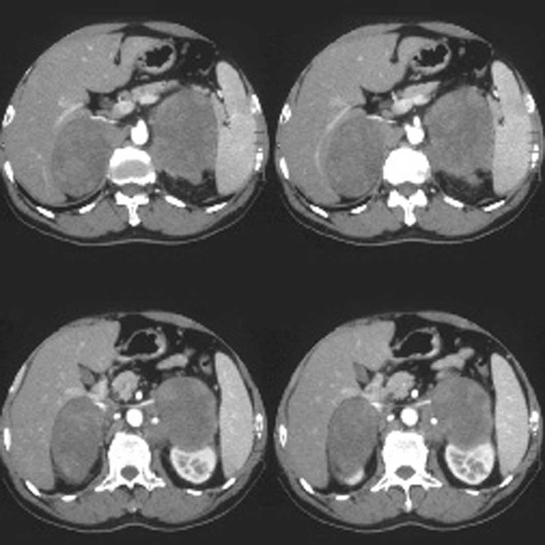
Routine laboratory tests of blood, urine and feces were normal. CT scan of the abdomen demonstrated large bilateral adrenal masses with heterogenous enhancement. The left adrenal gland measured 6.81×8.78 cm and the right adrenal gland measured 8.22×6.15 cm in its greatest dimension (Fig. 1). Swollen lymph nodes were detected in the abdomen. Based on the CT findings, a provisional diagnosis of adrenal metastases or possible malignant tumor of the adrenals was made.

Figure 1.

Enhanced CT scan of adrenals.

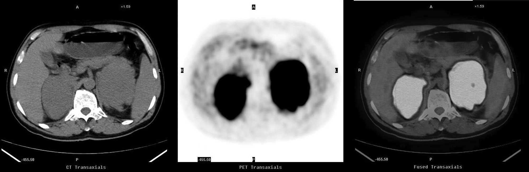
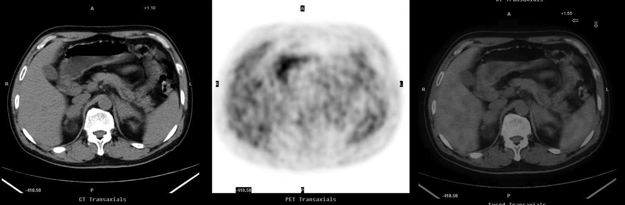
To further confirm the diagnosis and staging of the disease, an 18F-FDG PET/CT scan was obtained per physician’s request. The scan showed high FDG uptake in the bilateral adrenals with a max standard uptake value (SUV) of 29.0 
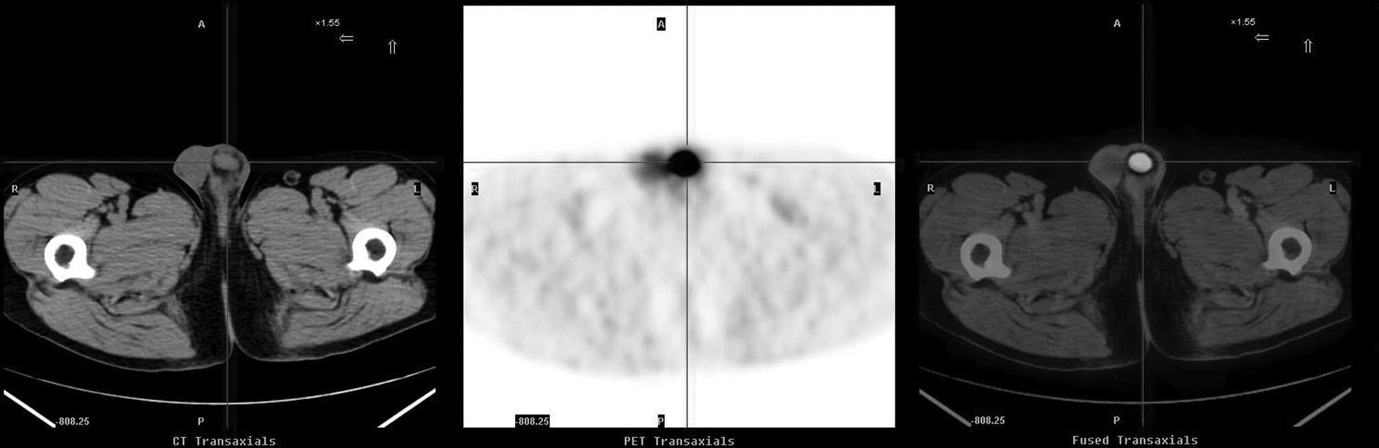
(Fig. 2), high uptake in the retroperitoneal swollen lymph nodes with a max SUV of 24.0 and high focal uptake in the left testicle with a max SUV of 21.0 (Fig. 3), corresponding to a soft tissue nodule on CT. Based on the PET/CT findings, the diagnosis of a primary malignant tumor, e.g., lymphoma, was made and further work-up was recommended.

Figure 2.

First 18F-FDG PET/CT scan. High uptake of tracer in the bilateral adrenals with max SUV 19.5 and 29.0, respectively.

Figure 3.

First 18F-FDG PET/CT scan. High uptake of tracer in the left testicle with max SUV 21.0.

An ultrasound examination of the testis was subsequently performed and showed a hypoechoic area above the left epididymis, suspicious of a space-occupying lesion.

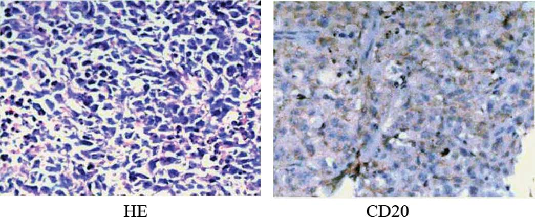
Finally, several days later, the patient underwent a left orchiectomy. Histopathological examination of the left testicle revealed diffuse large B-cell lymphoma (DLBCL); immunohistochemistry tested positive for cytoplasmic CD20, CD79α and CD117 (Fig. 4). Following the orchiectomy, the patient was treated with two cycles of CHOP chemotherapy (cyclophosphamide, doxorubicin, vincristine and prednisone) which was well tolerated. A mid-cycle 18F-FDG PET/CT was performed 21 days after the first two chemotherapy cycles and demonstrated no abnormal FDG uptake in the regions of the bilateral adrenals and retroperitoneal lymph nodes, which were much smaller or disappeared on CT (Fig. 5). This was followed by another four cycles of chemotherapy.

Figure 4.

Immunohistochemistry of the left testicle: CD3(−), CD20(+), CD79α(+), PLAP(−), CD117(+), CD30(−) and CK(weak +).

Figure 5.

Second 18F-FDG PET/CT scan after treatment: No high uptake of tracer in bilateral adrenals.

The final clinical diagnosis of the patient was DLBCL stage IV E.

Discussion

Lymphomas are classified into two main groups, non-Hodgkin’s lymphoma (NHL) and Hodgkin’s lymphoma (HL). NHL is more common than HL and represents 85% of lymphomas (1). DLBCL constitutes approximately 30% of all NHL cases and typically presents with rapidly enlarging symptomatic masses, most usually due to nodal enlargement. Lymphoma, particularly NHL, may occur in sites other than the lymph nodes, i.e., extranodal lymphoma in approximately 40% of total lymphoma cases (2). Extranodal involvement may appear in various sites, including the lungs, thymus, chest wall, pericardium breast, spleen, pleura, liver, pancreas, gastrointestinal tract, peritoneum, omentum, genitourinary tract, adrenal gland, bone marrow or bone, and the central nervous system. Concurrent detection of multiple extranodal involvements at presentation is quite rare, and the majority of such cases are characterized by gastric or intestinal disease localization (3).

In the present study, the patient was found to have extranodal involvement of both the left testicle and bilateral adrenals, which is exceptionally rare.

A variety of neoplastic processes involve the testes. The most common germ-cell tumors include seminoma, embryonal carcinoma, teratoma, yolk-sac tumor and choriocarcinoma. Germ-cell tumors are the most common solid tumors in men between 20 and 35 years of age. Seminomas represent 50% of all germ-cell tumors and usually occur in men in their 30s. Non-seminomatous germ-cell tumors include Soki Leydig cell tumor, fibroma, fibrosarcoma, lymphoma and leukemia. Primary testicular NHL was first described as a clinical entity in 1866 (4). It is a rare disease and accounts for 1% of all NHLs, 2% of all extranodal lymphomas and 5% of all testicular neoplasms (5,6).

Bilateral adrenal masses are commonly metastatic, but are also found in adrenal tuberculosis, pheochromocytoma and adrenal cortical carcinoma. NHL arising from endocrine glands represents only 3% of extranodal lymphomas (7). Secondary involvement of the adrenal gland with NHL has been reported to occur in up to 25% of patients during the course of the disease (8). However, primary adrenal lymphoma (PAL) is extremely rare. Most of the patients with PAL usually present with bilateral adrenal masses with no disease elsewhere.

In the present study, the patient was initially found to have bilateral adrenal masses by CT. CT scanning is considered the most important anatomic imaging modality for evaluating adrenal masses or masses in most other sites. The CT attenuation value has been helpful in differentiating a benign lesion from a malignant lesion (9). The sensitivity and specificity for characterizing a lesion display wide variability depending on the density of the lesion. A meta-analysis reported that the sensitivity for benign lesions ranged from 47% at a threshold of 2 HU to 88% at a threshold of 20 HU (10). Delayed enhanced CT can aid in analyzing the washout patterns noted in adrenal lesions (11). Mean CT attenuation of adrenal masses on contrast-enhanced CT has limited usefulness due to too much overlap between the benign and malignant lesions (9,12).

Unlike CT, FDG/PET measures glucose metabolism. It has been shown to play an important role in diagnosis, staging, monitoring response to treatment and detecting recurrence of various types of cancers. The first reports of 18F-FDG PET for lymphoma imaging were published more than 20 years ago (13). As most lymphomas show high levels of 18F-FDG uptake, subsequent studies investigating the value of 18F-FDG PET in the diagnosis and staging of lymphomas have been carried out. These studies almost invariably demonstrated an extremely high sensitivity for HL and high-grade or aggressive NHL (14). 18F-FDG PET consistently identified nodal and extranodal disease sites that were failed to be detected by conventional staging methods, including CT. 18F-FDG PET also improved the characterization of lesions that were equivocal on other types of imaging. Thus, FDG-PET has become an established imaging modality for staging, restaging and monitoring therapy. PET/CT imaging is more accurate than PET or CT alone. PET/CT has evolved to become the modality of choice for the staging of nodal and extranodal lymphoma, for assessing therapeutic response and for establishing patient prognosis (15).

In the present study, simultaneous involvement of the bilateral adrenals and the left testicle was detected by 18F-FDG PET/CT; therefore, the diagnosis of a primary malignant tumor, e.g., lymphoma, was made. It is well known that the final diagnosis of lymphoma is based on the pathological diagnosis. However, for ethical reasons, pathological diagnosis may not be possible for all lesions and abnormalities found. Thus, PET or PET/CT is useful for the diagnosis of lymphoma and usually detects unusual extranodal involvement (16,17), as illustrated in the present report. Assessment of early therapeutic response using PET/CT was also performed in our case.

In 2007, the consensus recommendations regarding the use of FDG/PET for the assessment of response after the treatment of patients with lymphoma were published by the Imaging Subcommittee of the International Harmonization Project (18). 18F-FDG PET/CT is currently the standard procedure for assessment of the post-treatment response for most lymphoma subtypes. In addition, early response monitoring with 18F-FDG PET is an effective tool for risk-adapted lymphoma therapy. This method may improve the outcome of patients who exhibit a poor respond to the initial therapy and who may benefit from an early modification of the treatment. 18F-FDG PET may also allow individualized therapy for patients who have a low risk of treatment failure and who would otherwise be unnecessarily administered toxic treatment (19).

Additionally, the National Comprehensive Cancer Network has incorporated FDG-PET in the evaluation and management algorithm for most HL and NHL patients (20). The utilization of FDG-PET (PET/CT where available) is recommended in clinical settings as a baseline for lymphomas that are potentially curative (HL and DLBCL), as a baseline to exclude systemic disease in clinically localized lymphomas (HL, DLBCL, follicular lymphoma, mantle cell lymphoma, AIDS-related B-cell lymphoma, nodal and splenic marginal zone lymphoma, peripheral T-cell lymphoma and mucosa-associated lymphoid tumors), to evaluate residual masses and to monitor treatment of aggressive lymphomas (HL and DLBCL).

Over the last two decades, FDG PET or PET/CT has been successfully used to detect nodal and extranodal sites, to stage, restage, monitor therapy, detect recurrent lymphomas and establish patient prognosis (21).

In conclusion, a diagnosis of lymphoma should be considered when a patient shows a high metabolism of glucose in FDG PET or PET/CT imaging in two remote extranodal sites, e.g., bilateral adrenals and the testicle in our case. 18F-FDG PET/CT is useful for the detection and assessment of the therapeutic response of lymphoma in addition to staging, restaging and detection of recurrent lymphomas.

References

1. 

Lu P: Staging and classification of lymphoma. Semin Nucl Med. 35:160–164. 2005. View Article : Google Scholar

2. 

Metser U, Goor O, Lerman H, Naparstek E and Even-Sapir E: PET-CT of extranodal lymphoma. AJR Am J Roentgenol. 182:1579–1586. 2004. View Article : Google Scholar : PubMed/NCBI

3. 

Economopoulos T, Papageorgiou S, Rontogianni D, et al: Multifocal extranodal non-Hodgkin lymphoma: a clinicopathological study of 37 cases in Greece, a Hellenic Cooperative Oncology Group Study. The Oncologist. 10:734–738. 2005. View Article : Google Scholar : PubMed/NCBI

4. 

Shahab N and Doll DC: Testicular lymphoma. Semin Oncol. 26:259–269. 1999.

5. 

Touroutoglou N, Dimopoulos MA, Younes A, et al: Testicular lymphoma: late relapses and poor outcome despite doxorubicin-based therapy. J Clin Oncol. 13:1361–1367. 1995.PubMed/NCBI

6. 

Ballen KK and Hasserjian RP: Case records of the Massachusetts General Hospital. Weekly clinicopathological exercises. Case 15-2004 A 31-year-old man with bilateral testicular enlargement. N Engl J Med. 350:2081–2087. 2004.

7. 

Kubo M, Koga M, Fujii T, Kaneko T, Yamashita K and Kokubu T: Bilateral adrenal lymphoma with neoplastic angioendotheliosis. Intern Med. 36:47–52. 1997. View Article : Google Scholar : PubMed/NCBI

8. 

Rosenberg SA, Diamond HD, Jaslowitz B and Craver LF: Lymphosarcoma: a review of 1269 cases. Medicine. 40:31–84. 1961. View Article : Google Scholar : PubMed/NCBI

9. 

Lee MJ, Hahn PF, Papanicolaou N, Egglin TK, Saini S, Mueller PR and Simeone JF: Benign and malignant adrenal masses: CT distinction with attenuation coefficients, size, and observer analysis. Radiology. 179:415–418. 1991. View Article : Google Scholar : PubMed/NCBI

10. 

Boland GW, Lee MJ, Gazelle GS, Halpern EF, McNicholas MM and Mueller PR: Characterization of adrenal masses using unenhanced CT: an analysis of the CT literature. AJR Am J Roentgenol. 171:201–204. 1998. View Article : Google Scholar : PubMed/NCBI

11. 

Caoili EM, Korobkin M, Francis IR, Cohan RH, Platt JF, Dunnick NR and Raghupathi KI: Adrenal masses: characterization with combined unenhanced and delayed enhanced CT. Radiology. 222:629–633. 2002. View Article : Google Scholar : PubMed/NCBI

12. 

Korobkin M, Brodeur FJ, Yutzy GG, Francis IR, Quint LE, Dunnick NR and Kazerooni EA: Differentiation of adrenal adenomas from nonadenomas using CT attenuation values. AJR Am J Roentgenol. 166:531–536. 1996. View Article : Google Scholar : PubMed/NCBI

13. 

Paul R: Comparison of fluorine-18-2-fluorodeoxyglucose and gallium-67 citrate imaging for detection of lymphoma. J Nucl Med. 28:288–292. 1987.PubMed/NCBI

14. 

Elstrom R, Guan L, Baker G, et al: Utility of FDG-PET scanning in lymphoma by WHO classification. Blood. 101:3875–3876. 2003. View Article : Google Scholar : PubMed/NCBI

15. 

Allen-Auerbach M, de Vos S and Czernin J: The impact of fluorodeoxyglucose-positron emission tomography in primary staging and patient management in lymphoma patients. Radiol Clin North Am. 46:199–211. 2008. View Article : Google Scholar

16. 

Lam WW and Osmany S: Biliary non-Hodgkin lymphoma detected by F-18 FDG PET/CT. Clin Nucl Med. 34:791–792. 2009. View Article : Google Scholar : PubMed/NCBI

17. 

Bosch-Barrera J, Arbea L, García-Velloso MJ, Gil-Bazo I, García-Foncillas J and Panizo C: Primary bone lymphoma of the mandible and thyroid incidentaloma identified by FDG PET/ CT: a case report. Cases J. 26:63842009.PubMed/NCBI

18. 

Juweid ME, Stroobants S, Hoekstra OS, et al: Use of positron emission tomography for response assessment of lymphoma: Consensus of the Imaging Subcommittee of International Harmonization Project in Lymphoma. J Clin Oncol. 25:571–578. 2007. View Article : Google Scholar

19. 

Hutchings M and Barrington SF: PET/CT for therapy response assessment in lymphoma. J Nucl Med. 50:S21–S30. 2009. View Article : Google Scholar

20. 

Podoloff DA, Advani RH, Allred C, et al: NCCN task force report: positron emission tomography (PET)/computed tomography (CT) scanning in cancer. J Natl Compr Canc Netw. 5(Suppl 1): 1–22. 2007.PubMed/NCBI

21. 

Juweid ME: FDG-PET/CT in lymphoma. Methods Mol Biol. 727:1–19. 2011. View Article : Google Scholar : PubMed/NCBI

Related Articles

  • Abstract
  • View
  • Download
  • Twitter
Copy and paste a formatted citation
Spandidos Publications style
Yin Y, Qing F, Li X, Du B, Li N and Li Y: Non-Hodgkin's lymphoma of the testicle and bilateral adrenals detected by 18F-FDG PET/CT. Exp Ther Med 2: 817-820, 2011.
APA
Yin, Y., Qing, F., Li, X., Du, B., Li, N., & Li, Y. (2011). Non-Hodgkin's lymphoma of the testicle and bilateral adrenals detected by 18F-FDG PET/CT. Experimental and Therapeutic Medicine, 2, 817-820. https://doi.org/10.3892/etm.2011.300
MLA
Yin, Y., Qing, F., Li, X., Du, B., Li, N., Li, Y."Non-Hodgkin's lymphoma of the testicle and bilateral adrenals detected by 18F-FDG PET/CT". Experimental and Therapeutic Medicine 2.5 (2011): 817-820.
Chicago
Yin, Y., Qing, F., Li, X., Du, B., Li, N., Li, Y."Non-Hodgkin's lymphoma of the testicle and bilateral adrenals detected by 18F-FDG PET/CT". Experimental and Therapeutic Medicine 2, no. 5 (2011): 817-820. https://doi.org/10.3892/etm.2011.300
Copy and paste a formatted citation
x
Spandidos Publications style
Yin Y, Qing F, Li X, Du B, Li N and Li Y: Non-Hodgkin's lymphoma of the testicle and bilateral adrenals detected by 18F-FDG PET/CT. Exp Ther Med 2: 817-820, 2011.
APA
Yin, Y., Qing, F., Li, X., Du, B., Li, N., & Li, Y. (2011). Non-Hodgkin's lymphoma of the testicle and bilateral adrenals detected by 18F-FDG PET/CT. Experimental and Therapeutic Medicine, 2, 817-820. https://doi.org/10.3892/etm.2011.300
MLA
Yin, Y., Qing, F., Li, X., Du, B., Li, N., Li, Y."Non-Hodgkin's lymphoma of the testicle and bilateral adrenals detected by 18F-FDG PET/CT". Experimental and Therapeutic Medicine 2.5 (2011): 817-820.
Chicago
Yin, Y., Qing, F., Li, X., Du, B., Li, N., Li, Y."Non-Hodgkin's lymphoma of the testicle and bilateral adrenals detected by 18F-FDG PET/CT". Experimental and Therapeutic Medicine 2, no. 5 (2011): 817-820. https://doi.org/10.3892/etm.2011.300
Follow us
  • Twitter
  • LinkedIn
  • Facebook
About
  • Spandidos Publications
  • Careers
  • Cookie Policy
  • Privacy Policy
How can we help?
  • Help
  • Live Chat
  • Contact
  • Email to our Support Team