Introduction
Core needle biopsy (CNB) has become widely used in
diagnosing breast disease (1,2). CNB
is a uncomplicated examination, which is easier for patients to
accept than excisional biopsy. Papillary lesions are a
heterogeneous group of breast lesions and include papilloma,
papillomatosis, atypical papilloma, non-invasive ductal carcinoma
and invasive ductal carcinoma. Sometimes, distinguishing malignant
from benign papillary lesions using CNB can be difficult since only
a small portion of the lesion is examined. This study evaluated the
follow-up methods and results of excisional biopsies for patients
with papillary lesions initially diagnosed using CNB.
Patients and methods
We retrospectively reviewed 31 papillary lesions of
the breast diagnosed using CNB between 2004 and 2007. All cases
were reviewed regarding the follow-up methods, distance from the
nipple to the tumor and final pathological diagnosis.
Patient background
The median patient age was 48.9 years (range 22–81).
Twelve patients came to our hospital due to nipple discharge, 10
patients presented with a lump, 8 patients had an abnormal check-up
and 1 patient presented with another complaint. Ultimately, 14
patients developed nipple discharges: 8 were bloody and 6 were
serous.
Percutaneous biopsy method
Ultrasonographically guided procedures were
performed with the patient in the supine or supine oblique
position. Imaging was performed with a high-resolution 8–12 MHz
linear array transducer (Acuson Sequoia 512; Siemens®).
The biopsy was performed using a freehand technique with a 14-gauge
needle and spring-loaded biopsy gun (Bard Magnum®). The
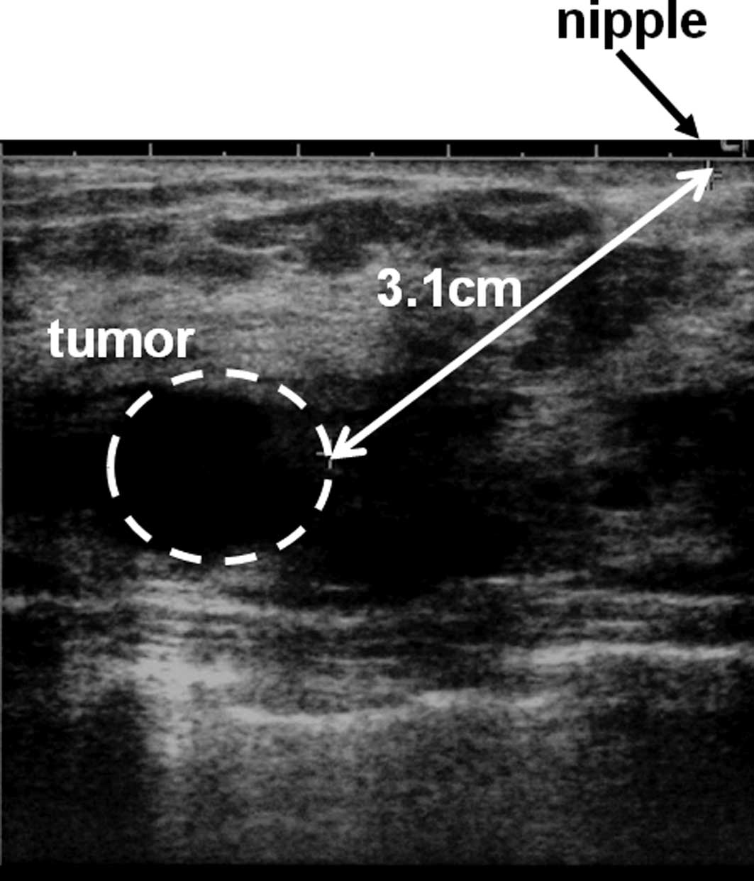
distance from the nipple to the tumor was measured using
ultrasonography at the time of CNB (Fig. 1). The biopsy specimen was evaluated
by an experienced breast pathologist.
Results
The initial diagnosis was intraductal papilloma in
25 patients (80%), intracystic papilloma in 4 (13%) and adenoma in
2 (7%). After CNB, excisional biopsies were performed in 23
patients and Mammotome® biopsies in 2 patients. The CNB
was repeated in 2 patients. Four patients did not undergo any
further biopsy, but were followed up with ultrasonography alone at
the’ request of the patients. Five (16%) of the 31 patients with
papillary lesions were ultimately diagnosed with breast cancer.
Five malignant cases
The mean patient age was 48.8 years (range 37–72).
All cases were diagnosed as intraductal papilloma initially. Two
patients presented with a lump, 2 with abnormal check-up results
and only 1 with a bloody nipple discharge. The average distance
from the nipple to the tumor was 2.46 cm. To obtain the final
diagnosis, 4 patients underwent excisional biopsies and 1 a repeat
CNB. The final diagnosis was invasive ductal carcinoma in 4 cases
and non-invasive ductal carcinoma in the others. After the final
diagnosis, 4 patients underwent breast-conserving surgery and 1
underwent a total mastectomy. Sentinel lymph node biopsies were
performed at the time of surgery in all cases, and there was no
cases of lymph node metastasis (Table
I).
 | Table I.Five malignant cases. |
Table I.
Five malignant cases.
| Case | Age (years) | Initial
diagnosis | Clinical
presentation | Discharge | TND (cm) | Follow-up | Final diagnosis | Op |
|---|
| 1 | 72 | Intraductal
papilloma | Discharge | Bloody | 0.50 | CNB | IDC | Bt |
| 2 | 44 | Intraductal
papilloma | Lump | No | 4.00 | Excisional
biopsy | IDC | Bp |
| 3 | 37 | Intraductal
papilloma | Lump | No | 3.20 | Excisional
biopsy | IDC | Bp |
| 4 | 41 | Intraductal
papilloma | Asymptomatic | No | 1.10 | Excisional
biopsy | DCIS | Bp |
| 5 | 50 | Intraductal
papilloma | Asymptomatic | No | 3.50 | Excisional
biopsy | IDC | Bp |
| Average | 48.8 | | | | 2.46 | | | |
Discussion
We examined the difference between the initial
diagnosis, based on CNB, and the final diagnosis, based on
excisional biopsy, for papillary lesions. Comparing the distance
from the nipple to the tumor, it was 2.46 cm (range 0.5–4.0) for
those diagnosed as malignant and 1.76 cm (range 0.5–4.5) for those
diagnosed as benign. Malignant lesions were located farther from
the nipple than benign lesions (Table
II), although the difference was not statistically significant.
There was no significant difference in the age of the patients with
benign and malignant lesions (Table
III). Distinguishing malignant from benign papillary lesions
can be problematic. The presence or absence of a myoepithelial cell
layer in the papillary component of the lesion is the most
important feature for differentiating a benign papilloma from a
papillary carcinoma.
| Table II.TND of malignant lesions and benign
lesions. |
Table II.
TND of malignant lesions and benign
lesions.
| Lesion | TND mean (range) |
|---|
| Benign | 1.76 (0.5–4.5) |
| Malignant | 2.46 (0.5–4.0) |
| Table III.Age of the patients with malignant and
benign lesions. |
Table III.
Age of the patients with malignant and
benign lesions.
| Lesion | Age, in years mean
(range) |
|---|
| Benign | 48.4 (22–78) |
| Malignant | 48.8 (37–72) |
We reviewed 21 studies (including ours); 10 studies
supported the need for surgical excision and 10 did not (Table IV) (3–22).
There were 643 cases in these studies; 424 cases (65.9%) underwent
excisional biopsies: 334 cases (78.8%) were diagnosed as benign; 27
were diagnosed as atypical ductal hyperplasia; 21 were non-invasive
ductal carcinoma; and 27 were invasive ductal carcinoma.
Ultimately, an excisional biopsy was required in 75 cases (11.7%).
Ten studies stated that an excisional biopsy was necessary to
diagnose papillary lesions, while the other 10 studies concluded
that it was not necessary.
| Table IV.Papillary lesions of the breast
diagnosed with core biopsy; summary of the literature. |
Table IV.
Papillary lesions of the breast
diagnosed with core biopsy; summary of the literature.
| Author/(Refs.) | Year | No. of cases | No. of excisions | Benign | ADH | DCIS | IDC | Other | Conclusions |
|---|
| Ioffe et al
(3) | 2000 | 28 | 8 | 8 | | | | | No excision |
| Philpotts et
al (4) | 2000 | 16 | 6 | 4 | | | | 2 | No excision |
| Liberman (5) | 2000 | 7 | 4 | 4 | | | | | No excision |
| Mercado et al
(6) | 2001 | 12 | 6 | 5 | | | 1 | | Excise |
| Rosen et al
(7) | 2002 | 44 | 14 | 11 | 2 | 1 | | | No excision |
| Irfan and Brem
(8) | 2002 | 6 | 3 | 1 | 1 | | | 1 | - |
| Ivan et al
(9) | 2004 | 30 | 6 | 6 | | | | | No excision |
| Puglisi et
al (10) | 2003 | 31 | 31 | 29 | | | 2 | | Excise |
| Agoff and Lawton
(11) | 2004 | 25 | 11 | 11 | | | | | No excision |
| Renshaw et
al (12) | 2004 | 8 | 8 | 8 | | | | | No excision |
| Gendler et
al (13) | 2004 | 13 | 13 | 9 | 2 | | 2 | | Excise |
| Carder et al
(14) | 2005 | 2 | 1 | 1 | | | | | No excision |
| Liberman et
al (15) | 2006 | 50 | 25 | 20 | | 4 | 1 | | Excise |
| Mercado et
al (16) | 2006 | 43 | 36 | 14 | 8 | 2 | | 12 | Excise |
| Valdes et al
(17) | 2006 | 36 | 36 | 30 | | | 6 | | Excise |
| Plantade et
al (18) | 2006 | 86 | 37 | 32 | | 5 | | | No excision |
| Skandarajah et
al (19) | 2007 | 80 | 80 | 54 | 11 | 8 | 7 | | Excise |
| Arora et al
(20) | 2007 | 18 | 18 | 18 | | | | | Excise |
| Sydnor et al
(21) | 2007 | 38 | 38 | 37 | | | 1 | | No excision |
| Askenazi et
al (22) | 2007 | 39 | 20 | 13 | 3 | | 4 | | Excise |
| Present study | | 31 | 23 | 19 | | 1 | 3 | | Excise |
| Total | | 643 | 424 | 334 | 27 | 21 | 27 | 15 | Ten studies
recommend excision |
It remains controversial whether papillary lesions
of the breast diagnosed using CNB need a further excisional biopsy.
An excisional biopsy has some merits. The pathologist can diagnose
the papillary lesion as benign or malignant from the entire lesion,
the nipple discharge will stop after an excisional biopsy and no
repeat CNB is necessary. Conversely, there are certain
disadvantages to an excisional biopsy. Local anesthesia (e.g.,
allergy and toxicity) and surgery convey various risks, an
excisional biopsy may alter the breast shape and an accurate
sentinel lymph node biopsy may be impossible as the lymphatic flow
is altered. Several reports supporting CNB alone for distinguishing
benign and malignant papillary lesions are based on
immunohistochemical studies, which suggest that different cell
surface markers help differentiate the two. Saddik and Lai proposed
that CD44 is a marker for benign papillary lesions (23). Recently, high-molecular-weight
cytokeratins, particularly CK5 and 6, have been studied as markers
(24). Shah et al added CK5
and 6 to calponin and p63 and investigated their effect on the
accuracy of CNB (25). The overall
accuracy increased from 84.5 to 92.8%. Moreover, Moriya et
al suggested that certain immunohistochemical markers are quite
useful in diagnosing various breast lesions, particularly for
separating benign lesions and malignant neoplasms; however, the
situations in which these markers are valuable are not universal,
and their application and methods for evaluation are limited
(26).
Ultimately, 5 papillary lesions (16%) initially
diagnosed as benign papillary lesions were diagnosed as a breast
cancer. The average distance from the nipple to the tumors
diagnosed as malignant was longer than that for those diagnosed as
benign. For papillary lesions, including intraductal papilloma,
located far from the nipple, it is also necessary to consider a
carcinoma which may be hidden near them. To avoid overlooking a
malignancy, surgical excision is advantageous for papillary
lesions, particularly those located far from the nipple.
References
|
1
|
Parker SH, Lovin JD, Jobe WE, Burke BJ,
Hopper KD and Yakes WF: Nonpalpable breast lesions: stereotactic
automated large-core biopsies. Radiology. 180:403–407. 1991.
View Article : Google Scholar : PubMed/NCBI
|
|
2
|
Parker SH, Lovin JD, Jobe WE, et al:
Stereotactic breast biopsy with a biopsy gun. Radiology.
176:741–747. 1990. View Article : Google Scholar : PubMed/NCBI
|
|
3
|
Ioffe O, Berg WA and Silverberg SG:
Analysis of papillary lesions diagnosed on core needle biopsy:
management implications. Mod Pathol. 13:23A2000.
|
|
4
|
Philpotts LE, Shaheen NA, Jain KS, Carter
D and Lee CH: Uncommon high risk lesions of the breast diagnosed at
stereotactic core-needle biopsy: clinical importance. Radiology.
216:831–837. 2000. View Article : Google Scholar : PubMed/NCBI
|
|
5
|
Liberman L: Clinical management issues in
percutaneous core breast biopsy. Radiol Clin North Am. 38:791–807.
2000. View Article : Google Scholar : PubMed/NCBI
|
|
6
|
Mercado CL, Hamele-Bena D, Singer C, et
al: Papillary lesions of the breast: evaluation with stereotactic
directional vacuum-assisted biopsy. Radiology. 221:650–655. 2001.
View Article : Google Scholar : PubMed/NCBI
|
|
7
|
Rosen EL, Bentley RC, Baker JA and Soo MS:
Imaging-guided core needle biopsy of papillary lesions of the
breast. AJR Am J Roentgenol. 179:1185–1192. 2002. View Article : Google Scholar : PubMed/NCBI
|
|
8
|
Irfan K and Brem RF: Surgical and
mammographic follow-up of papillary lesions and atypical lobular
hyperplasia diagnosed with stereotactic vacuum-assisted biopsy.
Breast J. 8:230–233. 2002. View Article : Google Scholar
|
|
9
|
Ivan D, Selinko V, Sahin AA, Sneige N and
Middleton LP: Accuracy of core needle biopsy diagnosis in assessing
papillary breast lesions: histologic predictors of malignancy. Mod
Pathol. 17:165–171. 2004. View Article : Google Scholar : PubMed/NCBI
|
|
10
|
Puglisi F, Zuiani C, Bazzocchi M, et al:
Role of mammography, ultrasound and large core biopsy in the
diagnostic evaluation of papillary breast lesions. Oncology.
65:311–315. 2003. View Article : Google Scholar : PubMed/NCBI
|
|
11
|
Agoff SN and Lawton TJ: Papillary lesions
of the breast with and without atypical ductal hyperplasia: can we
accurately predict benign behavior from core needle biopsy? Am J
Clin Pathol. 122:440–443. 2004. View Article : Google Scholar : PubMed/NCBI
|
|
12
|
Renshaw AA, Derhagopian RP, Tizol-Blanco
DM and Gould EW: Papillomas and atypical papillomas in breast core
needle biopsy specimens: risk of carcinoma in subsequent excision.
Am J Clin Pathol. 122:217–221. 2004. View Article : Google Scholar : PubMed/NCBI
|
|
13
|
Gendler LS, Feldman SM, Balassanian R, et
al: Association of breast cancer with papillary lesions identified
at percutaneous image-guided breast biopsy. Am J Surg. 188:365–370.
2004. View Article : Google Scholar : PubMed/NCBI
|
|
14
|
Carder PJ, Garvican J, Haigh I and Liston
JC: Needle core biopsy can reliably distinguish between benign and
malignant papillary lesions of the breast. Histopathology.
46:320–327. 2005. View Article : Google Scholar : PubMed/NCBI
|
|
15
|
Liberman L, Tornos C, Huzjan R, Bartella
L, Morris EA and Dershaw DD: Is surgical excision warranted after
benign, concordant diagnosis of papilloma at percutaneous breast
biopsy? AJR Am J Roentgenol. 186:1328–1334. 2006. View Article : Google Scholar : PubMed/NCBI
|
|
16
|
Mercado CL, Hamele-Bena D, Oken SM, Singer
CI and Cangiarella J: Papillary lesions of the breast at
percutaneous core-needle biopsy. Radiology. 238:801–808. 2006.
View Article : Google Scholar : PubMed/NCBI
|
|
17
|
Valdes EK, Tartter PI, Genelus-Dominique
E, Guilbaud DA, Rosenbaum-Smith S and Estabrook A: Significance of
papillary lesions at percutaneous breast biopsy. Ann Surg Oncol.
13:480–482. 2006. View Article : Google Scholar : PubMed/NCBI
|
|
18
|
Plantade R, Gerard F and Hammou JC:
[Management of non-malignant papillary lesions diagnosed on
percutaneous biopsy]. J Radiol. 87:299–305. 2006.(In French).
|
|
19
|
Skandarajah AR, Field L, Yuen Larn Mou A,
et al: Benign papilloma on core biopsy requires surgical excision.
Ann Surg Oncol. 15:2272–2277. 2008. View Article : Google Scholar : PubMed/NCBI
|
|
20
|
Arora N, Hill C, Hoda SA, Rosenblatt R,
Pigalarga R and Tousimis EA: Clinicopathologic features of
papillary lesions on core needle biopsy of the breast predictive of
malignancy. Am J Surg. 194:444–449. 2007. View Article : Google Scholar : PubMed/NCBI
|
|
21
|
Sydnor MK, Wilson JD, Hijaz TA, Massey HD
and Shaw de Paredes ES: Underestimation of the presence of breast
carcinoma in papillary lesions initially diagnosed at core-needle
biopsy. Radiology. 242:58–62. 2007. View Article : Google Scholar : PubMed/NCBI
|
|
22
|
Ashkenazi I, Ferrer K, Sekosan M, et al:
Papillary lesions of the breast discovered on percutaneous large
core and vacuum-assisted biopsies: reliability of clinical and
pathological parameters in identifying benign lesions. Am J Surg.
194:183–188. 2007. View Article : Google Scholar
|
|
23
|
Saddik M and Lai R: CD44s as a surrogate
marker for distinguishing intraductal papilloma from papillary
carcinoma of the breast. J Clin Pathol. 52:862–864. 1999.
View Article : Google Scholar : PubMed/NCBI
|
|
24
|
Tan PH, Aw MY, Yip G, et al: Cytokeratins
in papillary lesions of the breast: is there a role in
distinguishing intraductal papilloma from papillary ductal
carcinoma in situ? Am J Surg Pathol. 29:625–632. 2005. View Article : Google Scholar : PubMed/NCBI
|
|
25
|
Shah VI, Flowers CI, Douglas-Jones AG,
Dallimore NS and Rashid M: Immunohistochemistry increases the
accuracy of diagnosis of benign papillary lesions in breast core
needle biopsy specimens. Histopathology. 48:683–691. 2006.
View Article : Google Scholar : PubMed/NCBI
|
|
26
|
Moriya T, Kanomata N, Kozuka Y, et al:
Usefulness of immunohistochemistry for differential diagnosis
between benign and malignant breast lesions. Breast Cancer.
16:173–178. 2009. View Article : Google Scholar : PubMed/NCBI
|