Papillary lesions of the breast diagnosed using core needle biopsies

  • Authors:
    • Hideaki Tokiniwa
    • Jun Horiguchi
    • Daisuke Takata
    • Mami Kikuchi
    • Nana Rokutanda
    • Rin Nagaoka
    • Ayako Sato
    • Hiroki Odawara
    • Katsunori Tozuka
    • Tetsunari Oyama
    • Izumi Takeyoshi
  • View Affiliations

  • Published online on: August 11, 2011     https://doi.org/10.3892/etm.2011.332
  • Pages: 1069-1072
Metrics: Total Views: 0 (Spandidos Publications: | PMC Statistics: )
Total PDF Downloads: 0 (Spandidos Publications: | PMC Statistics: )


Abstract

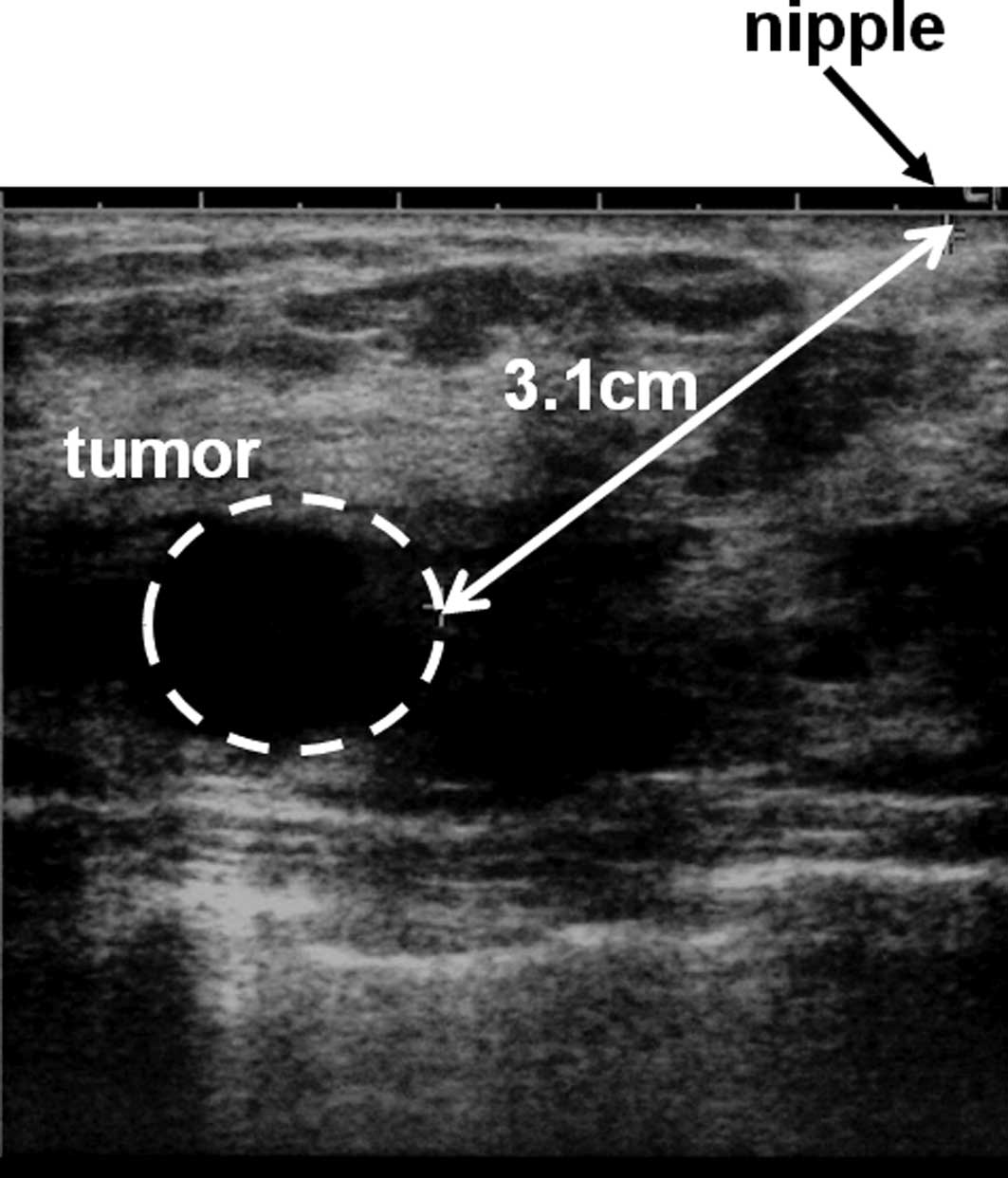
Papillary lesions of the breast include a broad spectrum of lesions, from benign papillomas to papillary carcinomas. It is difficult to determine whether a lesion is benign or malignant based on the fragmented material of a core needle biopsy (CNB). This study evaluated patients with papillary lesions examined using CNB. We retrospectively reviewed 31 papillary lesions diagnosed using CNB between 2004 and 2007. The clinical findings of benign and malignant papillary lesions were compared. The average patient age was 48.9 years. Twelve patients presented with a discharge and 10 patients presented with a lump. Eight patients were asymptomatic. The initial diagnoses by CNB of the 31 lesions were 25 intraductal papillomas, 4 intracystic papillomas and 2 adenomas. After CNB, excisional biopsies were performed in 23 patients and biopsies with a Mammotome® in 2 patients. Seven patients underwent regular follow-up. Five (16%) of the 31 patients with papillary lesions were ultimately diagnosed with breast cancer. The average distance from the nipple to a tumor diagnosed as malignant was 2.46 cm, which was longer than for a tumor diagnosed as benign. Ultimately, 5 papillary lesions (16%) were diagnosed as breast cancer. To avoid overlooking a malignancy, surgical excision is advantageous for papillary lesions, particularly those located far from the nipple.
View Figures
View References

Related Articles

Journal Cover

November-December 2011
Volume 2 Issue 6

Print ISSN: 1792-0981
Online ISSN:1792-1015

Sign up for eToc alerts

Recommend to Library

Copy and paste a formatted citation
x
Spandidos Publications style
Tokiniwa H, Horiguchi J, Takata D, Kikuchi M, Rokutanda N, Nagaoka R, Sato A, Odawara H, Tozuka K, Oyama T, Oyama T, et al: Papillary lesions of the breast diagnosed using core needle biopsies. Exp Ther Med 2: 1069-1072, 2011
APA
Tokiniwa, H., Horiguchi, J., Takata, D., Kikuchi, M., Rokutanda, N., Nagaoka, R. ... Takeyoshi, I. (2011). Papillary lesions of the breast diagnosed using core needle biopsies. Experimental and Therapeutic Medicine, 2, 1069-1072. https://doi.org/10.3892/etm.2011.332
MLA
Tokiniwa, H., Horiguchi, J., Takata, D., Kikuchi, M., Rokutanda, N., Nagaoka, R., Sato, A., Odawara, H., Tozuka, K., Oyama, T., Takeyoshi, I."Papillary lesions of the breast diagnosed using core needle biopsies". Experimental and Therapeutic Medicine 2.6 (2011): 1069-1072.
Chicago
Tokiniwa, H., Horiguchi, J., Takata, D., Kikuchi, M., Rokutanda, N., Nagaoka, R., Sato, A., Odawara, H., Tozuka, K., Oyama, T., Takeyoshi, I."Papillary lesions of the breast diagnosed using core needle biopsies". Experimental and Therapeutic Medicine 2, no. 6 (2011): 1069-1072. https://doi.org/10.3892/etm.2011.332