Introduction
Breast cancer represents the most common malignancy
affecting women in developed countries, with more than 200,000 new
cases diagnosed yearly in the US (1). Moreover, the incidence rates have
increased rapidly in previously low-incidence areas, such as China
(2), partly due to changes in life
style and professional habits, as well as the progression of
urbanization.
The last decade has witnessed significant
achievements in the management of advanced breast cancer, including
the introduction of novel chemotherapeutic agents (3,4), the
use of aromatase inhibitors in post-menopausal women (5,6), and
the benefits derived from molecular-targeted agents, e.g.,
trastuzumab (7,8) and lapatinib (9), in patients with epidermal growth
factor receptor 2 (HER2)-overexpressing tumors. Despite aggressive
multidisciplinary treatment approaches, the prognosis of metastatic
breast cancer remains poor, with a median survival of 20 months
(10).
HER2, a member of the epidermal growth factor
receptor family, is a major target for molecular-targeted therapy
in breast cancer. HER2 locates at human chromosome 17q11.2–12,
encoding a transmembrane tyrosine kinase that is composed of three
distinct regions: an extracellular region containing a
ligand-binding domain, a transmembrane domain and an intracellular
region harboring a tyrosine kinase domain. Ligand binding leads to
receptor dimerization, autophosphorylation and subsequent
activation of intrinsic tyrosine kinase activity. Activation of
HER2 receptors initiates a series of downstream signaling pathways
that regulate various cellular functions, including cell
proliferation, apoptosis, angiogenesis and motility.
Although HER2 is not expressed on the cell surface
of many normal tissues (11), HER2
gene amplification and protein overexpression are present in 20–30%
of breast cancers (12–14). HER2 receptor has become an
important target for targeted cancer therapy with trastuzumab
(Herceptin®). Trastuzumab, a humanized monoclonal
antibody (15), has revolutionized
therapy for patients with metastatic breast cancers. Studies have
indicated that trastuzumab is particularly effective in the
treatment of HER2-positive metastatic breast cancer.
Overexpression of HER2 has been identified in human
breast cancers (12–14). Although similar HER2 receptor
expression between primary breast cancers and metastatic lymph
nodes has also been reported (16–18),
there are only a few reports regarding the comparison of the HER2
status between the primary breast cancer and the distant metastatic
lesions (19,20). To date, the literature regarding
the concordance of HER2 receptor expression between primary and
local-regional recurrences is sparse. It remains uncertain whether
recurrences have the identical or similar HER2 receptor expression
pattern as the primary breast cancer.
Receptor overexpression, together with a similar
expression in both primary tumors and disseminated lesions, is
considered necessary for the success of targeted therapy,
particularly targeted nuclide radiotherapy. In receptor-mediated
tumor targeting nuclide radiotherapy, tumor cells are killed with
delivered radiation, and therapeutic efficiency is mainly dependent
on the receptor expression (21).
However, in most studies, samples for analysis are usually obtained
from the primary lesion, and the status of the targeted molecules
is determined based only on the primary tumor. In the present
study, the expression of HER2 was investigated
immunohistochemically in a series of primary breast cancer samples
and corresponding local-regional recurrent lesions.
Materials and methods
Patients and samples
Thirty-five breast cancer patients with
formalin-fixed, paraffin-embedded tumor samples available from
untreated primary tumors and later clinically manifested local or
regional recurrent tumor deposits were identified in the specimen
database of the Department of Pathology, Second Affiliated Hospital
Zhejiang University and Shaoxing Hospital. The time period between
surgical removal of the primary tumor and surgery or biopsy of the
recurrent tumor lesion ranged from 5 to 61 months (median 20). Two
separate metastatic lesions (chest wall and supraclavicular) were
available for this study from 2 patients. The site of recurrence
was chest wall in 32 cases, supraclavicular recurrence in 1 case
and 2 were both chest wall and supraclavicular recurrences. The
patients were diagnosed between the years 1998 and 2010, and the
patient age at diagnosis ranged from 31 to 74 years (median 51).
Twenty-five (71.4%) cases were invasive ductal carcinomas, 1 (2.8%)
was invasive lobular carcinoma, 2 (5.7%) were mucinous
adenocarcinomas, 6 (17.1%) were carcinoma simplex and 1 (2.8%) was
medullary cancer. Six cases had no lymph node metastasis, 9 cases
had 1–3 metastatic lymph nodes, 15 cases had ≥4 metastatic lymph
nodes, and the lymph node status was not available in the other 5
cases. All patients had no distant metastasis at the time of
diagnosis.
HER2 staining
The study was carried out under approval of the
Institutional Review Board in accordance with the Declaration of
Helsinki. The tissues were fixed in 4% buffered formalin, processed
and embedded in paraffin. Sections (4-μm) were then cut and
deparaffinized in xylene and hydrated through graded concentrations
of ethanol to distilled water. The HER2 immunohistochemical
staining was carried out as previously described (22). After deparaffinization, the
sections were incubated in methanol and hydrogen peroxide for 30
min to quench the endogenous peroxidase. Antigen retrieval was
performed in a waterbath at 98°C, pH 6.0 for 40 min. Thereafter,
the slides were cooled at room temperature and then washed in
distilled water. Immunohistochemical staining was performed using
the Elite ABC kit (Vectastain; Vector Laboratories, Burlingame, CA,
USA). Blocking serum was applied for 15 min followed by incubation
with rabbit anti-human c-erbB-2 oncoprotein (code no. A 0485;
Dako), diluted 1:350. Sections were then incubated with the
biotinylated secondary antibody and were visualized using the
peroxidase substrate 3-amino-9-ethyl-carbazole (AEC) (A-5754;
Sigma) as chromogen. Finally, the sections were counterstained with
Mayer's hematoxylin and mounted.
HER2 scores
HER2 expression was scored using the HercepTest
scoring criterion. The HER2 score was based on a scale where 0
corresponded to tumor cells that were completely negative, 1+
corresponded to faint perceptible staining of the tumor cell
membranes, 2+ corresponded to moderate staining of the entire tumor
cell membranes and 3+ was strong circumferential staining of the
entire tumor cell membranes creating a fishnet pattern. The
Canadian and the Dako HercepTest guidelines were applied, which
require >10% of the tumor cells to be stained (23). Cytoplasmic staining was considered
non-specific and was not included in the scoring. As positive
controls, in-house positive control tissue sections were used, as
well as positive control sections supplied by Dako. As negative
controls, normal tissues were used, which are expected not to
express HER2, such as connective tissue observed in the same
sections as the tumor cells. In sections of lymph node metastases,
lymphocytes and the surrounding capsule of the lymph nodes were
used as negative internal controls.
Excluded cases
In 10 cases, no primary tumor blocks were-found in
the specimen database, as these cases were previously treated at
other hospitals. In another case, there were no tumor cells in the
sections supposed to be recurrent breast cancer. Thus, 46 patient
cases with local/regional recurrences were intitialy identified,
but finally 35 cases with high-quality material of both primary
tumors and the corresponding recurrences were investigated in the
study.
Results
Table I shows the
HER2 scores for the analyzed 35 primary breast cancers and the
corresponding local-regional recurrences. HER2 overexpression (2+
or 3+) was found in 48.57% (17/35) of the primary breast cancers
and 45.71% (16/35) of the corresponding local-regional recurrences.
There was a good agreement between the primary tumors and the
paired asynchronous local-regional recurrences in the majority of
cases. A concordance of HER2 overexpression between the primary
lesions and matching regional recurrences was observed in 85.71% of
the breast cancer cases. Five changes were observed. However, there
were only 3 patients who had HER2 overexpression (all 3 cases had
2+ HER2 staining) in the primary tumors which changed to 1+ in the
chest wall recurrences; in another 2 patients, the score of 1+ in
the primary tumors switched to 2+ or 3+ in local-regional
recurrences. Moreover, all cases with 3+ HER2 staining in the
primary lesions retained HER2 overexpression in the recurrences.
The major results from the HER2-score analyses are summarized in
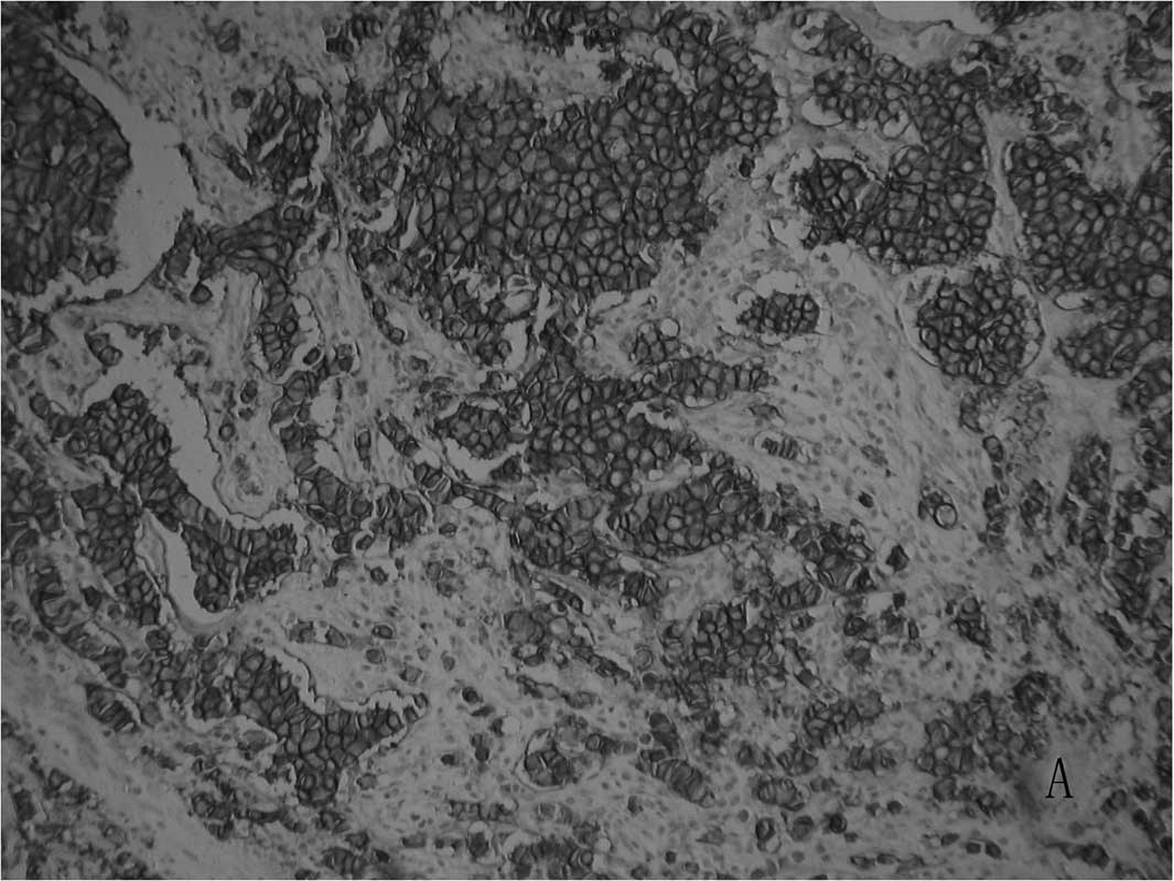
Tables II and III. Examples of staining patterns for a
primary tumor and the corresponding metastases (which were both
scored as 3+) are shown in Fig. 1A and
B.
 | Table I.HER2 scores for the analyzed primary
breast cancers and the paired asynchronous local-regional
recurrences (n=35). |
Table I.
HER2 scores for the analyzed primary
breast cancers and the paired asynchronous local-regional
recurrences (n=35).
| Local-regional
recurrences HER2-scores
|
|---|
| 0 | 1+ | 2+ | 3+ |
|---|
| Primary tumor | | | | |
| HER2 scores | | | | |
| 0 | 7 | 0 | 0 | 0 |
| 1+ | 2 | 7 | 1 | 1 |
| 2+ | 0 | 3 | 3 | 2 |
| 3+ | 0 | 0 | 5 | 4 |
| Table II.Discordant data between the primary
lesions and paired recurrences. |
Table II.
Discordant data between the primary
lesions and paired recurrences.
| Case no. | Primary tumor
(HER2) | Date | Metastasis
(HER2) | Metastatic site | Date |
|---|
| 1 | Negative | August, 2005 | Positive | Chest wall and
supraclavicular | April, 2008 |
| 2 | Positive | November, 1998 | Negative | Supraclavicular | May, 2001 |
| 3 | Positive | May, 1999 | Negative | Chest wall | July, 2000 |
| 4 | Negative | February, 2003 | Positive | Chest wall | November, 2004 |
| 5 | Positive | Jamuary, 2003 | Negative | Chest wall | January, 2004 |
| Table III.Major results from the HER2-score
analyses of breast cancer (n=35). |
Table III.
Major results from the HER2-score
analyses of breast cancer (n=35).
| HER2-score
characteristics | Cases (%) |
|---|
| Primary tumors with
2+ or 3+ | 17 (48.57) |
| Local-regional
recurrences with 2+ or 3+ | 16 (45.71) |
| Unchanged HER2 scores
in local-regional recurrences vs. the primary tumors | 30 (85.71) |
| Changed EGFR scores
in local-regional recurrences vs. the primary tumors | 5 (14.29) |
| Patients who had a
score of 0 or 1+ in primary tumors which changed to 2+ or 3+ in
local-regional recurrences | 2 (5.71) |
| Patients who had a
score of 2+ or 3+ in primary tumors which changed to 0 or 1+ in
local-regional recurrences | 3 (8.57) |
Two cases had both chest wall and supraclavicular
recurrent samples. The same HER2 scores were noted between the
chest wall and supraclavicular lesions; 1 case was scored as 2+ and
the other had negative staining.
Discussion
Anti-HER2-targeted therapy, e.g., trastuzumab, for
recurrent or metastatic breast cancer is generally thought to be
reasonable when the primary lesions of the breast overexpress HER2
receptor. In several cases, however, the recurrent lesions show no
response to trastuzumab treatment, even though the primary breast
cancer exhibits strong HER2 expression (8). Heterogeneity of the receptor status
in primary and metastatic breast cancer has been confirmed, and a
loss of HER2 receptor in recurrent lesions, which may be affected
by the intervening treatment, is known to be associated with a poor
response to anti-HER2-targeted therapy. Yet, knowledge of HER2
expression in local-regional recurrences of breast cancer is
relatively limited. For a receptor to be of interest for targeting,
a similar expression in both the primary tumors and the
disseminated lesions is required. Investigation of the concordance
of the receptor status between recurrences or metastases and the
primary tumors will provide valuable information on whether the
receptor is suitable as a target for diagnostic and/or therapeutic
procedures. In the present study, the expression of HER2 was
investigated immunohistochemically in paired samples from a series
of primary breast lesions and corresponding local-regional
recurrences.
HER2 overexpression (2+/3+) was found in 48.57% of
the primary lesions and 45.71% of the local-regional recurrences.
HER2 expression in breast cancer has been commonly reported to
range from 20 to 30% (12–14). Our result showed a higher
percentage since our analyzed cases represented a more malignant
subgroup which developed recurrences. Studies reporting a higher
HER2 expression rate were also noted in the literature. Carlsson
et al (17), using the same
scoring criteria, found HER2 expression in 55% of the analyzed
primary breast cancers and lymph node metastases. Braun et
al (24) reported HER2
overexpression in 60% of breast cancers with bone marrow
metastases.
Furthermore, a good agreement was noted between the
primary tumors and the paired asynchronous recurrences in the
majority of our studied cases. A concordance of HER2
over-expression between the primary lesions and matching regional
recurrences was observed in 85.71% of the breast cancer cases.
Previous studies mainly focused on the concordance of the HER2
status between primary breast cancer and synchronous lymph node
metastases, or between primary tumors and distant metastases, while
reports concerning local-regional recurrences are relatively
limited. The reported prevalence of concordance of the HER2 status
between primary tumors and synchronous lymph node metastases ranges
from 82 to 94% (16,25), and that between primary tumors and
distant metastases ranges from 92.4 to 97% (19,26,27).
Our data of local-regional recurrences are consistent with these
former findings; only 3 patients with HER2 overexpression (scored
as 2+) in the primary tumors had lower HER2 scores in the
corresponding recurrences, and in another 2 patients the score of
1+ in the primary tumors switched to 2+ or 3+ in the local-regional
recurrences. Moreover, all cases with 3+ HER2 staining in the
primary lesions retained HER2 overexpression in the
recurrences.
Although trastuzumab-based therapy is commonly used
to treat metastatic disease, HER2 status is generally evaluated in
the primary lesions since, in most cases, the metastatic lesions
are not removed or biopsied prior to treatment. With regards to
more recent clinical trials (8,28),
only 50% of the metastatic breast cancer patients with HER2
overexpression respond to trastuzumab treatment. There may be many
reasons for the poor response to trastuzumab (29). One of the explanations may be the
heterogeneity of expression of HER2 between the primary lesions and
metastatic tumors, as receptor characteristics change with time and
may be affected by anticancer treatment. However, based on our
result and other reports, it appears that heterogeneity is an
unlikely explanation.
The HER2 is commonly expressed in breast cancer, and
its expression in primary tumors and the corresponding recurrences
was concordant in the majority of cases. Our results add to the
body of data on the subject. As the receptor expression may lose or
gain in recurrences at a probability of approximately 10%,
assessment of the receptor status in recurrent lesions is
encouraged.
Acknowledgements
The authors acknowledge financial
support from grants from the Science and Technology Project of
Zhejiang (no. 2009C34018), the Outstanding Young Investigator fund
from the Health Bureau of Zhejiang China (no. 2008QN020), and the
National Natural Science Foundation of China to Q. Wei (no.
81071823).
References
|
1
|
Jemal A, Siegel R, Xu J and Ward E: Cancer
statistics 2010. CA Cancer J Clin. 60:277–300. 2010. View Article : Google Scholar
|
|
2
|
Xiang YB, Zhang W, Gao LF, Liu ZW, Xu WH,
Liu EJ and Ji BT: Methods for time analysis of cancer incidence
rates. Chin J Epidemiol. 25:173–177. 2004.
|
|
3
|
Thomas ES, Gomez HL, Li RK, Chung HC, Fein
LE, Chan VF, Jassem J, Pivot XB, Klimovsky JV, de Mendoza FH, Xu B,
Campone M, Lerzo GL, Peck RA, Mukhopadhyay P, Vahdat LT and Roché
HH: Ixabepilone plus capecitabine for metastatic breast cancer
progressing after anthracycline and taxane treatment. J Clin Oncol.
25:5210–5217. 2007. View Article : Google Scholar : PubMed/NCBI
|
|
4
|
O'Shaughnessy J, Twelves C and Aapro M:
Treatment for anthracycline-pretreated metastatic breast cancer.
Oncologist. 7(Suppl 6): 4–12. 2002.
|
|
5
|
Bonneterre J, Buzdar A, Nabholtz JM,
Robertson JF, Thürlimann B, von Euler M, Sahmoud T, Webster A and
Steinberg M; Arimidex Writing Committee; Investigators Committee
Members: Anastrozole is superior to tamoxifen as first-line therapy
in hormone receptor positive advanced breast carcinoma. Cancer.
92:2247–2258. 2001. View Article : Google Scholar : PubMed/NCBI
|
|
6
|
Mouridsen H, Gershanovich M, Sun Y,
Perez-Carrion R, Boni C, Monnier A, Apffelstaedt J, Smith R,
Sleeboom HP, Jaenicke F, Pluzanska A, Dank M, Becquart D, Bapsy PP,
Salminen E, Snyder R, Chaudri-Ross H, Lang R, Wyld P and Bhatnagar
A: Phase III study of letrozole versus tamoxifen as first-line
therapy of advanced breast cancer in postmenopausal women: analysis
of survival and update of efficacy from the International Letrozole
Breast Cancer Group. J Clin Oncol. 21:2101–2109. 2003. View Article : Google Scholar
|
|
7
|
Marty M, Cognetti F, Maraninchi D, Snyder
R, Mauriac L, Tubiana-Hulin M, Chan S, Grimes D, Antón A, Lluch A,
Kennedy J, O'Byrne K, Conte P, Green M, Ward C, Mayne K and Extra
JM: Randomized phase II trial of the efficacy and safety of
trastuzumab combined with docetaxel in patients with human
epidermal growth factor receptor 2-positive metastatic breast
cancer administered as first-line treatment: the M77001 Study
Group. J Clin Oncol. 23:4265–4274. 2005. View Article : Google Scholar
|
|
8
|
Slamon DJ, Leyland-Jones B, Shak S, Fuchs
H, Paton V, Bajamonde A, Fleming T, Eiermann W, Wolter J, Pegram M,
Baselga J and Norton L: Use of chemotherapy plus a monoclonal
antibody against HER2 for metastatic breast cancer that
overexpresses HER2. N Engl J Med. 344:783–792. 2001. View Article : Google Scholar : PubMed/NCBI
|
|
9
|
Geyer CE, Forster J, Lindquist D, Chan S,
Romieu CG, Pienkowski T, Jagiello-Gruszfeld A, Crown J, Chan A,
Kaufman B, Skarlos D, Campone M, Davidson N, Berger M, Oliva C,
Rubin SD, Stein S and Cameron D: Lapatinib plus capecitabine for
HER2-positive advanced breast cancer. N Engl J Med. 355:2733–2743.
2006. View Article : Google Scholar : PubMed/NCBI
|
|
10
|
Dawood S, Broglio K, Gonzalez-Angulo AM,
Buzdar AU, Hortobagyi GN and Giordano SH: Trends in survival over
the past two decades among white and black patients with newly
diagnosed stage IV breast cancer. J Clin Oncol. 26:4891–4898. 2008.
View Article : Google Scholar : PubMed/NCBI
|
|
11
|
Press MF, Cordon-Cardo C and Slamon DJ:
Expression of the HER-2/neu proto-oncogene in normal human adult
and fetal tissues. Oncogene. 5:953–962. 1990.PubMed/NCBI
|
|
12
|
Schechter AL, Stern DF, Vaidyanathan L,
Decker SJ, Drebin JA, Greene MI and Weinberg RA: The neu oncogene:
an erb-B-related gene encoding a 185,000-Mr tumour antigen. Nature.
312:513–516. 1984. View
Article : Google Scholar : PubMed/NCBI
|
|
13
|
Zhang D, Salto-Tellez M, Do E, Putti TC
and Koay ES: Evaluation of HER-2/neu oncogene status in breast
tumors on tissue microarrays. Hum Pathol. 34:362–368. 2003.
View Article : Google Scholar : PubMed/NCBI
|
|
14
|
Shimizu C, Fukutomi T, Tsuda H,
Akashi-Tanaka S, Watanabe T, Nanasawa T and Sugihara K: c-erbB-2
protein overexpression and p53 immunoreaction in primary and
recurrent breast cancer tissues. J Surg Oncol. 73:17–20. 2000.
View Article : Google Scholar : PubMed/NCBI
|
|
15
|
Vogel CL and Franco SX: Clinical
experience with trastuzumab (herceptin). Breast J. 9:452–462. 2003.
View Article : Google Scholar : PubMed/NCBI
|
|
16
|
Cho EY, Han JJ, Choi YL, Kim KM and Oh YL:
Comparison of Her-2, EGFR and cyclin D1 in primary breast cancer
and paired metastatic lymph nodes: an immunohistochemical and
chromogenic in situ hybridization study. J Korean Med Sci.
23:1053–1061. 2008. View Article : Google Scholar : PubMed/NCBI
|
|
17
|
Carlsson J, Nordgren H, Sjöström J, Wester
K, Villman K, Bengtsson NO, Ostenstad B, Lundqvist H and Blomqvist
C: HER2 expression in breast cancer primary tumours and
corresponding metastases. Original data and literature review. Br J
Cancer. 90:2344–2348. 2004.PubMed/NCBI
|
|
18
|
Tsutsui S, Ohno S, Murakami S, Kataoka A,
Kinoshita J and Hachitanda Y: EGFR, c-erbB2 and p53 protein in the
primary lesions and paired metastatic regional lymph nodes in
breast cancer. Eur J Surg Oncol. 28:383–387. 2002. View Article : Google Scholar : PubMed/NCBI
|
|
19
|
Gancberg D, di Leo A, Cardoso F, Rouas G,
Pedrocchi M, Paesmans M, Verhest A, Bernard-Marty C, Piccart MJ and
Larsimont D: Comparison of HER-2 status between primary breast
cancer and corresponding distant metastatic sites. Ann Oncol.
13:1036–1043. 2002. View Article : Google Scholar : PubMed/NCBI
|
|
20
|
Vincent-Salomon A, Jouve M, Genin P,
Fréneaux P, Sigal-Zafrani B, Caly M, Beuzeboc P, Pouillart P and
Sastre-Garau X: HER2 status in patients with breast carcinoma is
not modified selectively by preoperative chemotherapy and is stable
during the metastatic process. Cancer. 94:2169–2173. 2002.
View Article : Google Scholar : PubMed/NCBI
|
|
21
|
Carlsson J, Forssell Aronsson E, Hietala
SO, Stigbrand T and Tennvall J: Tumour therapy with radionuclides:
assessment of progress and problems. Radiother Oncol. 66:107–117.
2003. View Article : Google Scholar : PubMed/NCBI
|
|
22
|
Wei Q, Sheng L, Shui Y, Hu Q, Nordgren H
and Carlsson J: EGFR, HER2 and HER3 expression in laryngeal primary
tumours and corresponding metastases. Ann Surg Oncol. 15:1193–1201.
2008. View Article : Google Scholar : PubMed/NCBI
|
|
23
|
Bilous M, Dowsett M, Hanna W, Isola J,
Lebeau A, Moreno A, Penault-Llorca F, Rüschoff J, Tomasic G and van
de Vijver M: Current perspectives on HER2 testing: a review of
National Testing Guidelines. Mod Pathol. 16:173–182. 2003.
View Article : Google Scholar : PubMed/NCBI
|
|
24
|
Braun S, Schlimok G, Heumos I, Schaller G,
Riethdorf L, Riethmüller G and Pantel K: ErbB2 overexpression on
occult metastatic cells in bone marrow predicts poor clinical
outcome of stage I–III breast cancer patients. Cancer Res.
61:1890–1895. 2001.PubMed/NCBI
|
|
25
|
Cardoso F, di Leo A, Larsimont D, Gancberg
D, Rouas G, Dolci S, Ferreira F, Paesmans M and Piccart M:
Evaluation of HER2, p53, bcl-2, topoisomerase II-alpha, heat shock
proteins 27 and 70 in primary breast cancer and metastatic
ipsilateral axillary lymph nodes. Ann Oncol. 12:615–620. 2001.
View Article : Google Scholar : PubMed/NCBI
|
|
26
|
Thompson AM, Jordan LB, Quinlan P,
Anderson E, Skene A, Dewar JA and Purdie CA; Breast Recurrence in
Tissues Study Group: Prospective comparison of switches in
biomarker status between primary and recurrent breast cancer: the
Breast Recurrence In Tissues Study (BRITS). Breast Cancer Res.
12:R922010. View
Article : Google Scholar
|
|
27
|
Tapia C, Savic S, Wagner U, Schönegg R,
Novotny H, Grilli B, Herzog M, Barascud AD, Zlobec I, Cathomas G,
Terracciano L, Feichter G and Bubendorf L: HER2 gene status in
primary breast cancers and matched distant metastases. Breast
Cancer Res. 9:R312007. View
Article : Google Scholar : PubMed/NCBI
|
|
28
|
Untch M, Muscholl M, Tjulandin S, Jonat W,
Meerpohl HG, Lichinitser M, Manikhas AG, Coumbos A, Kreienberg R,
du Bois A, Harbeck N, Jackisch C, Müller V, Pauschinger M, Thomssen
C, Lehle M, Catalani O and Lück HJ: First-line trastuzumab plus
epirubicin and cyclophosphamide therapy in patients with human
epidermal growth factor receptor 2-positive metastatic breast
cancer: cardiac safety and efficacy data from the Herceptin,
Cyclophosphamide, and Epirubicin (HERCULES) trial. J Clin Oncol.
28:1473–1480. 2010.
|
|
29
|
Spector NL and Blackwell KL: Understanding
the mechanisms behind trastuzumab therapy for human epidermal
growth factor receptor 2-positive breast cancer. J Clin Oncol.
27:5838–5847. 2009. View Article : Google Scholar : PubMed/NCBI
|