Introduction
The guinea pig, a widely used experimental animal,
plays an important role in medical and biological experiments. The
structure of the guinea pig eyeball is similar to humans and
therefore guinea pig eyes have been used in myopia research in
recent years. For example, Howlett and McFadden showed that changes
in a guinea pig eyeball and refraction after defocusing are similar
to other mammals (1). Lu et
al (2) and Zhou et al
(3) induced myopia by applying a
facemask to intervene in the developing guinea pig eyes. The
formation of myopia in humans is significantly related to the
development of extraocular muscles (4). Von Graefe cited the activity of the
extraocular muscles, specifically the horizontal recti and the
medials, as myopigenic owing to their compression of the eye in
convergence (5). More recently,
Greene reviewed the potential effects of accommodation, vitreous
pressure, convergence and the extraocular muscles upon the human
eye, and concluded that convergence and the tension in the
extraocular muscles are of greatest importance because of their
affect upon vitreous pressure. They specifically implicated the
oplique muscles in this regard (6,7). The
structure and function of the extraocular muscles is important in
research on eyeball movement, regulation of movement, binocular
vision and surgical intervention. Investigation of extraocular
muscle anatomy and structure is important in the research of myopia
development. To our knowledge, there are only a few publications on
the microscopic anatomy of the extraocular muscles of guinea pigs
(8). Determining the anatomy of
the extraocular muscles of the guinea pig will set the foundation
for ophthalmological and optometric research that uses guinea pig
eyes as models.
Materials and methods
The present study used 10-week-old British guinea
pigs (n=10) weighing between 400 and 450 g. Those with significant
maldevelopment were eliminated. The guinea pigs were obtained from
the Shanghai Experimental Animal Center of the Chinese Academy of
Sciences. Rearing and treatment protocol was approved by the Animal
Ethics Committee of the Wenzhou Medical College.
The guinea pigs were sacrificed by an overdose of
anesthesia with an intraperitoneal injection of 1.5 ml of a mixture
of 1.2 ml 10% ketamine hydrochloride, 0.8 ml 2% xylazine
hydrochloride and 8.0 ml sterile saline. After decapitation, the
guinea pig heads were placed under a microscope. The eye fissure
was expanded by 1 cm on the nasal and temporal side. The skin
around the eye was cut along the conjunctiva leaving the eye fully
exposed. The membrane of the eyeball was removed close to the
corneal limbus, and the membrane bag was separated exposing the
connective tissue on the eyeball surface by pulling on the eyeball.
The extraocular muscles were isolated by marking the original
points and the points of insertion, and then removing the orbital
septum and fat. The heads of the remaining five pigs were removed
and their skin was removed after being sacrificed. The skull was
split along the midline and placed into 10% neutral formaldehyde
solution for 1 week. The optic nerve from the optic chiasm was
identified under the microscope and the bone tissue was peeled off
with a hemostat. The bony orbital wall was removed, the periosteum
opened and the muscle tissue was identified. The fat tissue and
orbital septum were removed from the eye socket, and the number of
muscles and the points of origin and insertion of the extraocular
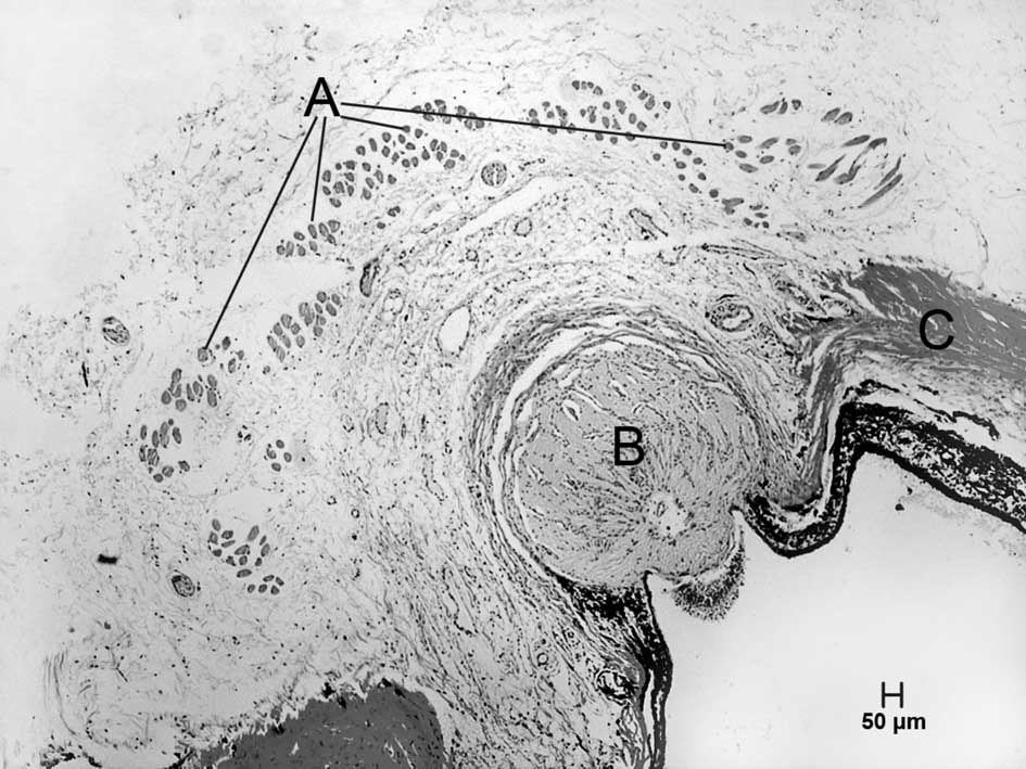
muscles were determined. The retractor bulbi muscle was confirmed
by paraffin sections and tissue H&E staining.
Results
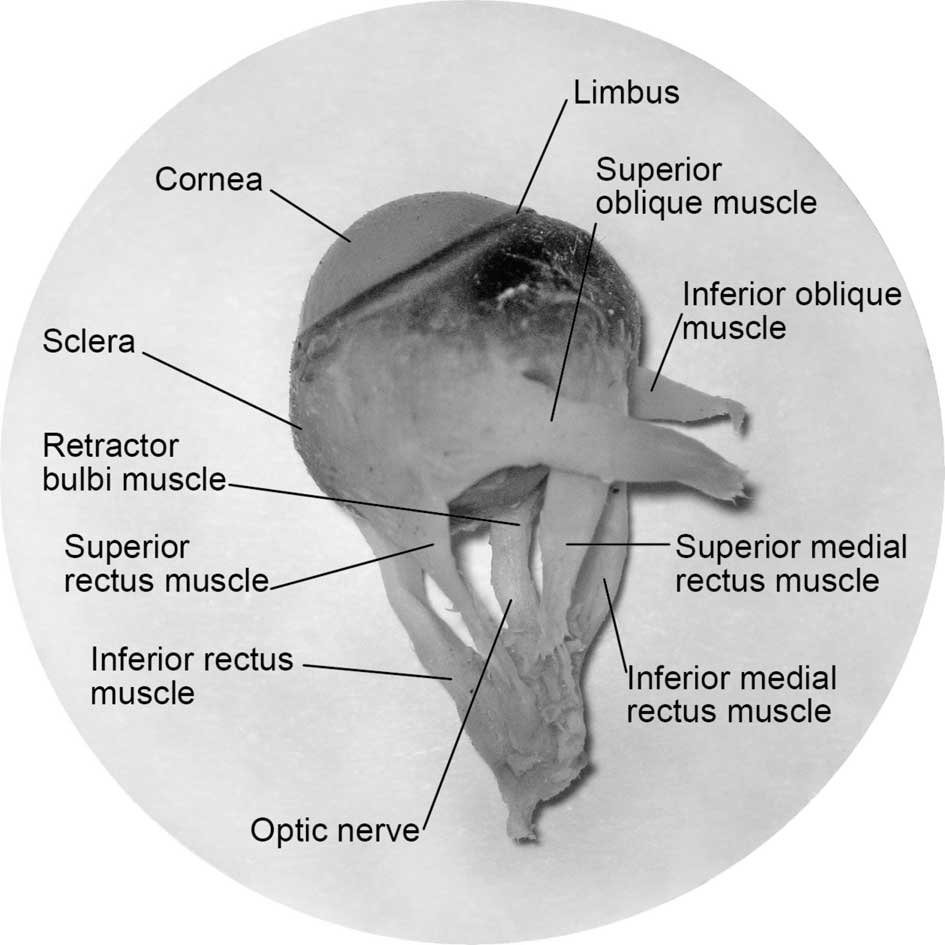
The guinea pig has seven extraocular muscles: two
medial rectus muscles, one superior rectus muscle, one inferior
rectus muscle, one superior oblique muscle, one inferior oblique
muscle and one retractor bulbi muscle (Figs. 1–6). Two medial rectus muscles originate
from the optic foramen around the optic nerve, located on the back
temporal side of the orbit (Fig.
4). They are parallel with separate origins and insertions. Two
medial rectus muscles are located on the nasal side of the eyeball
after passing around the equator of the eyeball from the nasal side
along the wall. The insertion lines are situated 1.5 mm from the
nasal limbus and parallel to the limbus. The superior rectus muscle
travels from the back temporal side of the optic foramen, while the
inferior rectus muscle travels from the back temporal side of the
optic foramen and inserts 1.5 mm inferior to the limbus. The
superior and inferior oblique muscles both originate from the
medial orbital wall following a straight course and are behind the
insertions of the superior rectus muscles and inferior rectus
muscles. The insertion line of the superior rectus muscles and
superior oblique muscle creates a 45° angle. The insertion of the
inferior rectus and inferior oblique muscles creates a 20° angle.
Among the six muscles that insert on the aspect near the limbus,
the length of the two medial rectus muscles and tendons were the
longest of all the extraocular muscles, while the superior rectus
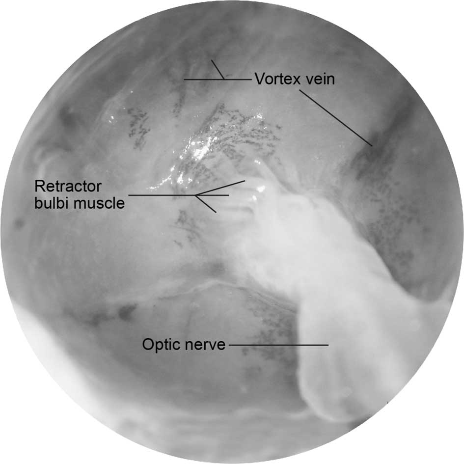
muscle was relatively short. The retractor bulbi muscle originates
from the optic foramen within the origins of the rectus muscles. It
surrounds the optic nerve longitudinally and is inserted
circumferentially into the posterior pole of the eyeball around the
optic nerve head (Figs. 5 and
6).
Discussion
The data revealed that the three color breed of
British guinea pigs has two medial rectus muscles, but no lateral
rectus muscle. The superior oblique muscle track does not have a
trochlea structure, which is present in the human eye. The
specimens in our study confirmed the tracks of the extraocular
muscles and the relative location to the eyeball. It is generally
agreed that most mammals, such as tigers and lions, have four
rectus and two oblique muscles (10). The origins of the four rectus
muscles are close to the optic nerve behind the orbit. The location
of the eye shank and two oblique muscles originate from the front
of the orbit. The results of this study indicate that the number
and origins of guinea pig extraocular muscles have the
characteristics of most vertebrates. The reports by Cooper and
Schiller suggest that the guinea pig has four rectus muscles
(medial, lateral, superior and inferior), two oblique muscles and
one retractor bulbi muscle (8).
The four rectus muscles originate from the optic nerve around the
orbit's apex; the two oblique muscles originate from the medial
wall of the orbit, but without the pulley structure observed in the
human eye on the superior oblique muscle (8). Their descriptions are different from
the results presented here. There are five types of guinea pigs and
accounting for the five most common strains of guinea pigs
[short-haired (British and American species), Dunkan-Hartley,
Hartly, 2 strain and 13 strain], we do not consider that the
disparities between previous reports and the results of this study
are explained by species differences.
Typically, the eyes of certain lower level animals
are located on the sides of the head, thus giving a large visual
field, but no three-dimensional vision. During evolution, the eyes
of higher level animals have moved to the front of the head
allowing for three-dimensional vision. The optical axis angle of
the guinea pig ranges from 103 to 110°; binocular vision ranges
from 20 to 63° and the combined visual field is between 325 and
340°. By contrast, the optical axis angle of the human eye is 0°;
binocular vision is between 140 to 160° and the combined visual
field is between 180 and 190°. The guinea pig's combined visual
field is close to 360° (9,10), and therefore the lateral rectus
muscle is not required. The optic nerve of the guinea pig is
located near the temporal side from the anatomical structure of the
superior and inferior rectus muscles, and the origin is on the back
of the eyeball's temporal side. Therefore, the superior and
inferior rectus muscles provide sufficient movement of the eyeball.
Guinea pig eyes are located on two sides of the head; therefore,
binocular vision is relatively narrow. The two medial rectus
muscles are conducive to broadening binocular vision and achieving
three-dimensional vision.
In conclusion, the anatomy of the guinea pig
extraocular muscles is different from that of humans. There are two
medial rectus muscles without a lateral rectus muscle, which is
consistent with eyeball location and guinea pig living habits.
Acknowledgments
This study was supported by the Shanghai Leading
Academic Discipline Project (S30205) and the Science and Technology
Foundation of Shanghai Jiaotong University School of Medicine.
References
|
1
|
Howlett MH and McFadden SA:
Form-deprivation myopia in the guinea pig (Cavia porcellus).
Vision Res. 46:267–283. 2006. View Article : Google Scholar : PubMed/NCBI
|
|
2
|
Lu F, Zhou X, Zhao H, Wang R, Jia D, Jiang
L, Xie R and Qu J: Axial myopia induced by a monocularly-deprived
facemask in guinea pigs: a non-invasive and effective model. Exp
Eye Res. 82:628–636. 2005. View Article : Google Scholar : PubMed/NCBI
|
|
3
|
Zhou X, Lu F, Xie R, Jiang L, Wen J, Li Y,
Shi J, He T and Qu J: Recovery from axial myopia induced by a
monocularly deprived facemask in adolescent (7-week-old) guinea
pigs. Vision Res. 47:1103–1111. 2007. View Article : Google Scholar : PubMed/NCBI
|
|
4
|
Curtin BJ: The Myopias. Harper & Row
Publishers; Philadelphia: pp. 104pp. 1985
|
|
5
|
Von Graefe A: Beitrage zur Physiologie und
Pathologie der schiefen Augenmuskeln. Albrecht Von Graefes Arch
Ophthalmol. 3:2771857.
|
|
6
|
Greene PR: Myopia and the extraocular
muscles. Doc Ophthalmol. 8:1631981.
|
|
7
|
Greene PR: Mechanical aspects of myopia
Master's thesis. Harvard University; 1978
|
|
8
|
Cooper G and Schiller AL: Anatomy of the
Guinea Pig. Harvard University Press; Cambridge, MA: pp. 369–389.
1975
|
|
9
|
Duke-Elder S: The Eye in Evolution System
of Ophthalmology. 1. Henry Kimpton Press; London: pp. 6721976
|
|
10
|
Prince Jack H: Comparative Anatomy of the
Eye. Charles C Thomas Publisher; Illinois: pp. 27–29. pp. 286–298.
1956
|