Article
Comparative study of multi-slice CT angiography with digital subtraction angiography in the blood supply of meningiomas
- Authors:
- Xiangjun Han
- Yuefu Zhan
- Jianqiang Chen
-
-
Pages:
31-36
|
Published online on:
September 21, 2011
https://doi.org/10.3892/etm.2011.354
- Expand metrics +
Metrics:
Total
Views: 0
(Spandidos Publications: | PMC Statistics:
)
Metrics:
Total PDF Downloads: 0
(Spandidos Publications: | PMC Statistics:
)
This article is mentioned in:
Abstract
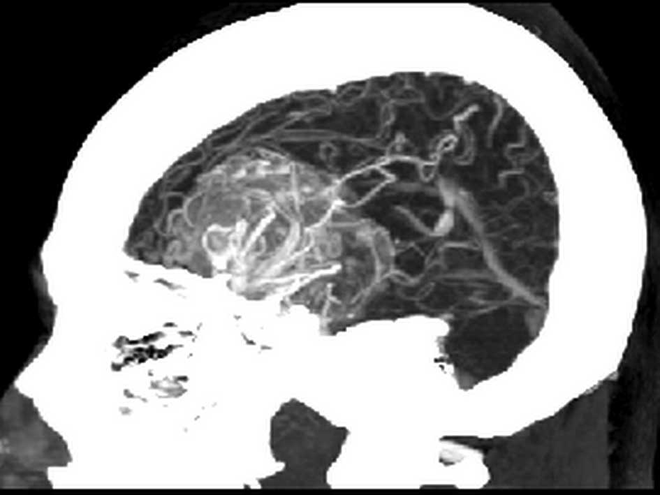
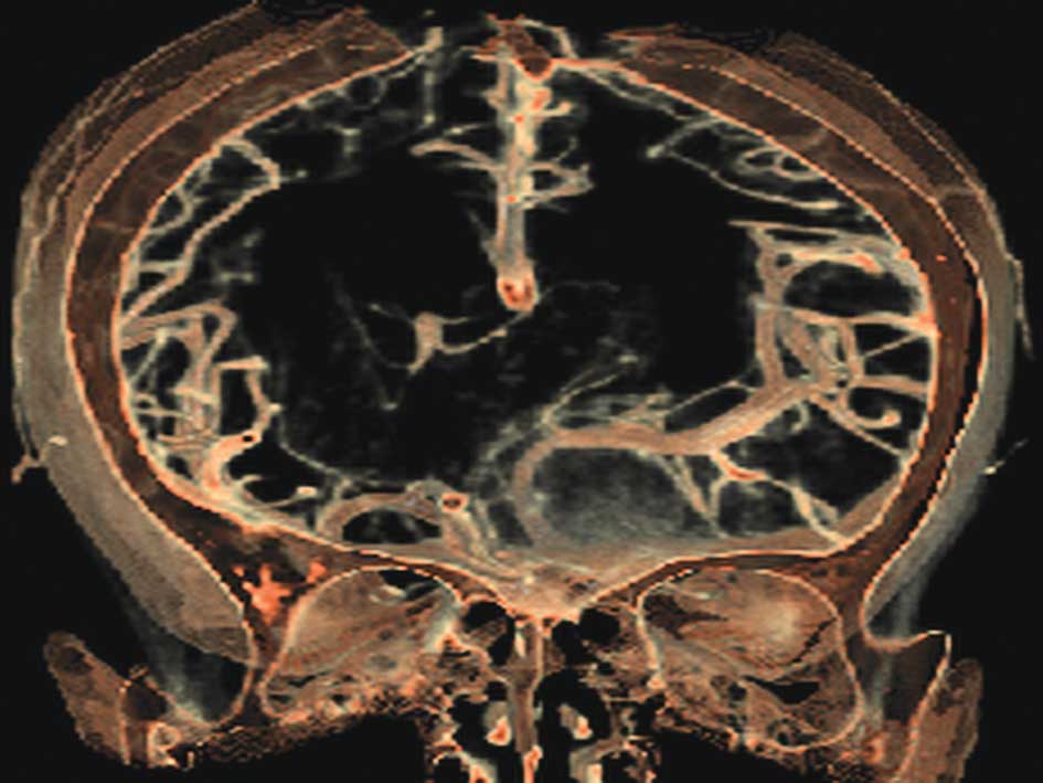
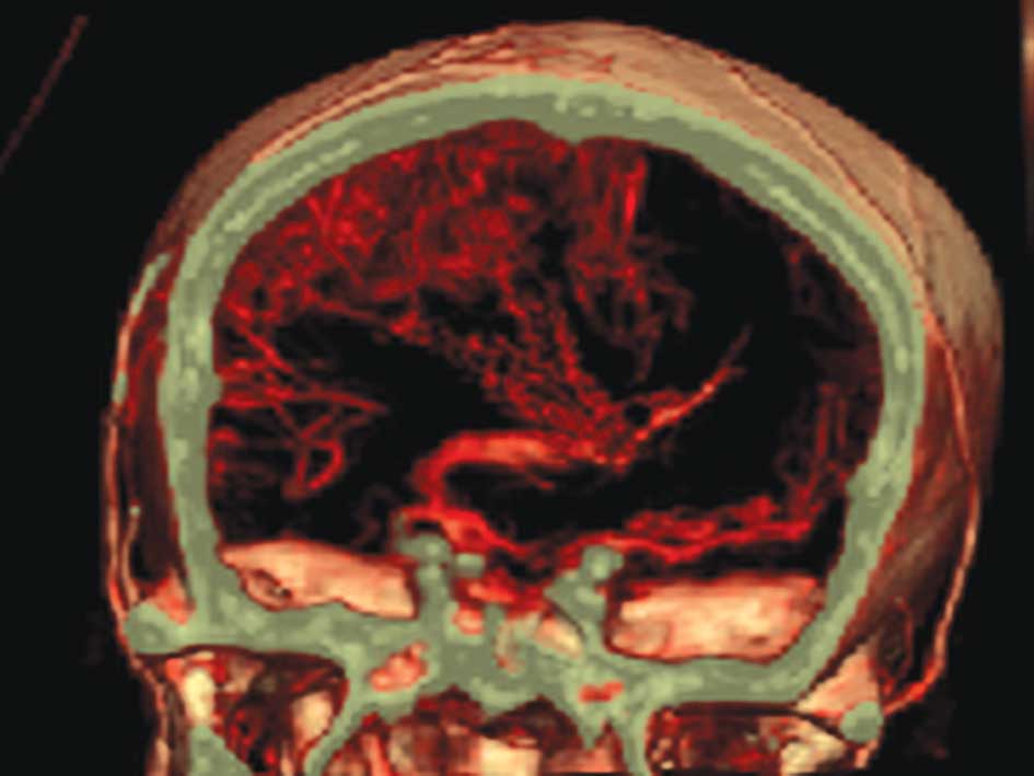
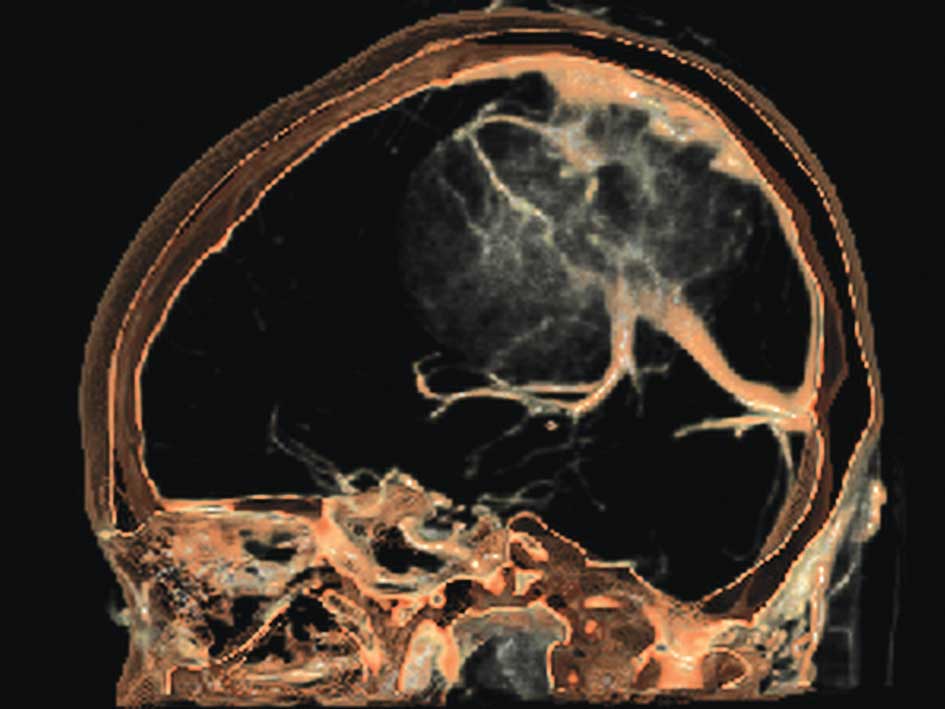
Evaluation of multi-slice CT angiography (MSCTA) and digital subtraction angiography (DSA) was made through the comparison of the two methods in detecting the blood supply of meningiomas, providing further clues for the improved application of MSCTA and DSA in the blood supply of meningiomas. In this study, 20 patients with meningiomas underwent CT angiography by 16-slice spiral CT and inspection 1 week later using DSA. The blood supply of the meningiomas (including the tumor feeding arteries, draining veins, venous sinuses, vessels around the tumor and skull-shaped images, such as comparative evaluation) was compared. The results showed that MSCTA and DSA inspection clearly depicted tumor feeding arteries, draining veins and venous sinuses in all 20 patients. DSA was slightly more effective than MSCTA in displaying fine branches, and DSA had obvious advantages over MSCTA in displaying vessels near the skull. In the display of the tumor's compression and pushing over its peripheral vasculature, MSCTA had the three-dimensional advantage, although in displaying parasagittal sinus meningiomas, it may overestimate the presence of venous sinus invasion due to the impact of bone structure. This does not affect DSA. MSCTA accurately assesses the relationship between the tumor and the bone, and provides three-dimensional images for the pre-operative simulation of surgical approaches. Taken together, MSCTA and DSA are both able to provide important information, such as accurate visualization of tumor-feeding arteries, draining veins and venous sinuses, with high consistency. MSCTA is more advantageous in the depiction of three-dimensional relationships between the tumor and peripheral vessels and skull. Compared to DSA, MSCTA is non-invasive, faster and less costly.
View References
|
1.
|
C ManelfeP LasjauniasJ
RuscalledaPreoperative embolization of intracranial meningiomasAm J
Neuroradiol796319863096121
|
|
2.
|
X FanQ FangL HuangThe meningioma
diagnostic value with digital subtraction cerebral angiographyJ
Intervent Radiol154304312006
|
|
3.
|
J MaW RenP ChenThe diagnostic value of
digital subtraction angiography of intracranial meningiomaJ
Intervent Radiol134834852006
|
|
4.
|
R LiD WangZ ZengDigital subtraction
angiography of meningioma and preoperative embolotherapyJ Practical
Radiol219499512005
|
|
5.
|
X HanT WangH GaoClinical study on
nurturing vessels of menigioma by multi-slice CT angiography and 3D
reconstructionChin J Intervent Imaging Ther31721742006
|
|
6.
|
M LellK AndersE KlotzH DittW BautzBF
TomandlClinical evaluation of bone-subtraction CT angiography
(BSCTA) in head and neck imagingEur
Radiol16889897200610.1007/s0330-005-0032-116267665
|
|
7.
|
SG WetzelE KirschKW StockCerebral veins:
comparative study of CT venography with intraarterial digital
subtraction angiographyAm J Neuroradiol20249255199910094346
|
|
8.
|
MM LellK AndersU MichaelNew techniques in
CT
angiographyRadioGraphics26S45S62200610.1148/rg.26si06550817050518
|