Introduction
Ultrasound therapy is a safe and non-invasive
therapeutic approach which shows great promise in the medical
field, particularly for tumor therapy. Therapeutic ultrasound
mainly includes high- and low-intensity ultrasound. At present,
high-intensity focal ultrasound has been applied in the clinical
management of tumors since it effectively eradicates tumor tissues
through high temperatures and mechanical effects (1,2).
Low-intensity ultrasound activates a sensitizer to produce
sonodynamic action, which kills tumor cells directly or indirectly
(3–5). Sonodynamic therapy has been
considered as a feasible strategy for treating malignancies. Our
previous studies also confirmed that low-intensity ultrasound
activated 5-ALA, hypocrellin B and mematoporphyrin monomethyl ether
to induce the cell death of tumor cells (6–9).
Curcumin is a naturally ocurring spice, often prescribed in India
and China to treat various diseases, such as skin, rheumatic and
digestive diseases (10,11). Recently, our studies found that
low-intensity ultrasound enhanced the cell death of nasopharyngeal
carcinoma cells in the presence of curcumin (12,13).
However, the underlying mechanisms are unclear.
Necrosis, apoptosis and autophagy are the main modes
of cell death which occur when tumor cells respond to chemotherapy
and physical therapy (14–16). Our previous studies showed that
ultrasound exposure in the presence of curcumin significantly
increased the necrotic or late apoptotic rates of nasopharyngeal
carcinoma cells (12). In the
present study, we observed mitophagy in nasopharyngeal carcinoma
CNE2 cells under transmission electron microscopy (TEM) after
ultrasound treatment in the presence of curcumin.
Materials and methods
Sensitizer
A stock solution of curcumin from Sigma (USA) was
constructed in dimethyl sulfoxide (DMSO) at a concentration of 100
mM and maintained in the dark at −20°C until it was used.
Cell culture
Nasopharyngeal carcinoma CNE2 cells were purchased
from the Shanghai Biology Institute and approval was obtained by
the Ethics Committee of Chongqing Medical University prior to the
study. The cells were cultured in RPMI-1640 medium supplemented
with 10% fetal calf serum (Gibco), 50 μg/ml penicillin, 50
μg/ml streptomycin and 10 μg/ml neomycin. The cells
were incubated at 37°C in a humidified CO2 (5%)
incubator.
Ultrasound treatment
CNE2 cells (1.0×10⁵/ml) grown overnight
in a 24-well culture plate with a flat bottom (Corning, USA) were
placed on a platform containing a 1-cm diameter plane transducer,
and then exposed to ultrasound 1 h after incubation with curcumin
(10 μM). The plane transducer with a central frequency of
1.7 MHz was used to generate continuous ultrasound energy. The
spatial average intensity of the ultrasound was 0.46
W/cm2 and exposure time was set at 8 sec. The plane
transducer was placed in a water tank filled with degassed water
during ultrasonic exposure and the experiment was randomly divided
into four groups as described in our previous studies (12,13).
Transmission electron microscopy
(TEM)
TEM was used to observe mitophagy and
ultrastructural changes in the CNE2 cells 6 h after curcumin and
ultrasound treatment. Fixed cells were post-fixed in 2%
OsO4, dehydrated in graded alcohol and flat-embedded in
Epon 812 (Electron Microscopy Sciences, Fort Washington, PA, USA).
Ultra-thin sections (100-nm) were prepared, stained with uranyl
acetate and lead citrate, and examined under an electron microscope
(H-600; Hitachi, Japan).
Results
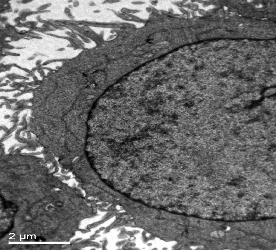
Conventional TEM is a standard method for monitoring
cellular ultrastructure and autophagy (17). In the TEM observation, the
untreated CNE2 cells showed integrity of the cell membrane and
mitochondria, with well-developed cristae and a variety of cell
nuclei (Fig. 1A). Slight swelling
of mitochondria was observed in the CNE2 cells treated by
ultrasound alone or curcumin alone. Severely swollen and disrupted
mitochondria, and more extensive mitophagy was observed in the CNE2
cells treated with ultrasound radiation in the presence of curcumin
(Fig. 1B). This demonstrated that
ultrasound treatment in the presence of curcumin markedly damaged
mitochondria and initiated mitophagy in the CNE2 cells.
Discussion
Our previous studies demonstrated that ultrasound
treatment in the presence of curcumin increased the necrotic or
late apoptotic rates of nasopharyngeal carcinoma cells (12). In our previous TEM study, apoptotic
features, such as the disappearance of microvillin, membrane
blebbing and chromatin condensation, were observed in CNE2 cells
treated with both ultrasound and curcumin (13). These findings indicated that
ultrasound in the presence of curcumin markedly induced the
necrosis and apoptosis of CNE2 cells. Necrosis is a passive form of
cell death usually accompanied by rigorous inflammation; apoptosis
is an active form of cell death through programmed self-destruction
of a single cell characterized by specific nuclear condensation and
fragmentation (18,19). Autophagy is a mode of cell death
distinct from necrosis and apoptosis. Autophagy is usually
considered to be a genetically programmed cell death involving the
degradation of cellular proteins and organelles often accompanied
by the formation of an autophagosome (20,21).
In the process of autophagy, cytoplasmic components such as
mitochondria and peroxisomes are encapsulated by an isolation
membrane to cause the formation of an autophagosome (20,21).
Increasing evidence shows that the autophagic and apoptotic
pathways may be regulated by common factors (21,22).
Mitochondria are regarded as an important convergent point of
apoptotic and autophagic signals, which initiate apoptotic and
autophagic cell death (23). In
our previous studies, a significant collapse of mitochondrial
membrane potential (MMP) and ultrastructural changes in
mitochondria were observed after the combined treatment of curcumin
and ultrasound (12,16). In the present study, we found
severely swollen and disrupted mitochondria, and more extensive
mitophagy in cells after the combined treatment of ultrasound and
curcumin, which demonstrated that the treatment significantly
damaged mitochondria and initiated mitophagy. Mitophagy is a
selective form of cell death to degrade damaged mitochondria
through autophagy in mammalian cells (24). Our findings suggest that ultrasound
treatment in the presence of curcumin initiates autophagy to
degrade damaged mitochondria.
It is well known that autophagy plays dual roles in
determining the fate of cells, such as death and survival. Certain
studies have demonstrated that the induction of autophagy enhances
cell death, whereas other reports reveal that autophagy sustains
metabolism for promoting cell survival through the degradation of
damaged proteins and organelles (25–27).
Therefore, the exact mechanisms of mitophagy and autophagy
regarding the bioeffects induced by ultrasound treatment in the
presence of curcumin need to be determined in future
investigations.
In summary, the present study revealed that
ultrasound treatment in the presence of curcumin initiated
mitophagy in CNE2 cells. Our results highlight that mitophagy may
be an important event involving cancer cell death induced by
ultrasound treatment in the presence of curcumin.
Acknowledgements
This study was supported by grants
from the National Nature Science Foundation of China (30973168) and
the Affiliated Hospital of Xi’an Medical University (XYFY10-02).
The authors express sincere thanks to Professor Faqi Li, Mr Eric
Chuck Hey Pun, Mr Jianyong Wu, Mr Dejiang Chao and Ms. Qinglin Li
for the helpful assistance.
References
|
1.
|
C ObynF MambourgAssessment of high
intensity focused ultrasound for the treatment of prostate
cancerActa Chir Belg109581586200919994799
|
|
2.
|
C ChaussyS ThüroffHigh-intensity focused
ultrasound in the management of prostate cancerExpert Rev Med
Devices7209217201010.1586/erd.09.6620214427
|
|
3.
|
W HiraokaH HondaLB Feril JrN KudoT
KondoComparison between sonodynamic effect and photodynamic effect
with photosensitizers on free radical formation and cell
killingUltrason
Sonochem13535534200610.1016/j.ultsonch.2005.10.00116325451
|
|
4.
|
I RosenthalJZ SostaricP RieszSonodynamic
therapy – a review of the synergistic effects of drugs and
ultrasoundUltrason Sonochem113493632004
|
|
5.
|
M KurokiK HachimineH AbeH ShibaguchiM
KurokiS MaekawaJ YanagisawaT KinugasaT TanakaY YamashitaSonodynamic
therapy of cancer using novel sonosensitizersAnticancer
Res2736733677200717970027
|
|
6.
|
P WangCS XuJ XuX WangAW LeungHypocrellin B
enhances ultrasound-induced cell death of nasopharyngeal carcinoma
cellsUltrasound Med
Biol36336342201010.1016/j.ultrasmedbio.2009.09.00720018428
|
|
7.
|
P WangC XuX XiaJ XuX WangJ XiangAW
LeungMitochondrial damage in nasopharyngeal carcinoma cells induced
by ultrasound radiation in the presence of Hypocrellin BJ
Ultrasound Med294350201020040774
|
|
8.
|
Y HeX XiaC XuH YuD BaiJ XiangAW
Leung5-Aminolaevulinic acid enhances ultrasound-induced
mitochondrial damage in K562
cellsUltrasonics50777781201010.1016/j.ultras.2010.03.00420381823
|
|
9.
|
Z TianX QuanC XuL DanH GuoW
LeungHematoporphyrin monomethyl ether enhances sonokilling action
of ultrasound on osteosarcoma in vivoJ Ultrasound
Med2816951702200919933484
|
|
10.
|
P AnandSG ThomasAB KunnumakkaraC
SundaramKB HarikumarB SungST TharakanK MisraIK PriyadarsiniKN
RajasekharanBB AggarwalBiological activities of curcumin and its
analogues (Congeners) made by man and mother natureBiochem
Pharmacol7615901611200810.1016/j.bcp.2008.08.00818775680
|
|
11.
|
C Alarcón de la LastraCurcumin: a
promising spice for therapeuticsMol Nutr Food
Res52985200818792006
|
|
12.
|
X WangX XiaC XuJ XuP WangJ XiangD BaiAW
LeungUltrasound-induced cell death of nasopharyngeal carcinoma
cells in the presence of curcuminIntegr Cancer
Ther107076201110.1177/153473541037719720702493
|
|
13.
|
X WangX XiaAW LeungJ XiangY JiangP WangJ
XuH YuD BaiC XuUltrasound induces cellular destruction of
nasopharyngeal carcinoma cells in the presence of
curcuminUltrasonics51165170201110.1016/j.ultras.2010.07.00620728195
|
|
14.
|
A SasnauskieneJ KadziauskasN VezelyteV
JonusieneV KirvelieneApoptosis, autophagy and cell cycle arrest
following photodamage to mitochondrial
interiorApoptosis14276286200910.1007/s10495-008-0292-819165602
|
|
15.
|
CS XuAWN LeungL LiuXS XiaLED-activated
pheophorbide a induces cellular destruction of colon cancer
cellsLaser Physics Lett7544548201010.1002/lapl.201010008
|
|
16.
|
V Garcia-EscuderoR GarginiAutophagy
induction as an efficient strategy to eradicate
tumorsAutophagy4923925200810.4161/auto.671418716458
|
|
17.
|
A ApelI HerrH SchwarzHP RodemannS
MayerBlocked autophagy sensitizes resistant carcinoma cells to
radiation therapyCancer
Res6814851494200810.1158/0008-5472.CAN-07-056218316613
|
|
18.
|
MC JäckelMA DorudianD MarxU BrinckA
SchauerW SteinerSpontaneous apoptosis in laryngeal squamous cell
carcinoma is independent of bcl-2 and bax protein
expressionCancer85591599199910091732
|
|
19.
|
GM LaMuragliaJ SchiereckJ HeckenkampG
NigriP WatermanD LeszczynskiS KossodoPhotodynamic therapy induces
apoptosis in intimal hyperplastic arteriesAm J
Pathol157867875200010.1016/S0002-9440(10)64600-710980126
|
|
20.
|
A ApelI HerrH SchwarzHP RodemannS
MayerBlocked autophagy sensitizes resistant carcinoma cells to
radiation therapyCancer
Res6814851494200810.1158/0008-5472.CAN-07-056218316613
|
|
21.
|
LK SySC YanCN LokRYK ManCM CheTimosaponin
A-III induces atuophagy preceding mitochondria apoptosis in HeLa
cancer cellsCancer
Res681022910237200810.1158/0008-5472.CAN-08-198319074891
|
|
22.
|
M LiX JiangD LiuY NaGF GaoZ XiAutophagy
protects LNCaP cells under androgen deprivation
conditionsAutophagy45460200810.4161/auto.520917993778
|
|
23.
|
CH YanZQ LiangZL GuYP YangP ReidZH
QinContributions of autophagic and apoptotic mechanisms to
CrTX-induced death of K562
cellsToxicon47521530200610.1016/j.toxicon.2006.01.01016542694
|
|
24.
|
EH KimKS ChoiA critical role of superoxide
anion in selenite-induced mitophagic cell
deathAutophagy47678200810.4161/auto.511917952022
|
|
25.
|
P GeJ ZhangX WangF MengW LiY LuanF LingY
LuoInhibition of autophagy induced by proteasome inhibition
increases cell death in human SHG-44 glioma cellsActa Pharmacol
Sin3010461052200910.1038/aps.2009.7119575007
|
|
26.
|
D KesselMGH Vicente JrJJ ReinersInitiation
of apoptosis and autophagy by photodynamic
therapyAutophagy2289290200610.4161/auto.279216921269
|
|
27.
|
Y KondoT KanzawaR SawayaS KondoThe role of
autophagy in cancer development and response to therapyNat Rev
Cancer9726734200510.1038/nrc169216148885
|