Introduction
Stroke is an acute and progressive neurodegenerative
disorder. Ischemic stroke is the result of a transient or permanent
reduction in cerebral blood flow caused by the occlusion of a
cerebral artery via an embolus or local thrombosis (1). The major pathobiological mechanisms
of ischemia/reperfusion injury include excitotoxicity, oxidative
stress, inflammation, and apoptosis (2,3).
These changes are associated with mitochondrial dysfunction and
rapid decreases in the ATP level, resulting in free radical
generation and lipid peroxidation.
Oxidative stress is a crucial factor in cerebral
ischemic injury, since the brain consumes a large quantity of
oxygen. Reactive oxygen species (ROS) have been indicated as one of
the earliest and most significant components of tissue injury
following reperfusion of the ischemic organ (4,5). The
brain is quite vulnerable to oxidative stress due to its high
polyunsaturated fatty acid content, which is particularly
susceptible to ROS damage (6,7).
Korean red ginseng (KRG, the steamed root of
Panax ginseng C.A. Meyer, Araliaceae) is used frequently as
a crude substance that is taken orally as a traditional medicine in
Asian countries. It has been reported to show a range of biological
activities, such as antitumor, anti-inflammatory, and antistress
activities (8–10). These activities have been
attributed mainly to saponin, which contains a variety of
ginsenosides. There is evidence that the medicinal efficacy of
ginseng is closely linked to its protective effects against free
radical attack (11). The
administration of ginseng to rats has been reported to prevent
myocardial ischemia-reperfusion damage induced by hyperbaric oxygen
(11). Ginseng has been reported
to have protective effects against hepatic oxidative stress caused
by exhaustive exercise (12) and
against muscle injury and inflammation after eccentric exercise
(13). Moreover, it has been
reported that ginseng extracts scavenge superoxide radicals,
inhibit lipid peroxidation and reduce the level of oxidative DNA
damage caused by the Fenton agent (14,15).
However, few in vivo studies have examined the
neuroprotective effects of KRG on the rat brain affected by
ischemia/reperfusion injury. In a previous study, we reported that
KRG treatment prevented cerebral ischemic injury induced by middle
cerebral artery occlusion/reperfusion (MCAO/R) in rats, which was
assessed by staining brain tissue with 2%
2,3,5-triphenyltetrazolium chloride (16). This study investigated the
neuroprotective effect of KRG following a focal brain
ischemia/reperfusion injury and focused on the antioxidant
activities of KRG.
Materials and methods
Animals and treatments
Thirty Sprague-Dawley rats weighing 300±10 g (mean ±
standard deviation, 9 weeks old) were used. The animals were
purchased from Samtako Bio Korea Co., Ltd. (Kyung Gi-Do, Korea).
The rats were housed in an environmentally controlled room at
22±2°C, with a relative humidity of 55±5%, a 12-h light/dark cycle,
and food and water ad libitum. The study was carried out in
accordance with the Korean Academy of Medical Sciences. The animals
were placed under general anesthesia and monitored in accordance
with the corresponding standard procedures (Laboratory Animal
Manual 2000, Korean Academy of Medical Sciences, Seoul, Korea). All
experimental procedures were approved by the Kyung Hee University
Medical Center Institutional Animal Care and Use Committee, Seoul,
Korea.
The animals were divided randomly into three groups:
i) the sham operation group (n=9), in which the animal was
anesthetized with the left common and external carotid artery
exposed but with no arterial ligation or opening; ii) the saline
group (n=12), in which the animal underwent the MCAO/R procedure
and was treated with normal saline (6 ml/kg/day perorally) for 7
days; and iii) the KRG group (n=9), in which the animal underwent
the MCAO/R procedure and was treated with KRG (100 mg/kg/day
perorally) for 7 days.
Feeding with normal saline or KRG was
initiated after the onset of reperfusion injury
The rats were administered with either saline or KRG
once daily, perorally, for 7 days. They were treated between 10:00
and 12:00 to avoid any nonspecific effects due to the circadian
rhythm.
KRG extract
KRG extract in concentrated form was purchased
commercially from the Korea Ginseng Corp. (Daejeon, Korea). It was
produced from the roots of fully matured six-year-old Korean
ginseng plants. KRG compound contains various ginseng saponin
fractions: 30.1% ginsenoside (G)-Rb1, 13.9% G-Rb2, 14.4% G-Rc, 6.1%
G-Rd, 13.9% G-Re, 4.7% G-Rf, 11.5% G-Rg1, 2.6% G-Rg2, and 2.8%
G-Rg3.
Measurement of regional cerebral blood
flow
Each rat was anesthetized with isoflurane (initiated
with 5% and maintained at 2%) in a mixture of 30% oxygen and 70%
nitrous oxide using a vaporizer and face mask. The rat was mounted
in the prone position on a dual small animal stereotaxic instrument
(David Kopf Instruments, Los Angeles, CA, USA). After a cranial
mid-line incision, a 1-mm burr hole was made with a microdrill at
both sides, 2-mm-posterior and 5-mm-lateral to the bregma. The rat
was then placed in the supine position. A laser Doppler probe was
placed on the burr hole under the rat skull, almost contacting the
dura mater of the brain surface avoiding the large vessels. The
parietal cortical perfusion in the territory of MCA was measured.
The laser Doppler probe was connected to a BLF21D laser Doppler
flowmeter (Transonic systems Inc., Ithaca, NY, USA), and the values
were obtained from 10 min before the ischemic event until 15 min
after reperfusion. The changes in regional cerebral blood flow
(rCBF) are expressed as the percentage of the baseline values. The
signals were digitalized and sent to a computer for recording,
storage and analysis.
MCAO/R
After the laser Doppler probe had been placed on the
skull, a left MCAO was induced using the method reported by Longa
et al (17) and revised by
Lourbopoulos et al (18).
The left common carotid artery (CCA) and external carotid artery
(ECA) were exposed under an operation microscope followed by
electrical coagulation of the ECA branches. The internal carotid
artery (ICA) was then dissected to the level of the pterygopalatine
artery. After the distal part of the ECA was ligated permanently,
silk thread (6-0) was placed loosely around the ECA stump. The CCA
and ICA were occluded temporarily using microvascular clips. A
small incision was made on the ECA, and 25 mm nylon thread (4-0)
with a rounded tip and a distal cylinder of silicon rubber (0.30 mm
in diameter) was inserted through the incision. The silk thread
around the ECA stump was held tightly to prevent bleeding, and a
microvascular clip on the ICA was removed. The nylon thread within
ECA was advanced gently through the ICA until laser Doppler
flowmetry revealed a sharp decrease in the regional blood flow in
the MCA to approximately 20% of the baseline value, as determined
by monitoring. The microvascular clip on the CCA was removed, and
the incised skin was closed. Two hours after the induction of
ischemia, reperfusion was performed by removing the nylon thread
from the ICA under isoflurane anesthesia, as described above. The
restoration of blood flow was identified in the operation field
primarily, and was confirmed by laser Doppler flowmetry. The
animals in the sham operation group were subjected to the same
surgical procedure but without insertion of the nylon filament. The
rectal temperature was monitored continuously using a thermometer
and maintained at 37.0±0.5°C using an electrical blanket and
heating lamp throughout the experiment.
Neurological deficits
The neurological deficits were evaluated prior to
brain ischemia and at 1, 3, and 7 days after MCAO/R injury.
Modified neurological severity scores (mNSS).
The mNSS is a composite of motor, sensory (visual, tactile and
proprioceptive), reflex and balance tests. In the severity scores
of the injury, the neurological function was graded on a scale of
0–18 (normal score, 0; maximal deficit score, 18). One score point
was awarded for the inability to perform the test or for the lack
of a tested reflex. Thus, a higher score indicates a more severe
injury (19).
Corner test. A rat was placed between two
boards, each 30×20×1 cm in size. The edges of the two boards were
attached at a 30° angle with a small opening along the joint
between the two boards in order to encourage entry into the corner.
The rat was placed between the two angled boards facing the corner
and halfway to the corner. When the rat reached the wedge of the
corner, both sides of the body were stimulated simultaneously. The
animal usually reared and turned either to the right or left. Each
animal was tested for 10 trials, and the chosen sides for turning
were noted. In the case of ventral turning (i.e., when the animal
turned without rearing), the trial was not considered but was
performed again at the end of the session. In general, a
non-ischemic rat turns either left or right, whereas an ischemic
rat preferentially turns toward the non-impaired ipsilateral side.
The results were expressed as the percentage of right turns
compared to the total number of turns.
Determination of antioxidant enzyme
activity and lipid peroxidation
Following the behavioral tests, the animals were
sacrificed and their brains were removed quickly and homogenized.
The supernatant containing the crude membrane was used to estimate
the malondialdehyde (MDA) level. The post-mitochondrial supernatant
was used to examine antioxidant enzyme activities and estimate the
protein level. The protein concentration was determined according
to the method described by Lowry et al (20) using purified bovine serum albumin
as the standard.
The level of oxidative stress was assessed by
estimating the level of MDA, considered an indicator of lipid
peroxidation. The MDA concentration was assayed using a chromogenic
assay (Bioxytech LPO-586, Oxis International Inc., Portland, OR,
USA). This assay measures the free and protein-bound MDA without
undue interference from the other lipid peroxidation products. The
standard curves for the 0–20 μmol/l range were prepared for each
assay using the chromogen supplied in the kits. The assay has a
detection limit of 0.1 μmol/l and an interassay variation of
<5%. The results are expressed as nanomoles of MDA per milligram
of protein tissue (nmol mg−1 protein).
The glutathione peroxidase (GPx) activity was
estimated using the procedure reported in the study by Mohandas
et al (21). The reaction
mixture consisted of phosphate buffer (0.05 M, pH 7.0), EDTA (1
mM), sodium azide (1 mM), glutathione reductase (1 enzyme
units/ml), glutathione (1 mM), NADPH (0.2 mM),
H2O2 (0.25 mM) and 0.1 ml of the
post-mitochondrial supernatant in a final volume of 2 ml. The
optical density was recorded spectrophotometrically at 340 nm. One
unit of the enzyme was defined as the number of micromoles of
reduced NADPH oxidized per minute. The GPx activity was expressed
as units per milligram of protein (units mg−1
protein).
The superoxide dismutase (SOD) activity was measured
using the Beauchamp and Fridovich method (22). The total reaction mixture
consisting of phosphate buffer (0.5 M, pH 7.4), post-mitochondrial
supernatant, xanthine (1 mM), and NBT (57 µM) was incubated for 15
min at room temperature, and the reaction was initiated by adding
xanthine oxidase (50 mU). The reaction rate was measured by
recording the change in the absorbance at 550 nm. The SOD activity
is presented as units mg−1 protein.
The catalase (CAT) activity was examined using the
method reported by Greenwald (23). Briefly, the assay mixture consisted
of 0.05 M phosphate buffer (pH 7.0), 0.019 M
H2O2, and post-mitochondrial supernatant in a
total volume of 3.0 ml. The rate of H2O2
decomposition was measured spectrophotometrically at 240 nm. The
CAT activity was expressed as units mg−1 protein.
Statistical analysis
All data were presented as the means ± standard
error of the mean (SEM). The differences between groups were
evaluated by analysis of variance (ANOVA) followed by
Student-Newman-Keuls multiple comparison test. Statistical analyses
were performed using SPSS for Windows 13.0 software (SPSS Inc.,
Chicago, IL, USA). The differences between the two groups were
examined using a Student's t-test. P-values <0.05 were
considered to indicate a statistically significant difference.
Results
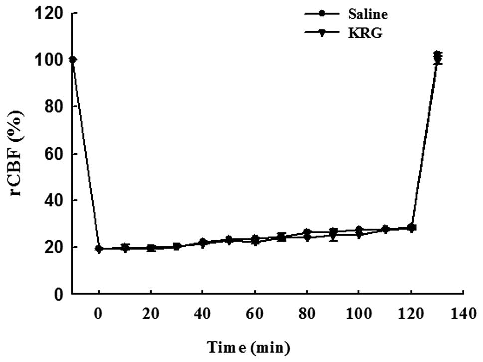
Effect of KRG on rCBF
Fig. 1 shows the
relative changes in rCBF by laser Doppler flowmetry during and
after 2 h of MCAO. MCAO immediately reduced the rCBF value to
approximately 20% of the baseline level. The rCBF measurements
showed similar patterns in all groups. There was no significant
difference between the saline and KRG group.
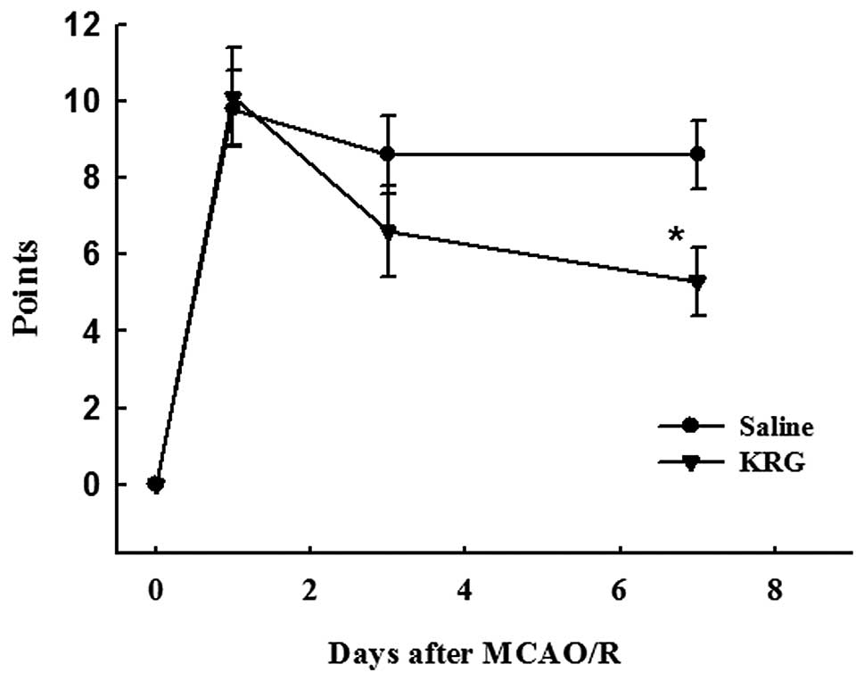
Effect of KRG on the neurological
deficits
mNSS. At day 1 after the MCAO/R injury, the
rats in the operation groups showed neurological deficits: Saline
group (9.8±1.0 mNSS points) and KRG group (10.1±1.3 mNSS points).
There were no significant differences between the operation groups
until 3 days after surgery. However, at day 7 after surgery, the
mNSS points of the KRG group (5.3±0.9 points) decreased
significantly compared to the saline (8.6±0.9 points) group
(p<0.05) (Fig. 2A).
Corner test. The rats in the operation groups
showed higher percentages of right turns at one day after MCAO/R
injury; saline group (97.1±1.1%) and KRG group (97.1±1.3%).
However, there was no significant difference between them. Three
days after surgery, the percentage of right turns in the KRG group
(80.0±3.1%) was significantly lower than that in the saline group
(89.9±2.5%) (p<0.05). The percentage of right turns in the KRG
group (75.3±2.7%) was also significantly lower at 7 days than that
in the saline (87.8±2.5%) group (p<0.01) (Fig. 2B).
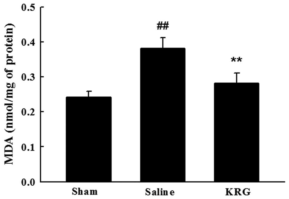
Effect of KRG on lipid
peroxidation
The effect of KRG on lipid peroxidation was
demonstrated by the brain MDA content. The MDA levels were
significantly higher in the brain of the saline group (0.24±0.12
vs. 0.38±0.03, p<0.01). The KRG-treated group followed by MCAO/R
showed significant decreases in the elevated MDA levels (0.28±0.03,
p<0.01) (Fig. 3).
Effect of KRG on antioxidant
enzymes
Table I lists the
changes in the endogenous antioxidant enzymes activities after
MCAO/R injury. The GPx activity was reduced significantly in the
saline groups compared to the sham group (8.71±1.06 vs. 4.46±0.69,
p<0.01) and recovered to 7.40±0.76 in the KRG group (p<0.05).
The CAT and SOD levels were also significantly lower (p<0.01) in
the saline group than those in the sham group, and their activities
were restored significantly in the KRG group compared to the saline
group (p<0.01).
 | Table I.Effect of KRG on the antioxidant
enzymes in the MCAO/R rats. |
Table I.
Effect of KRG on the antioxidant
enzymes in the MCAO/R rats.
| Groups | GPx (units
mg−1 protein) | SOD (units
mg−1 protein) | CAT (units
mg−1 protein) |
|---|
| Sham | 8.71±1.06 | 2.52±0.34 | 34.98±3.07 |
| Saline | 4.46±0.69a | 0.79±0.06a | 24.08±1.51a |
| KRG | 7.40±0.76b | 2.34±0.36c | 32.40±1.69c |
Discussion
Thrombolytic therapy with intravenous recombinant
tissue plasminogen activator (rt-PA) has been widely administered
for ischemic stroke (must be administered within 3 h of symptom
onset), but its extended implementation is limited (24). It may result in high risk of
intracerebral hemorrhage. Therefore, it has been a major challenge
to develop effective therapeutics for stroke. In this study, we
examined the potential role for clinical applications of KRG in the
treatment of ischemic stroke. We used the MCAO model with
reperfusion, which mimics many features of stroke in humans
(17).
Oxidative stress promotes lipid peroxidation and
alters the antioxidant defense system in the brain tissue under
ischemic conditions (25). The
brain is quite susceptible to oxidative stress due to its
environment with non-heme iron, which is involved catalytically in
the production of free radicals (26). The close relationship between
oxidative stress and cerebral ischemia has generated considerable
interest in the development of antioxidant therapies to combat
ischemia-induced damage (27,28).
Our results showed that the administration of KRG ameliorated brain
injury subsequent to MCAO/R in rats. The antioxidant and
neuroprotective potential of KRG may be responsible for this effect
by reducing the level of oxidative stress. These findings are in
agreement with those from previous studies (29,30).
Lipids are the macromolecules most susceptible to
oxidative stress, which play a major role in the production of MDA
(31). The level of MDA in the
homogenate of the whole brain of rats was measured to evaluate the
oxidative stress induced by MCAO/R. Our results showed that
treatment with KRG extract markedly reduced the MDA level in the
brain tissue affected by MCAO/R injury. This indicates that KRG is
effective in decreasing the oxidative stress of MCAO/R.
The presence of numerous antioxidant enzymes in the
brain, including GPx, SOD, and CAT, protects these tissues from
oxidative damage caused by the formation of free radicals (6). GPx plays a key role in removing the
excessive free radicals and hydroperoxides, and is a major defense
system against oxidative stress in the brain (32). SOD reacts with superoxide radicals
to form H2O2 and CAT, which is found at very
low levels in the brain and detoxifies H2O2
into H2O. Oxidative stress reduces the activity of these
enzymes. Therefore, this study examined the endogenous antioxidant
enzymes, such as GPx, SOD, and CAT, which were decreased
significantly following ischemia/reperfusion injury. Administration
of the KRG extract increased the activity of endogenous antioxidant
enzymes to the control levels. The activity of the KRG extract
appears to work by restoring the altered antioxidant enzyme
activity and decreasing the rate of lipid peroxidation in the brain
induced by MCAO/R. Lee et al (33) reported that the long-term
administration of ginseng protected the liver cytosolic SOD, GPx,
and CAT from age-related deterioration, and antioxidant properties
have been reported in ischemia/reperfusion injury in the rat brain
(34).
MCAO/R causes behavioral deficits in the rat brain,
possibly by generating free radicals. The mNSS and corner test were
used to measure the neurobehavioral changes. The mNSS is sensitive
to unilateral cortical injury as it reflects multiple asymmetries,
including postural, sensory and limb-use asymmetries. The corner
test was performed to detect the sensorimotor and postural
asymmetries. In this study, the rats in the operation groups showed
neurological deficits following MCAO/R injury. The impairment of
the neurological function reached a maximum at 24 h after MCAO/R,
and improved progressively at 7 days after MCAO/R. At day 7 after
MCAO/R, the neurological outcome of the KRG group was notably
ameliorated compared to the saline group. These results indicate
that KRG, which is a potent antioxidant, prevents the
neurobehavioral deficits in the animals by scavenging free
radicals. Several authors have also reported that certain
antioxidants, including oxyresveratrol and rutin, promote the
improvement of neurological deficits by diminishing the level of
oxidative stress (35,36). Moreover, it has been reported that
glutathione depletion in the rat brain affects neurobehavioral
activity (37), indicating a
correlation between oxidative stress and behavioral impairment
following MCAO injury.
Ginseng has antioxidant activity. Ginseng extract
has been reported to inhibit metal-induced lipid peroxidation
(38), the progression of renal
failure by scavenging radicals, human low-density lipoprotein
oxidation and nitric oxide synthase expression in the hippocampus
of streptozocin-induced diabetic rats (39). The administration of ginseng to
rats protected neurons in the rat brain from cerebral
ischemia-induced injury (40).
Shah et al (41) reported
that Korean ginseng tea offered marked protection against
ischemia/reperfusion-induced brain damage, as evidenced by
significant reversal of enzymatic alterations. However, these
studies focused on the effect of white ginseng and not red ginseng.
Of the two types of ginseng, white ginseng is air-dried ginseng,
and red ginseng is produced by steaming raw ginseng at 98-100°C for
2-3 h. Red ginseng has superior pharmacological activity to white
ginseng. The improved biological activities of red ginseng result
from changes in the chemical constituents caused by the steaming
treatment. Ginseng saponins, referred to as ginsenosides, are
believed to play a pharmacologically significant role. Several
studies have reported new ginsenosides from red ginsengs that are
not normally found in raw ginseng (42,43).
The major components of red ginseng are ginsenosides Rg3 and Rb1
(44). Rg3 protects the brain from
cerebral ischemia-induced injury by reducing lipid peroxidase,
scavenging free radicals and improving the energy metabolism
(45). It has been reported that
Rb1 prevents ischemic neuronal death induced by transient cerebral
ischemia through a mechanism related to the increased expression of
the antiapoptotic genes and modulated expression of the glial
cell-derived neurotrophic factor (GDNF) (46). Rh2 and Re also protect the brain
from ischemic and reperfusion injuries (47,48).
Therefore, the observed prevention of MCAO-induced cerebral
ischemic injury by the KRG extract may be due to the beneficial
effects of the major active components, such as ginsenosides Rg3
and Rb1.
In conclusion, KRG protects neurons from
MCAO-induced ischemic brain damage, which may be due to the
inhibition of neurological deficit and lipid peroxidation, as well
as to an increase in the endogenous antioxidant defense enzymes.
These results indicate that KRG may be a beneficial intervention in
the treatment of ischemic stroke, and highlight the clinical
applications of KRG in the treatment of ischemia/reperfusion
damage.
References
|
1.
|
Dirnagl U, Iadecola C and Moskowitz MA:
Pathobiology of ischaemic stroke: an integrated view. Trends
Neurosci. 22:391–397. 1999. View Article : Google Scholar : PubMed/NCBI
|
|
2.
|
Shin WH, Park SJ and Kim EJ: Protective
effect of anthocyanins in middle cerebral artery occlusion and
reperfusion model of cerebral ischemia in rats. Life Sci.
79:130–147. 2006. View Article : Google Scholar : PubMed/NCBI
|
|
3.
|
Mehta SL, Manhas N and Raghubir R:
Molecular targets in cerebral ischemia for developing novel
therapeutics. Brain Res Rev. 54:34–66. 2007. View Article : Google Scholar : PubMed/NCBI
|
|
4.
|
Kelly PJ, Morrow JD, Ning M, et al:
Oxidative stress and matrix metalloproteinase-9 in acute ischemic
stroke: the Biomarker Evaluation for Antioxidant Therapies in
Stroke (BEAT-Stroke) study. Stroke. 39:100–104. 2008. View Article : Google Scholar : PubMed/NCBI
|
|
5.
|
Wu HW, Li HF, Wu XY, Zhao J and Guo J:
Reactive oxygen species mediate ERK activation through different
Raf-1-dependent signaling pathways following cerebral ischemia.
Neurosci Lett. 432:83–87. 2008. View Article : Google Scholar
|
|
6.
|
Cui K, Luo X, Xu K and Ven Murthy MR: Role
of oxidative stress in neurodegeneration: recent developments in
assay methods for oxidative stress and nutraceutical antioxidants.
Prog Neuropsychopharmacol Biol Psychiatry. 28:771–799. 2004.
View Article : Google Scholar : PubMed/NCBI
|
|
7.
|
Halliwell B: Role of free radicals in the
neurodegenerative diseases: therapeutic implications for
antioxidant treatment. Drugs Aging. 18:685–716. 2001. View Article : Google Scholar : PubMed/NCBI
|
|
8.
|
Wu JY, Gardner BH, Murphy CI, et al:
Saponin adjuvant enhancement of antigen-specific immune responses
to an experimental HIV-1 vaccine. J Immunol. 148:1519–1525.
1992.PubMed/NCBI
|
|
9.
|
Sato K, Mochizuki M, Saiki I, Yoo YC,
Samukawa K and Azuma I: Inhibition of tumor angiogenesis and
metastasis by a saponin of Panax ginseng, ginsenoside-Rb2.
Biol Pharm Bull. 17:635–639. 1994. View Article : Google Scholar : PubMed/NCBI
|
|
10.
|
Kaneko H and Nakanishi K: Proof of the
mysterious efficacy of ginseng: basic and clinical trials: clinical
effects of medical ginseng, korean red ginseng: specifically, its
anti-stress action for prevention of disease. J Pharmacol Sci.
95:158–162. 2004. View Article : Google Scholar : PubMed/NCBI
|
|
11.
|
Maffei Facino R, Carini M, Aldini G, Berti
F and Rossoni G: Panax ginseng administration in the rat
prevents myocardial ischemia-reperfusion damage induced by
hyperbaric oxygen: evidence for an antioxidant intervention. Planta
Med. 65:614–619. 1999.
|
|
12.
|
Voces J, Alvarez AI, Vila L, Ferrando A,
Cabral de Oliveira C and Prieto JG: Effects of administration of
the standardized Panax ginseng extract G115 on hepatic
antioxidant function after exhaustive exercise. Comp Biochem
Physiol C Pharmacol Toxicol Endocrinol. 123:175–184.
1999.PubMed/NCBI
|
|
13.
|
Cabral de Oliveira AC, Perez AC, Merino G,
Prieto JG and Alvarez AI: Protective effects of Panax
ginseng on muscle injury and inflammation after eccentric
exercise. Comp Biochem Physiol C Toxicol Pharmacol. 130:369–377.
2001.
|
|
14.
|
Keum YS, Park KK, Lee JM, et al:
Antioxidant and anti-tumor promoting activities of the methanol
extract of heat-processed ginseng. Cancer Lett. 150:41–48. 2000.
View Article : Google Scholar : PubMed/NCBI
|
|
15.
|
Kitts DD, Wijewickreme AN and Hu C:
Antioxidant properties of a North American ginseng extract. Mol
Cell Biochem. 203:1–10. 2000. View Article : Google Scholar : PubMed/NCBI
|
|
16.
|
Lee JS, Choi HS, Kang SW, et al:
Therapeutic effect of Korean red ginseng on inflammatory cytokines
in rats with focal cerebral ischemia/reperfusion injury. Am J Chin
Med. 39:83–94. 2011. View Article : Google Scholar : PubMed/NCBI
|
|
17.
|
Longa EZ, Weinstein PR, Carlson S and
Cummins R: Reversible middle cerebral artery occlusion without
craniectomy in rats. Stroke. 20:84–91. 1989. View Article : Google Scholar : PubMed/NCBI
|
|
18.
|
Lourbopoulos A, Karacostas D, Artemis N,
Milonas I and Grigoriadis N: Effectiveness of a new modified
intraluminal suture for temporary middle cerebral artery occlusion
in rats of various weight. J Neurosci Methods. 173:225–234. 2008.
View Article : Google Scholar : PubMed/NCBI
|
|
19.
|
Chen J, Li Y, Wang L, Zhang Z, Lu D, Lu M
and Chopp M: Therapeutic benefit of intravenous administration of
bone marrow stromal cells after cerebral ischemia in rats. Stroke.
32:1005–1011. 2001. View Article : Google Scholar : PubMed/NCBI
|
|
20.
|
Lowry OH, Rosebrough NJ, Farr AL and
Randall RJ: Protein measurement with the Folin phenol reagent. J
Biol Chem. 193:265–275. 1951.PubMed/NCBI
|
|
21.
|
Mohandas J, Marshall JJ, Duggin GG,
Horvath JS and Tiller DJ: Low activities of glutathione-related
enzymes as factors in the genesis of urinary bladder cancer. Cancer
Res. 44:5086–5091. 1984.PubMed/NCBI
|
|
22.
|
Beauchamp C and Fridovich I: Superoxide
dismutase: improved assays and an assay applicable to acrylamide
gels. Anal Biochem. 44:276–287. 1971. View Article : Google Scholar : PubMed/NCBI
|
|
23.
|
Greenwald RA: CRC Handbook of methods for
oxygen radical research. Catalase activity. CRC Press; Boca Raton,
FL: pp. 283–284. 1985
|
|
24.
|
Semplicini A, Benetton V, Macchini L, et
al: Intravenous thrombolysis in the emergency department for the
treatment of acute ischaemic stroke. Emerg Med J. 25:403–406. 2008.
View Article : Google Scholar : PubMed/NCBI
|
|
25.
|
Thiyagarajan M and Sharma SS:
Neuroprotective effect of curcumin in middle cerebral artery
occlusion induced focal cerebral ischemia in rats. Life Sci.
74:969–985. 2004. View Article : Google Scholar : PubMed/NCBI
|
|
26.
|
Yousuf S, Salim S, Ahmad M, Ahmed AS,
Ansari MA and Islam F: Protective effect of Khamira Abresham Uood
Mastagiwala against free radical induced damage in focal cerebral
ischemia. J Ethnopharmacol. 99:179–184. 2005. View Article : Google Scholar : PubMed/NCBI
|
|
27.
|
Simonyi A, Wang Q, Miller RL, Yusof M,
Shelat PB, Sun AY and Sun GY: Polyphenols in cerebral ischemia:
novel targets for neuroprotection. Mol Neurobiol. 31:135–147. 2005.
View Article : Google Scholar : PubMed/NCBI
|
|
28.
|
Ikeda K, Negishi H and Yamori Y:
Antioxidant nutrients and hypoxia/ischemia brain injury in rodents.
Toxicology. 189:55–61. 2003. View Article : Google Scholar : PubMed/NCBI
|
|
29.
|
Kang KS, Kim HY, Pyo JS and Yokozawa T:
Increase in the free radical scavenging activity of ginseng by
heat-processing. Biol Pharm Bull. 29:750–754. 2006. View Article : Google Scholar : PubMed/NCBI
|
|
30.
|
Bae EA, Hyun YJ, Choo MK, Oh JK, Ryu JH
and Kim DH: Protective effect of fermented red ginseng on a
transient focal ischemic rats. Arch Pharm Res. 27:1136–1140. 2004.
View Article : Google Scholar : PubMed/NCBI
|
|
31.
|
Nazam Ansari M, Bhandari U, Islam F and
Tripathi CD: Evaluation of antioxidant and neuroprotective effect
of ethanolic extract of Embelia ribes Burm in focal cerebral
ischemia/reperfusion-induced oxidative stress in rats. Fundam Clin
Pharmacol. 22:305–314. 2008.PubMed/NCBI
|
|
32.
|
Imam SZ and Ali SF: Selenium, an
antioxidant, attenuates methamphetamine-induced dopaminergic
toxicity and peroxynitrite generation. Brain Res. 855:186–191.
2000. View Article : Google Scholar : PubMed/NCBI
|
|
33.
|
Lee H, Kim D and Chang C: Antioxidant
effects of Korean red ginseng components on the antioxidant enzymes
activity and liver peroxidation in the liver of mouse treated with
paraquat. J Ginseng Res. 23:182–189. 1999.
|
|
34.
|
Zhang Y and Liu T: Protective effects of
total saponins of P. ginseng on ischemia-reperfusion injury
in rat brains. Chin J Pharmacol Toxicol. 8:7–12. 1994.
|
|
35.
|
Khan MM, Ahmad A, Ishrat T, et al: Rutin
protects the neural damage induced by transient focal ischemia in
rats. Brain Res. 1292:123–135. 2009. View Article : Google Scholar : PubMed/NCBI
|
|
36.
|
Andrabi SA, Spina MG, Lorenz P, Ebmeyer U,
Wolf G and Horn TF: Oxyresveratrol
(trans-2,3′,4,5′-tetrahydroxystilbene) is neuroprotective and
inhibits the apoptotic cell death in transient cerebral ischemia.
Brain Res. 1017:98–107. 2004.
|
|
37.
|
Cruz-Aguado R, Almaguer-Melian W, Diaz CM,
Lorigados L and Bergado J: Behavioral and biochemical effects of
glutathione depletion in the rat brain. Brain Res Bull. 55:327–333.
2001. View Article : Google Scholar : PubMed/NCBI
|
|
38.
|
Zhang D, Yasuda T, Yu Y, Zheng P, Kawabata
T, Ma Y and Okada S: Ginseng extract scavenges hydroxyl radical and
protects unsaturated fatty acids from decomposition caused by
iron-mediated lipid peroxidation. Free Radic Biol Med. 20:145–150.
1996. View Article : Google Scholar
|
|
39.
|
Chang HK, Jang MH, Lim BV, et al:
Administration of Ginseng radix decreases nitric oxide
synthase expression in the hippo-campus of streptozotocin-induced
diabetic rats. Am J Chin Med. 32:497–507. 2004.
|
|
40.
|
Kim YO, Kim HJ, Kim GS, et al: Panax
ginseng protects against global ischemia injury in rat
hippocampus. J Med Food. 12:71–76. 2009. View Article : Google Scholar
|
|
41.
|
Shah ZA, Gilani RA, Sharma P and Vohora
SB: Cerebroprotective effect of Korean ginseng tea against global
and focal models of ischemia in rats. J Ethnopharmacol.
101:299–307. 2005. View Article : Google Scholar : PubMed/NCBI
|
|
42.
|
Kasai R, Besso H, Tanaka O, Saruwatari Y
and Fuwa T: Saponins of red ginseng. Chem Pharm Bull. 31:2120–2125.
1983. View Article : Google Scholar
|
|
43.
|
Baek NI, Kim DS, Lee YH, Park JD, Lee CB
and Kim SI: Ginsenoside Rh4, a genuine dammarane glycoside from
Korean red ginseng. Planta Med. 62:86–87. 1996. View Article : Google Scholar : PubMed/NCBI
|
|
44.
|
Kitagawa I, Yoshikawa M, Yoshihara M,
Hayashi T and Taniyama T: Chemical studies of crude drugs (1).
Constituents of Ginseng radix rubra. Yakugaku Zasshi.
103:612–622. 1983.PubMed/NCBI
|
|
45.
|
Tian J, Fu F, Geng M, et al:
Neuroprotective effect of 20(S)-ginsenoside Rg3 on cerebral
ischemia in rats. Neurosci Lett. 374:92–97. 2005. View Article : Google Scholar : PubMed/NCBI
|
|
46.
|
Yuan QL, Yang CX, Xu P, et al:
Neuroprotective effects of ginsenoside Rb1 on transient cerebral
ischemia in rats. Brain Res. 1167:1–12. 2007. View Article : Google Scholar : PubMed/NCBI
|
|
47.
|
Zhou XM, Cao YL and Dou DQ: Protective
effect of ginsenosideRe against cerebral ischemia/reperfusion
damage in rats. Biol Pharm Bull. 29:2502–2505. 2006. View Article : Google Scholar : PubMed/NCBI
|
|
48.
|
Park EK, Choo MK, Oh JK, Ryu JH and Kim
DH: Ginsenoside Rh2 reduces ischemic brain injury in rats. Biol
Pharm Bull. 27:433–436. 2004. View Article : Google Scholar : PubMed/NCBI
|