Introduction
Wound healing is a sequence of events involving
cellular and biochemical mechanisms that results in new granulation
tissue (1). Wound healing occurs
in all tissues through the same mechanisms, divided into three
phases: i) hemostasis and inflammation (from 1 to 4 days) that
occur immediately following injury and result in the aggregation
and activation of thrombocytes, migration of leukocytes into the
wound, vascular dilatation, and lymphatics surrounding the area;
ii) proliferation then occurs (days 4–21), whereby fibroblasts
enter the wound and the synthesis of collagen is initiated; and
iii) maturation and remodeling (day 21 to 1 year) occurs, resulting
in the production of type 1 collagen, which is then replaced by the
stronger type 3 collagen. A discontinuation of any of the three
phases delays wound healing (2,3).
Alanine and glutamine are amino acids that provide energy sources
for rapidly proliferating cells. Glutamine is an important
precursor for nucleic acid biosynthesis in all cells. Their use in
the body increases in situations of disease, trauma, or stress, and
thus exogenous dietary replacement may be helpful during these
periods. Alanine and glutamine are also considered to be important
in wound healing (4,5).
Few studies have investigated the potential problems
in wound healing in the postpartum period due to hormonal
imbalances (6). We previously
observed clinically that women in the postpartum period who develop
pneumothorax show delayed healing and delayed cessation of the air
leak occurring when a tube is introduced. We therefore suggest that
wound healing in the lung tissue may be decreased during the
postpartum period. In this study, we examined parameters related to
wound healing in injured lung tissue from rats in the postpartum
period.
Materials and methods
Animals and procedure
Prior to the main study, we performed preliminary
surgical experiments to identify the lung injury to be introduced.
A 15-gauge needle beneath the second nipple, 1.4 cm inwards, was
the confirmed method and the injury was visible macroscopically
when the lungs were removed. The study rats were female Wistar
albino rats weighting 220±20 g (n=42). These rats were divided into
six groups. Group IA included rats not in the postpartum period
that were sacrificed on the third day after lung injury, group IB
included rats not in the postpartum period that were sacrificed on
the tenth day after lung injury, group II included rats not in the
postpartum period that did not receive lung injury, group IIIA
included rats in the postpartum period that were sacrificed on the
third day after lung injury, group IIIB included rats in the
post-partum period that were sacrificed on the tenth day after lung
injury and group IV included rats in the postpartum period without
lung injury. Each group comprised seven rats. Prior to the injury
procedure, the rats were anesthetized with 90 mg/kg of subcutaneous
ketamine-HCl and sacrificed following injury with 200 mg/kg of
pentothal sodium, administered intraperitoneally. This study was
approved by the Cumhuriyet University Local Ethics Committee for
Experiments on Animals (B.30.2. CUM.0.01.00.00–50/307).
Measurement of lung tissue hydroxyproline
OH-P
Lung tissue was rinsed with distilled water, dried
with absorbent paper, weighed and sliced into small sections. The
specimens were then dried in open-mouthed beakers at 100°C for 72
h. Dried specimens were hydrolyzed in 6 M HCl at 110°C for 18 h.
The specimens were then washed three times with distilled water and
dissolved in 2 ml of buffer containing acetic acid 1.2%, sodium
acetate 12%, citric acid 5% and sodium hydroxide 3.4%, pH 6.
Chloramine-T (0.5 ml per 1 ml of specimen) was added to the
specimens, which were then incubated at room temperature for 20
min. A mixture of perchloric acid 15.6% and
dimethylaminobenzaldehyde, dissolved in 0.5 ml of propanol, was
then added. Following incubation at 60°C for 15 min, the absorbance
was read using a spectrophotometer at 550 nm. A standard curve was
plotted for the OH-P content (mg/g) of the specimens (7,8).
Serum alanine and glutamine
measurements
Plasma samples (7 μl) were spotted at 3-cm
intervals on 3MM Whatman chromatography paper. The amino acids were
then separated over 16 h in acetic acid-butanol-distilled water.
The chromatograms were removed from the solution, dried and stained
with ninhydrin-acetone dye. The amino acids moved depending on
their molecular weights and their positions were interpreted by
comparison with standards. Samples with elevated levels of amino
acids were analyzed with gas chromatography.
Histopathological evaluation of wound
healing
Lung specimens were fixed in 10% formaldehyde for a
minimum of 24 h and embedded in paraffin blocks. Slide sections (5
μm-thick) were taken from the paraffin blocks and stained
with hematoxylin and eosin (H&E) and Masson’s trichrome (MT)
stains. H&E- and MT-stained slides were examined in a blinded
manner according to the intensity of inflammation, the intensity of
fibroblastic proliferation, neovascularisation and the levels of
edema and collagen using a modified 0–4 Ehrlich and Hunt numerical
scale. These parameters were independently evaluated using a
histopathological grading scale (Table
I).
 | Table I.Histopathological grading scale. |
Table I.
Histopathological grading scale.
| Grade | Presence of
collagen |
|---|
| 0 | No evidence |
| 1 | Occasional
evidence |
| 2 | Light scattering |
| 3 | Abundant
evidence |
| 4 | Confluent cells or
fibers |
Statistical analyses
Data are presented as the means ± SD. A Mann-Whitney
U-test was performed for comparisons between groups.
Results
Histopathological findings
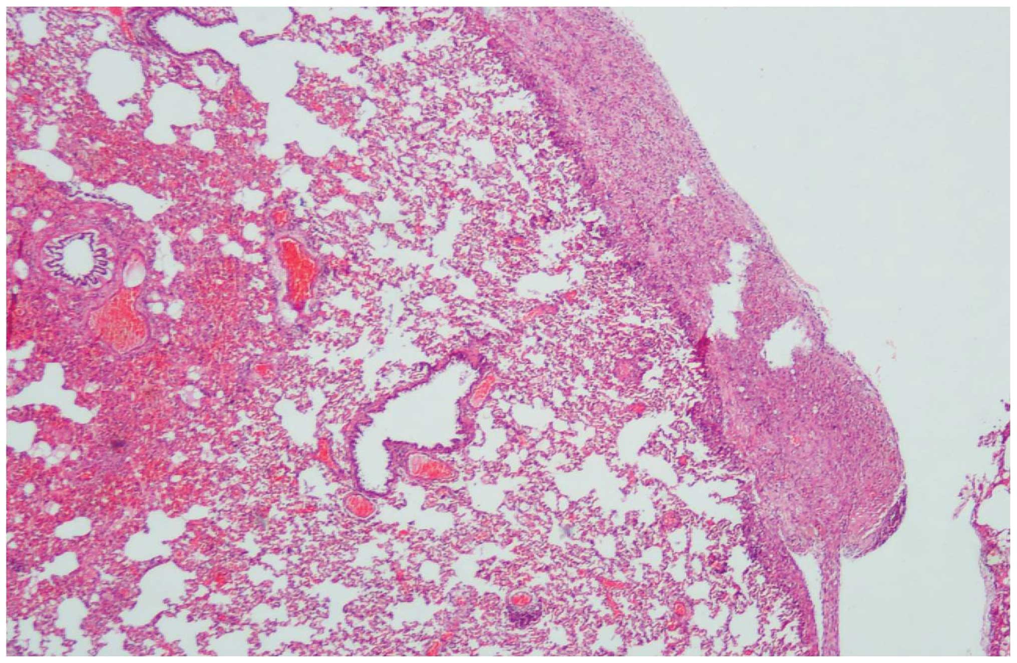
Upon histopathological examination, the lung tissues
showed minor differences among the groups. In the control groups
with no trauma or perforation, no inflammation or fibrosis was
evident. In the trauma groups, which were either pre- or
postpartum, histopathological changes were characterised by minimal
acute inflammation, hemorrhage, and almost no fibrosis in either
the pleura or lung parenchyma. In the two groups sacrificed 10 days
after trauma, chronic inflammation and fibrosis occurred, although
this was the case in relatively few specimens (Fig. 1). Regarding fibrosis, MT staining
was not different between the groups. With regard to wound healing,
the levels of OH-P in the lungs, and the mean serum values of
alanine and glutamine are presented in Table II.
| Table II.Mean values of wound healing
parameters in the study groups. |
Table II.
Mean values of wound healing
parameters in the study groups.
| Parameters | Study group
| P-value |
|---|
| IA | IB | II | IIIA | IIIB | IV |
|---|
| Inflammation score
(mean) | 1.29±0.8 | 0.71±0.5 | 0.29±0.5 | 1.00±0.0 | 1.00±0.6 | 0.14±0.4 | p>0.05 |
| Blood vessel
proliferation | 0.29±0.5 | 1.14±0.7 | 0.00±0.0 | 0.57±0.5 | 1.29±0.5 | 0.14±0.4 | p>0.05 |
| Intensity of
fibroblasts | 0.43±0.8 | 1.14±0.7 | 0.00±0.0 | 0.57±0.5 | 1.71±1.0 | 0.00±0.0 | p>0.05 |
| Collagen | 0.43±0.8 | 1.00±0.8 | 0.00±0.0 | 0.86±0.9 | 1.00±0.8 | 0.00±0.0 | p>0.05 |
| Lung OH-P (mg/g) | 7.60±0.3 | 7.94±0.6 | 7.91±0.3 | 7.78±0.6 | 7.10±1.2 | 7.85±0.3 | p>0.05 |
| Serum alanine
(mg/dl) | 5.11±0.6 | 5.27±0.6 | 3.46±0.7 | 4.11±0.9 | 3.88±1.1 | 4.50±0.8 | p<0.05 |
| Serum glutamine
(mg/dl) | 0.60±1.0 | 0.59±1.6 | 0.00±0.0 | 1.46±0.4 | 1.51±0.5 | 1.22±0.6 | p>0.05 |
When the groups and variables were compared using
the Mann-Whitney U-test, a significant difference in serum alanine
levels was observed between groups IA and IIIA. The differences
between the IB and IIIB groups were significant only for the serum
alanine and glutamine levels.
Discussion
The wound healing process involves a series of
events, including the production of collagen fibers,
re-epithelialization, and neovascularization of newly developed
connective tissue (1). The most
common cause of spontaneous pneumothorax is the rupture of the
apical subpleural blebs (9). In
pneumothorax, the cascade of wound healing should occur to prevent
air leakage. When a thorax tube is inserted accurately and
pulmonary expansion is properly ensured, the pleuron repairs itself
within 24–48 h and the air leak is stopped. However, a systemic
cause that affects wound healing may delay recovery from
pneumothorax.
In previous studies an experimental pneumothorax was
produced by damaging the parietal pleura alone (10–12).
As we considered wound healing occurring in the visceral pleura and
lung tissue, we created a minimal injury that damaged the lungs,
but did not lead to mortality.
Increased collagen production from fibroblasts is
important to the healing process. The newly produced fibers of
collagen account for the strength of the surrounding connective
tissue. Thus, wound healing involves a balance between the
synthesis and degradation of collagen. It is well known that in
newly formed connective tissue, the fibroblastic bridges do not
occur prior to the first 3–5 days after incision and that collagen
deposition occurs towards the end of the first week. Thereafter,
neovascularization of newly formed granulation tissue occurs
(1,13–15).
In this study, we independently evaluated rats on the third and
tenth days to allow individual assessment of the early phase of
wound healing, in which inflammatory cell infiltration occurs, and
the later phase of healing, in which collagen deposition occurs. In
this study, we observed no significant difference between the
postpartum period examined on days 3 and 10 in terms of wound
healing, inflammation score, the proliferation of blood vessels,
fibroblast density or collagen deposition.
OH-P, the most common amino acid found in the
collagen structure, is an important marker of collagen
concentration. In many studies, OH-P levels were measured to assess
wound healing and fibrosis (1,16,17).
In the study by O’Sullivan et al (18), higher OH-P levels were observed in
subjects showing improved wound healing. In studies of intestinal
anastomoses, Yamaguchi et al (16) reported elevated levels of OH-P in
subjects with better wound healing. In this study, although the
levels of OH-P were increased in rats not in the postpartum period,
no statistically significant difference between the rats in the
postpartum period was observed. This may indicate that OH-P levels
are somewhat lower in the postpartum period, leading to impaired
wound healing.
In states of trauma or stress, the body’s use of
alanine and glutamine increases (4). In this study, serum alanine levels
were lower in rats in the postpartum period compared with those not
in the postpartum period. This may indicate that alanine usage
increased during the postpartum period, meaning that insufficient
alanine was available for body wound repair, contributing to
impaired wound healing.
Placental growth factor (PlGF) is an important
angiogenic factor primarily secreted by the placenta, but also
present in the heart, lungs, thyroid, skeletal muscles and fatty
tissue (19). Despite the presumed
high PlGF levels during pregnancy and in the postpartum period,
which may be expected to lead to improved wound healing in lung
tissue, no differences were observed between the groups in terms of
wound healing in the present study.
In conclusion, we demonstrated that serum alanine
levels were decreased in postpartum rats, which may lead to
negative effects on wound repair. Additionally, our findings
suggest that exogenous alanine may contribute positively to wound
healing capacity in response to trauma in the postpartum
period.
Acknowledgements
This work was supported by the
Scientific Research Project Fund of Cumhuriyet University, under
project number T-406.
References
|
1.
|
Turan M, Saraydin SU, Bulut HE, et al: Do
vascular endothelial growth factor and basic fibroblast growth
factor promote phenytoin’s wound healing effects in rat? An
immunohistochemical and histopathologic study. Dermatol Surg.
30:1303–1309. 2004.PubMed/NCBI
|
|
2.
|
Barbul A: Wound healing. Schwartz’s
Principles Of Surgery. Brunicardi FC, Andersen DK, Billiar TR, Dunn
DL, Hunter JG and Pollock RE: 8th edition. McGraw-Hill; New York,
NY: pp. 223–248. 2005
|
|
3.
|
Erbil Y: Yara Iyilesmesi. Genel Cerrahi
Cilt-1. Istanbul Tıp Fakültesi Temel ve Klinik Bilimler Ders
Kitapları. Kalayci Göksel: Nobel Tıp Kitabevi; Istanbul: pp. 51–60.
2002
|
|
4.
|
Soeters PB, van de Poll MC, van Gemert WG
and Dejong CH: Amino acid adequacy in pathophysiological states. J
Nutr. 134:1575–1582. 2004.PubMed/NCBI
|
|
5.
|
Peng X and Wang SL: Glutamine and
immunonutrition for burn patients. Zhonghua Shao Shang Za Zhi.
25:321–324. 2009.PubMed/NCBI
|
|
6.
|
Andreassen TT, Fogdestam I and Rundgren A:
A biomechanical study of healing of skin incisions in rats during
pregnancy. Surg Gynecol Obstet. 145:175–178. 1977.PubMed/NCBI
|
|
7.
|
Edwards CA and O’Brien WD Jr: Modified
assay for determination of hydroxyproline in a tissue hydrolyzate.
Clinica Chimica Acta. 104:161–167. 1980. View Article : Google Scholar : PubMed/NCBI
|
|
8.
|
Bergman I and Loxley R: Two improved and
simplified methods for the spectrophotometric determination of
hydroxyproline. Anal Chem. 35:1961–1965. 1963. View Article : Google Scholar
|
|
9.
|
De Hoyos A and Fry WA: Pneumothorax.
General Thoracic Surgery. Shields MD, Thomas W, LoCicero J, Reed CE
and Feins RH: 7th edition. Lippincott Williams & Wilkins;
Philadelphia, PA: pp. 739–761. 2009
|
|
10.
|
Akkas Y, Sahin E, Celik B, et al: An
experimental model to study pneumothorax in rats. Sci Res Essays.
5:77–80. 2010.
|
|
11.
|
Hill RC, Decarlo DP, Hill JF, Beamer KC,
Hill ML and Timberlake GA: Resolution of experimental pneumothorax
in rabbits by oxygen therapy. Ann Thorac Surg. 59:825–828. 1995.
View Article : Google Scholar : PubMed/NCBI
|
|
12.
|
England GJ, Hill RC, Timberlake GA, et al:
Resolution of experimental pneumothorax in rabbits by graded oxygen
therapy. J Trauma. 2:333–334. 1998. View Article : Google Scholar : PubMed/NCBI
|
|
13.
|
Forrest L: Current concepts in soft
connective tissue wound healing. Br J Surg. 70:133–140. 1983.
View Article : Google Scholar : PubMed/NCBI
|
|
14.
|
Hardy MA: The biology of scar formation.
Phys Ther. 69:1014–1024. 1989.PubMed/NCBI
|
|
15.
|
Robbins S, Cotron R and Vinay K: Healing
and repair. Pathologic Basis of Disease. Robbins S and Cotron R:
3rd edition. Saunders; Philadelphia, PA: pp. 69–81. 1984
|
|
16.
|
Yamaguchi R, Terashima H, Yoneyama S,
Tadano S and Onkohchi N: Effects of platelet-rich plasma on
intestinal anastomotic healing in rats: PRP concentration is a key
factor. J Surg Res. View Article : Google Scholar
|
|
17.
|
Ohbayashi M, Suzuki M, Yashiro Y, et al:
Induction of pulmonary fibrosis by methotrexate treatment in mice
lung in vivo and in vitro. J Toxicol Sci. 35:653–661. 2010.
View Article : Google Scholar : PubMed/NCBI
|
|
18.
|
O’Sullivan JB and Hanson R: Tight
glycaemic control is a key factor in wound healing enhancement
strategies in an experimental diabetes mellitus model. Ir J Med
Sci. 180:229–236. 2011.PubMed/NCBI
|
|
19.
|
Ribatti D: The discovery of the placental
growth factor and its role in angiogenesis: a historical review.
Angiogenesis. View Article : Google Scholar
|