Introduction
Fibrosis is the process of forming fibrous tissue,
usually by degeneration. The process occurs normally in the
formation of scar tissue to replace normal tissue lost through
injury, infection or chronic liver insults (1–5). The
cells responsible for extracellular matrix (ECM) fiber formation
are hepatic stellate cells (HSCs), which enhance cell proliferation
at the onset of liver injury (6).
During our previous studies of the effect of green tea extract
(GTE) on the liver, kidney, and stomach, we presented various
observations on the role of GTE in altering the deleterious effects
of drugs such as reserpine within 30 days of administration
(7,8). This encouraged us to study the effect
of GTE on the amelioration of hepatic fibrosis caused by carbon
tetrachloride (CCl4) (9, Safer et al, Third
Kuwait International Pharmacy Conference, Kuwait, 2011), which
induces hepatic fibrosis through oxidative stress. This causes HSCs
to become over-active (6) and
trigger an increase in ECM synthesis; collagen fibers are then
deposited in the extracellular spaces of the liver cells, causing
them to lose their capacity for blood infusion and harden, leading
to liver fibrosis (4,6).
The present study aimed to highlight the curative
propensity of GTE towards hepatic fibrosis in rat liver through
CCl4 administration.
Materials and methods
Preparation of GTE
Dried Japanese tea leaves (100 g) were powdered in a
Waring blender and extracted with double-distilled water (1 liter),
at 80°C for one hour. The extract was filtered through a nylon
filter, and the filtrate was centrifuged at 3000 × g for 15 min.
The clear supernatant was removed and the residual pellet was
shaken with distilled water, warmed at 35°C, and centrifuged again.
The supernatants were pooled and lyophilized, and the resulting
material was stored at −20°C in a screw-capped bottle.
Animals
Male albino rats (n=20) weighing 200–250 g were used
in this study. They were divided into four groups: GI, control;
GII, administered 50 mg/kg GTE dissolved in pysiological saline
daily for four weeks; GIII, administered subcutaneous injection of
40% CCl4 (1 ml/kg body weight) thrice weekly for four
weeks; GIV, treated as GIII, followed by 50 mg/kg GTE dissolved in
physiological saline daily for four weeks.
Histology
Liver tissues were fixed by immersion in 10%
buffered neutral formalin for 18 h, then processed and stained with
hematoxylin and eosin.
Semi-thin sections
Semi-thin sections (1-μm) were cut and
stained with toluidine blue for light microscopic survey and
photography.
Masson’s trichrome stain
Liver sections immersed in 10% buffered neutral
formalin were processed for collagen fiber staining using Masson’s
trichrome stain.
Three-dimensional architecture
Sample blocks from all groups were prepared and
processed for scanning electron microscopy for 3-dimensional
architectural observation. Blocks were fixed in 3%
glutaraldehyde/cacodylate buffer, pH 7.2, using a tissue processor,
dehydrated in ethanol, and solid-dried in CO2 and
fractured inside-out of each block. Each half of the block was
mounted on a stub with the newly exposed surface face-up. The
latter was coated with platinum/gold using a sputter coater. A
scanning electron microscope (Carryscope JCM 5700; JEOL, Japan)
with a resolution of 5.0 nm was used for capturing the scanning
electron microscopy (SEM) images.
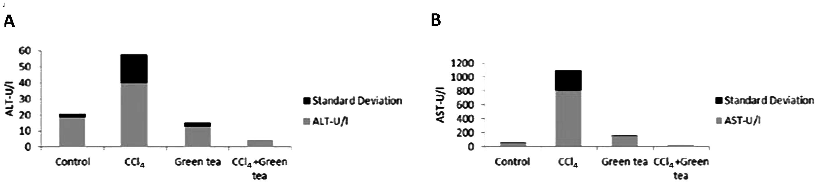
Biochemical analysis
Alanine aminotranferase (ALT) and aspartate
aminotransferase (AST) levels in rats treated with CCl4
and GTE were measured in serum samples using Randox kits. Sample
results were expressed as U/I.
Results
Rats treated with GTE only showed results similar to
those in the control groups with normal AST and ALT levels
(Fig. 1A and B). Serum AST and ALT
levels were significantly elevated in rats treated with
CCl4 only, indicating severe hepatic damage. The
CCl4/GTE group showed a significant decrease in these
enzyme levels.
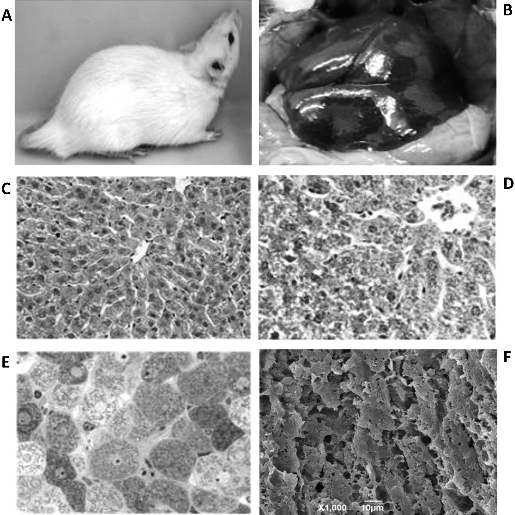
Hepatic fibrosis was evaluated by several criteria;
namely, external features of the rats. In the control rats the fur
color was bright white with a healthy-looking tail (Fig. 2A), and the gross anatomy at the
onset of postmortem and prior to organ excision showed the liver
with its normal brownish-red color with minimal loci of fat
(Fig. 2B). In H&E-stained
paraffin and toluidine blue-stained epon sections, the control
group showed normal tissue and cell architecture (Fig. 2C and D). This was observed by
3-dimensional architecture at SEM levels (Fig. 2E).
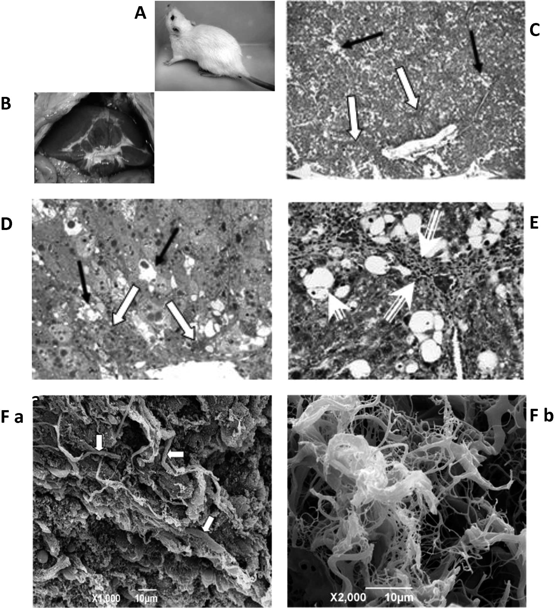
In the CCl4-treated group, the external
features of the rat showed fur with off-white to brownish
coloration with an abnormally dark-colored tail (Fig. 3A). At the onset of postmortem and
prior to organ excision, the liver was fibrous-orange and topped
with thick fat (Fig. 3B). H&E
paraffin and toluidine blue epon sections exhibited pathological
features, most notably the formation of an extensive amount of
extracellular fibrous materials in the parenchyma of the liver
(Fig. 3C and D). The fibrous
materials (collagen fibers) were clearly noted in Masson’s
trichrome-stained sections as shades of blue-green stained
structures (Fig. 3E). Profuse
collagen fiber deposits were found to fill numerous areas in the
extracellular spaces of the liver parenchyma of
CCl4-treated rats. The fibers varied in thickness from
250 to 1000 nm (Fig. 3F). Other
pathological features observed were destruction of lobular
architecture, inflammation, foamy vacuolated cytoplasm, necrosis,
fatty cells, steatosis, nuclear shrinkage, abnormal tri-polar and
tetra-polar divisions, nuclear karyorrhesis, nuclear karyolysis,
nuclear hyperchromatism, dead cells, thickening of portal vein and
portal triad, hypertension of arterioles, nuclear hyperchromatism,
nuclear fragmentation, condensed eosinophilic protein, hyperactive
Kuppfer cells and proliferation of HSCs.
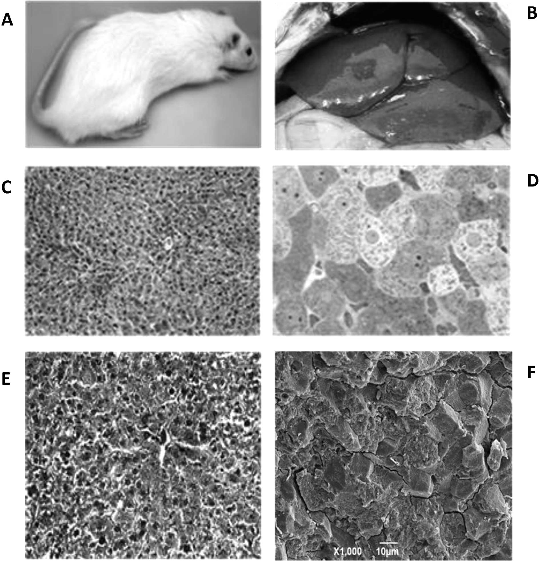
Rats treated with GTE after a month of
CCl4 treatment exhibited prominent restoration of liver
function. Externally the animals’ healthy, bright color, including
the appearance of the tail, was restored (Fig. 4A). In gross morphology, the liver
looked fairly normal: bright red color, and no fat present
(Fig. 4B). The fibrous materials
and lipid droplets were not present whether observed by H&E
paraffin or toludine blue epon sections (Fig. 4C and D). Masson’s trichrome-stained
liver tissue sections exhibited prominent restoration of liver
function. The fibrous materials were entirely absent from the ECM
and there were no signs of lipid droplets (Fig. 4E). This feature was also evident in
SEM images (Fig. 4F).
Discussion
Studies involving experimental animals and humans
have suggested that green tea and GTE may be beneficial in the
treatment of numerous health conditions including atherosclerosis,
high cholesterol and cancer of the bladder, breast, ovary,
colorectum, esophagus, lung, pancreas, prostate, skin and stomach.
Consumption of GTE has been reported to have a therapeutic effect
on inflammatory bowel disease, diabetes, weight loss, dental
caries, arthritis, cartilage breakdown and genital warts, and a
preventative effect on symptoms of colds and influenza and liver
disease (10–24; Safer et al, International Conference on
Free Radicals in Biosystems, Kuwait, 2007).
Hepatic fibrosis is a serious insult to the liver
due to accumulation of extracellular matrix (ECM) proteins,
specifically several types of collagen fibers (2,25).
The main source of hepatofibrosis occurs when hepatic stellate
cells proliferate to formulate ECM (6). In the present study, hepatofibrosis
was successfully induced by subcutaneous injection of 40%
CCl4 for a period of 4 weeks. Histological observation
of liver tissues in H&E and toluidine blue- and Masson’s
trichrome-stained sections all coincide with the external state of
the animals and the autopsy features as explained above.
Histopathological changes, such as destruction of lobular
architecture and extracellular fibrous materials scattered across
the extracellular matrix of liver parenchyma, were clear in H&E
and toluidine blue liver sections. Masson’s trichrome-stained liver
tissues clearly revealed the intermingled fibrous materials in the
liver of the GIII group (CCl4) as blue-green fibrous
structures among the cells and around the blood vessels. Such
fibers were not present in the GTE-treated groups (GIV), which
resembled the control group (GI). A more detailed cellular
pathology will be observed in our forthcoming publication.
3D-architecture and surface topography of the fractured surface of
the liver blocks was observed under SEM. This technique showed that
certain types of fibers of various thicknesses and directions
intermingled in the liver parenchyma. A number of studies have
stated that during hepatic fibrosis, collagen types I and III
mainly proliferate (26). A recent
western blot analysis revealed various fiber types present in the
liver, particularly type I, III, V, and VI collagens. However,
types I and III are the most profuse ECM components. When hepatic
fibrosis occurs, expression of type I and III collagen was found to
increase to account for 90–95% of total collagen, and the overall
balance of collagen types was disrupted (9).
Administration of GTE either simultaneously or
following CCl4 administration prevented or demolished
hepatic fibrosis. The rats were found to regain their normal body
weight and their fur color, which had earlier faded due to weight
loss. The autopsy results also showed the animal liver returning to
normal shape and color, and fibrotic lesions virtually disappeared
following 4 weeks of treatment with GTE, thus returning the
architecture of the liver tissue to its normal state. This
indicates that GTE inhibits the proliferation of HSCs (4,27) as
in the case of the CCl4/GTE-treated mice, or remained in
a state of ‘stand-by’ for the potential toxin attack, as in the
case of GTE treatment alone. Scanning electron microscopy
techniques for capturing detailed 3D-architecture of the liver
samples fractured-face inside-out was the optimal choice for
observing the morphological features, distribution and depth of
collagen fibers in hepatic fibrosis. The
CCl4/GTE-treated group exhibited marked efficacy in
removing almost all fibers observed in an area comparable to that
of the CCl4 group. A group of rats treated with
CCl4 and left untreated for a month (unpublished data)
exhibited minimal effects; connective tissue fibers were still
present. However, there was no marked difference in terms of fiber
formation between CCl4-damaged liver and that of
CCl4-treated rats who had subsequently been left
untreated for a month. Although this is a preliminary study, we
found GTE inhibited proliferation of HSCs (4) and several studies demonstrated a
causal relationship between green tea and reversal of damage caused
by oxidants, possibly through blocking the expression of TGF-B1
(27).
The hepatoprotective effects of green tea are mainly
attributed to its antioxidant properties and the ability of its
polyphenolic catechins to scavenge reactive oxygen species
(28), which were generated in the
present study by CCl4 (29). These properties are due to the
presence of the phenolic hydroxy groups on the B-ring in
ungalloylated catechins (epicatechin and epigallocatechin) and in
the B- and D-rings of the galloylated catechins
(epigallocatechin-3-gallate and epicatechin-3-gallate) (30). This possesses the ability to
inhibit several growth factor signaling cascades, either by direct
blockade of growth factor receptors or through downstream effects
(31). In addition to its
antioxidant effects, green tea exhibits effects on several cellular
and molecular targets in signal transduction pathways associated
with cell death and cell survival (32).
The low levels of ALT and AST in the GTE-treated
group (GIV) beyond the control (GI) may indicate that the hepatic
parenchyma was at its recovery stage from the trauma caused by
CCl4 and the marked loss of hepatocytes and their
organelles, as observed at the ultrastructural level (Safer et
al, Third Kuwait International Pharmacy Conference, Kuwait,
2011).
This study provides a clear indication, for the
first time, that Japanese GTE is a potent anti-fibrotic agent
against hepatic damage caused by CCl4. With these
histopathological studies, we hope to further advance and establish
the impact of GTE in providing a protective shield against the
deleterious effect of chemicals such as CCl4, and
possible other reactive oxygen species (ROS), on rat liver cells.
We anticipate that this will confirm GTE as a strong therapeutic
candidate and preventive measure against hepatic fibrosis.
This study demonstrates that GTE has an
anti-fibrotic effect in CCl4-induced fibrotic rats and
may be used as a therapeutic and preventive measure against hepatic
fibrosis. However, the anti-fibrotic mechanism of GTE requires
further investigation.
Acknowledgements
Our thanks to Mr Mahmoud El-Sayed, the
Nanoscopy Science Center Group, Department of Biological Sciences,
Faculty of Science, Kuwait University, and The Pharmaceutical
Research Institute at Albany College of Pharmacy and Health
Sciences, Albany, NY, USA for all the efforts carried out to make
this research possible.
References
|
1.
|
Paz Z and Shoenfeld Y: Anti-fibrosis: to
reverse the irreversible. Clin Rev Allergy Immunol. 38:276–286.
2010. View Article : Google Scholar : PubMed/NCBI
|
|
2.
|
Bateller R and Brenner DA: Liver fibrosis.
J Clin Invest. 115:209–218. 2005. View
Article : Google Scholar
|
|
3.
|
Bissell DM: Hepatic fibrosis as wound
repair: a progress report. J Gastroenterol. 33:295–302. 1998.
View Article : Google Scholar : PubMed/NCBI
|
|
4.
|
Kim HK, Yang TH and Cho HY: Antifibrotic
effects of green tea on in vitro and in vivo models of liver
fibrosis. World J Gastroenterol. 15:5200–5205. 2009. View Article : Google Scholar : PubMed/NCBI
|
|
5.
|
Li CH, Piao DM, Xu WX, Yin ZR, Jin JS and
Shen ZS: Morphological and serum hyaluronic acid, laminin and type
IV collagen changes in dimethylnitrosamine-induced hepatic fibrosis
of rats. World J Gastroenterol. 28:7620–7624. 2005.PubMed/NCBI
|
|
6.
|
Wu J and Zern MA: Hepatic stellate cells:
a target for the treatment of liver fibrosis. J Gastroenterol.
35:665–672. 2000. View Article : Google Scholar : PubMed/NCBI
|
|
7.
|
Al-Bloushi S, Safer AM, Afzal M and Shaker
M: Green tea modulates reserpine toxicity in animal model. J
Toxicol Sci. 34:77–87. 2009. View Article : Google Scholar : PubMed/NCBI
|
|
8.
|
Safer AM, Afzal M, Al-Bloushi S, Rafique M
and Mousa SA: Inhibition property of green tea extract in relation
to reserpine-induced ribosomal strips of rough endoplasmic
reticulum (rER) of the rat kidney proximal tubule cells. J Toxicol
Sci. 34:637–645. 2009. View Article : Google Scholar
|
|
9.
|
Wang XH, Zhao J, Zhang W, Zhang L, Ma R,
Wang L, Zhang S and Tian L: Scanning electron microscopic
observation: three-dimensional architecture of the collagen in
hepatic fibrosis rats. Chin Med J. 120:308–312. 2007.PubMed/NCBI
|
|
10.
|
Borrelli F, Capasso R, Russo A and Ernst
E: Systematic review: green tea and gastrointestinal cancer risk.
Aliment Pharmacol Ther. 19:497–510. 2004. View Article : Google Scholar : PubMed/NCBI
|
|
11.
|
Cooper R, Morre DJ and Morre DM: Medicinal
benefits of green tea: part I. Review of non-cancer health
benefits. J Altern Complement Med. 11:521–528. 2005. View Article : Google Scholar : PubMed/NCBI
|
|
12.
|
Fujita H and Yamagami T:
Anti-hypercholesterolemia effect of Chinese black tea extract in
human subjects with borderline hypercholesterolemia. Nutr Res.
28:450–456. 2008. View Article : Google Scholar : PubMed/NCBI
|
|
13.
|
Gross G, Meyer KG, Pres H, Thielert C,
Tawfik H and Mescheder A: A randomized, double-blind, four-arm
parallel-group, placebo-controlled Phase II/III study to
investigate the clinical efficacy of two galenic formulations of
Polyphenon E in the treatment of external genital warts. J Eur Acad
Dermatol Venereol. 2:1404–1412. 2007. View Article : Google Scholar
|
|
14.
|
Inoue M, Tajima K and Mizutani M: Regular
consumption of green tea and the risk of breast cancer recurrence:
follow-up study from the Hospital-based Epidemiologic Research
Program at Aichi Cancer Center (HERPACC), Japan. Cancer Lett.
167:175–182. 2001. View Article : Google Scholar
|
|
15.
|
Jian L, Xie LP, Lee AH and Binns CW:
Protective effect of green tea against prostate cancer: a
case-control study in southeast China. Int J Cancer. 108:130–135.
2004. View Article : Google Scholar : PubMed/NCBI
|
|
16.
|
Jin X, Zheng RH and Li YM: Green tea
consumption and liver disease: a systematic review. Liver Int.
28:990–996. 2008. View Article : Google Scholar : PubMed/NCBI
|
|
17.
|
Kato A, Minoshima Y, Yamamoto J, Adachi I,
Watson AA and Nash RJ: Protective effects of dietary chamomile tea
on diabetic complications. J Agric Food Chem. 56:8206–8211. 2008.
View Article : Google Scholar : PubMed/NCBI
|
|
18.
|
Kovacs EM, Lejeune MP, Nijs I and
Westerterp-Plantenga MS: Effects of green tea on weight maintenance
after body-weight loss. Br J Nutr. 91:431–437. 2004. View Article : Google Scholar : PubMed/NCBI
|
|
19.
|
Kuriyama S, Shimazu T, Ohmori K, Kikuchi
N, Nakaya N, Nishino Y, Tsubono Y and Tsuji I: Green tea
consumption and mortality due to cardiovascular disease, cancer and
all causes in Japan: the Ohsaki study. JAMA. 296:1255–1265. 2006.
View Article : Google Scholar : PubMed/NCBI
|
|
20.
|
Low Dog T, Riley D and Carter T:
Traditional and alternative therapies for breast cancer. Altern
Ther Health Med. 7:36–47. 2001.PubMed/NCBI
|
|
21.
|
Lyn-Cook BD, Rogers T, Yan Y, Blann EB,
Kadlubar FF and Hammons GJ: Chemopreventive effects of tea extracts
and various components on human pancreatic and prostate tumor cells
in vitro. Nutr Cancer. 35:80–86. 1999. View Article : Google Scholar : PubMed/NCBI
|
|
22.
|
Inoue M, Sasazuki S, Wakai K, Suzuki T,
Matsuo K, Shimazu T, Tsuji I, Tanaka K, Mizoue T, Nagata C, et al:
Green tea consumption and gastric cancer in Japanese: a pooled
analysis of six cohort studies. Gut. 58:1323–1332. 2009. View Article : Google Scholar : PubMed/NCBI
|
|
23.
|
Miura Y, Chiba T, Tomita I, Koizumi H,
Miura S, Umeqaki K, Hara Y, Ikeda M and Tomita T: Tea catechins
prevent the development of atherosclerosis in apoprotein
E-deficient mice. J Nutr. 131:27–32. 2001.PubMed/NCBI
|
|
24.
|
Nagao T, Hase T and Tokimitsu I: A green
tea extract high in catechins reduces body fat and cardiovascular
risks in humans. Obesity (Silver Spring). 15:1473–1483. 2007.
View Article : Google Scholar : PubMed/NCBI
|
|
25.
|
Lotersztajn S, Julien B, Teixeira-Clerc F,
Grenard P and Mallat A: Hepatic fibrosis molecular mechanisms and
drug targets (Review). Annu Rev Pharmacol Toxicol. 45:605–628.
2005. View Article : Google Scholar : PubMed/NCBI
|
|
26.
|
Gressner AM: The cell biology of liver
fibrogenesis – an imbalance of proliferation, growth arrest and
apoptosis of myofibroblasts. Cell Tissue Res. 292:447–452.
1998.
|
|
27.
|
Oh SW, Kim DH, Ha JR and Kim DY:
Anti-fibrotic effects of a methylenedioxybenzene compound, CW209292
on dimethylnitrosamine-induced hepatic fibrosis in rats. Biol Pharm
Bull. 32:1364–1370. 2009. View Article : Google Scholar : PubMed/NCBI
|
|
28.
|
Yang CS: Tea and health. Nutrition.
15:946–949. 1999.
|
|
29.
|
Ohta Y, Kongo M, Sasaki E, Nishida K and
Ishiguro I: Therapeutic effect of melatonin on carbon
tetrachloride-induced acute liver injury in rats. J Pineal Res.
28:119–126. 2000. View Article : Google Scholar : PubMed/NCBI
|
|
30.
|
Salah N, Miller NJ, Paganga G, Tijburg L,
Bolwell GP and Rice-Evans C: Polyphenolic flavanols as scavengers
of aqueous phase radicals and as chain-breaking antioxidants. Arch
Biochem Biophys. 322:339–346. 1995. View Article : Google Scholar : PubMed/NCBI
|
|
31.
|
Gouni-Berthold I and Sachinidis A:
Molecular mechanisms explaining the preventive effects of catechins
on the development of proliferative diseases. Curr Pharm Des.
10:1261–1271. 2004. View Article : Google Scholar : PubMed/NCBI
|
|
32.
|
Mandel S and Youdim MB: Catechin
polyphenols: neurodegeneration and neuroprotection in
neurodegenerative diseases. Free Radic Biol Med. 37:304–317. 2004.
View Article : Google Scholar : PubMed/NCBI
|