Introduction
Syphilis is a sexually transmitted disease caused by
the spirochete Treponema pallidum, which can invade any
mucous membrane and cause lymphadenopathy. The clinical
presentation can involve chancre formation, regional
lymphadenopathy, widespread mucocutaneous lesions, condyloma lata,
cardiovascular syphilis, neurosyphilis and syphilitic gummas,
according to the natural course of the disease. Chancres and
regional lymphadenopathy are common clinical manifestations of
syphilis. A chancre usually begins as a single firm, non-tender,
raised, painless lesion located at a site on the penis, cervix,
vaginal wall or anus; it is rarely found in the nasopharynx
(1–4). Regional lymphadenopathy, a notable
clinical manifestation of syphilis during its early stage, may
involve any of the lymph nodes, with the inguinal nodes being most
frequently involved; the disease is a rare cause of cervical
adenitis.
Cervical lymphadenopathy is associated with a number
of disease conditions; most often, these are tuberculosis, distant
metastasis and lymphoma. Differentiation among infectious,
metastatic and lymphomatous cervical lymph nodes is critical from a
therapeutic viewpoint. Arriving at the correct diagnosis as early
as possible is also important, as a delayed diagnosis can lead to
upstaging of the malignancy, possibly making a curable lesion
incurable. In South China, one of the most important diagnoses of
cervical lymphadenopathy is nasopharyngeal carcinoma, which usually
presents clinically as cervical lymphadenopathy, nasal blockage and
epistaxis (5).
In the assessment of patients with cervical
lymphadenopathy, a history and full clinical examination are
essential (6). This can be
supplemented by radiological diagnostic techniques such as computed
tomography (CT) and magnetic resonance imaging (MRI) and by an
examination under anesthesia (7,8).
Further assessment with a fine-needle aspiration biopsy or an open
biopsy can help to confirm or refute a diagnosis of head and neck
malignancy (9,10).
The present study reports a case of syphilis without
chancre formation that manifested as nasopharyngeal carcinoma with
enlarged cervical lymphadenopathy and a nasopharyngeal mass.
Case report
A 50-year-old male worker presented with a 6-month
history of enlarged and growing lymph nodes of the right upper neck
and a blood-tinged post-nasal drip. His mother had died from
nasopharyngeal carcinoma 10 years earlier.
Physical examination showed multiple enlarged lymph
nodes located in the right upper neck. These were painless, firm
and non-tender, with the largest measuring 4×3×2 cm. There were no
signs of genital involvement, skin eruption, or inguinal or
axillary lymphadenopathy.
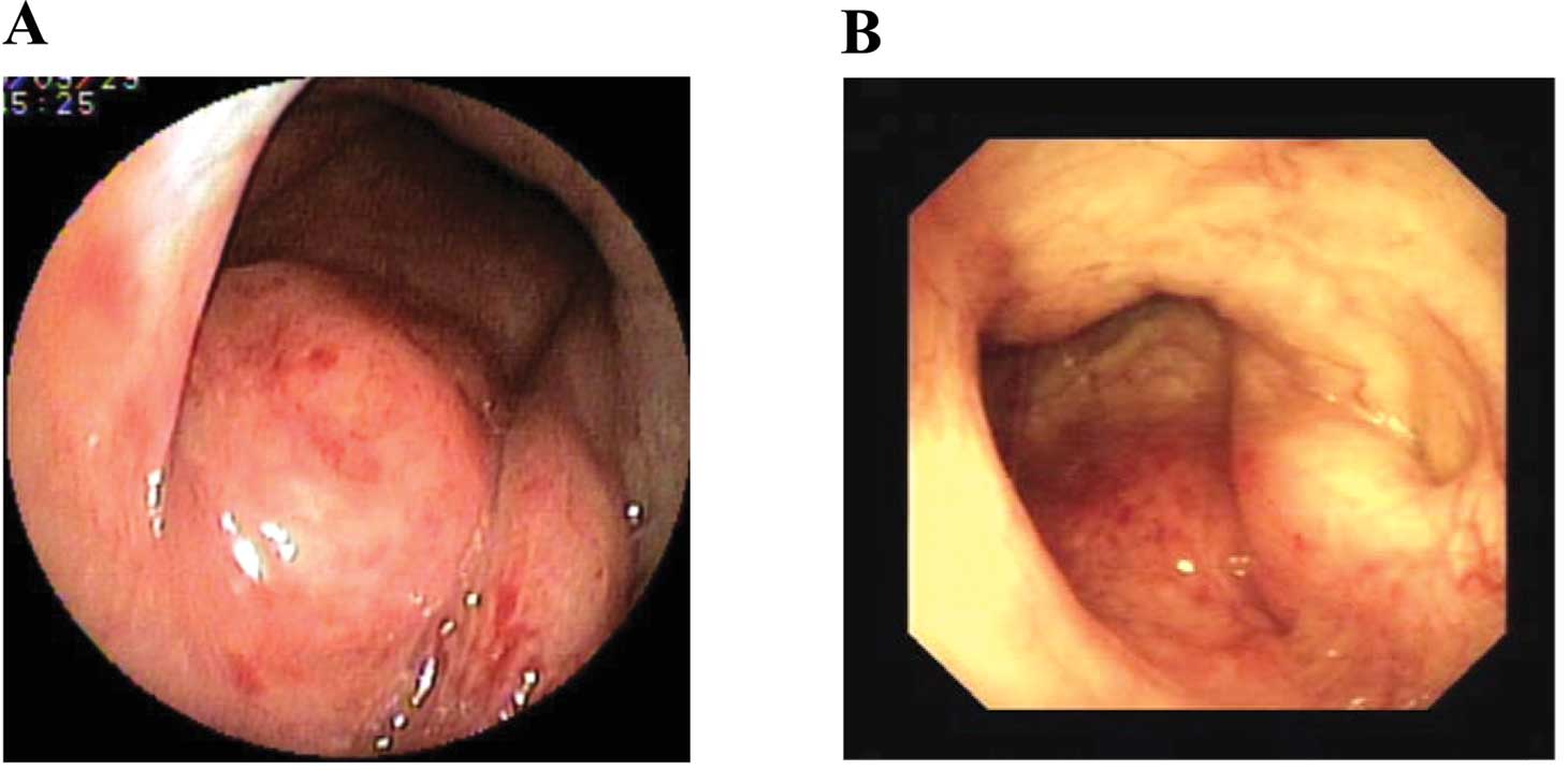
On nasopharyngoscopy, a mass was found in his
nasopharynx and was biopsied. No lesions were noted in the
oropharynx or larynx (Fig. 1A).
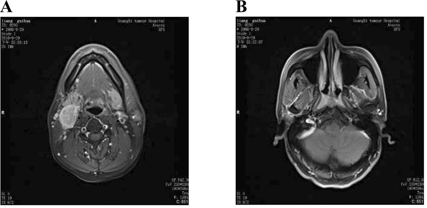
MRI of the head and neck showed a mass in the nasopharynx and
multiple lymph nodes at the II, III region. On T1-weighted images,
the mass and lymph nodes had homogenous intensity, similar to that
of muscle; high signal intensity was noted on T1-weighted enhanced
images (Fig. 2). The clinical
impression was nasopharyngeal carcinoma with cervical lymph node
metastasis.
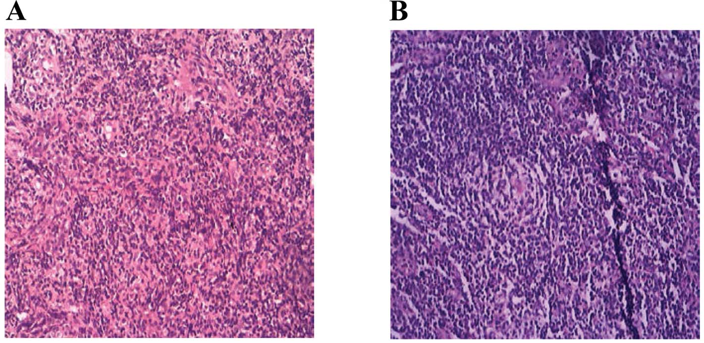
Histopathological examination of the nasopharyngeal
mass demonstrated chronic mucosal inflammation, which was confirmed
by immunohistochemistry showing that the mass was immunonegative
for leukocyte common antigen and cytokeratin (Fig. 3A). Histopathological examination of
the resected enlarged neck lymph node revealed lymphadenitis
(Fig. 3B). There were no signs of
nasopharyngeal carcinoma or lymphoma.
Rapid plasma reagin (RPR) test results (titer,
1:1280) and Treponema pallidum particle assay (TPPA) results
(titer, 1:2560) were positive. Neither human immunodeficiency virus
(HIV) nor hepatitis B virus was detected. A diagnosis of syphilis
was confirmed clinically and serologically.
The patient was treated with penicillin G
benzathine, 24 million units in one intramuscular injection
repeated at weekly intervals for 3 weeks, without a
Jarisch-Herxheimer or allergic reaction. At the 12-month follow-up
after the completion of treatment, a physical examination showed a
significant size reduction in the lymph nodes. The nasopharynx had
a normal appearance and no masses, upon nasopharyngoscopy (Fig. 1B). RPR test results were negative,
but a TPPA gave positive results.
Discussion
The prevalence of syphilis has risen over the last
decade (11). At the same time,
syphilis, known as a great masquerader, can be difficult to
diagnose because of its variable manifestations. Clinicians should
be aware of the possibility of syphilitic lymphadenopathy when
confronted with cervical masses, especially without chancre
formation or other manifestations of syphilis.
A sexual history is important for a syphilis
diagnosis, but patients often tend to conceal their sexual
histories from physicians. The patient in our case did not indicate
risky sexual behaviors in the initial clinical history. Further
careful history-taking after TPPA and RPR testing revealed that he
had experienced sexual promiscuity, although he denied oral
sex.
The differential diagnosis for lymph node
enlargement includes infections and neoplasms. The infectious
diseases include tuberculosis, toxoplasmosis, HIV infection,
cat-scratch disease, Lyme disease and rubella; the malignant
diseases include lymphomas, lymph node metastases and carcinomas
(12). Syphilitic cervical
lymphadenopathy is often misdiagnosed because it is thought to be a
primary or metastatic tumor. As in the present case, the
manifestations of a blood-tinged post-nasal drip, growing enlarged
right upper neck lymph nodes and nasopharyngeal mass were likely to
lead physicians to diagnose nasopharyngeal carcinoma with lymph
node metastasis. His positive family history for nasopharyngeal
carcinoma and MR imaging findings both further indicated the
misdiagnosis.
The use of MR to image the head and neck region has
become increasingly widespread, but the efficacy of MRI in
evaluating cervical lymphadenopathy has not been fully evaluated
(13). In our case, both the
nasopharyngeal mass and cervical lymph nodes had homogenous
intensity on T1-weighted images and high signal intensity on
T1-weighted enhanced images. The MRI findings strongly indicated a
nasopharyngeal carcinoma diagnosis, although it is non-specific for
malignancy. There is little information concerning imaging patterns
of syphilitic cervical lymphadenopathy, and findings of syphilitic
lymphadenopathy are limited. Thus, it is unclear how specific
and/or diagnostically helpful MR images would be.
18F-fluorodeoxyglucose positron emission
tomography/computed tomography (18FDG PET/CT) may be helpful for
the differential diagnosis of infections and neoplasms. However,
positive findings on 18FDG PET/CT may mimic many diseases. Lymph
node uptake is most often the result of malignant diseases, but
18FDG can accumulate with any inflammation, including mycobacterial
disease, for example. Therefore, abnormalities on MRI and 18FDG
PET/CT could be interpreted incorrectly.
We diagnosed the patient with syphilis based mainly
on RPR and TPPA serologic tests, after excluding malignant diseases
by histopathology. The TPPA, widely used as a confirmatory test for
syphilis, is specific and has a highly positive predictive value.
The RPR test is used primarily to assess the progression of
syphilis or the efficacy of treatment. In our case, both the RPR
and TPPA titers were high before therapy. The TPPA results remained
positive, while the RPR results were negative, following penicillin
G benzathine therapy. The titration and time courses of the RPR and
TPPA titers, and the patient’s clinical course strongly suggested
an acquired syphilis diagnosis.
In conclusion, we report the case of a patient with
syphilis manifesting as a nasopharyngeal carcinoma with cervical
lymphadenopathy and a nasopharyngeal mass. Clinicians should keep
syphilitic cervical lymphadenopathy in mind when making the
differential diagnosis of neck masses.
References
|
1.
|
Baarsma EA, Kazzaz B and Soei KI:
Secondary syphilis of the tonsils. J Laryngol Otol. 99:601–603.
1985. View Article : Google Scholar : PubMed/NCBI
|
|
2.
|
Fiumara NJ and Berg M: Primary syphilis in
the oral cavity. Br J Vener Dis. 50:463–464. 1974.PubMed/NCBI
|
|
3.
|
Kleidermacher P, Vito KJ and Strome M:
Otolaryngologic manifestations of acquired syphilis. Otolaryngol
Head Neck Surg. 119:399–402. 1998. View Article : Google Scholar : PubMed/NCBI
|
|
4.
|
Shimizu T, Shinogi J, Majima Y and
Sakakura Y: Secondary syphilis of the tonsil. Arch
Otorhinolaryngol. 246:117–120. 1989. View Article : Google Scholar
|
|
5.
|
Wei WI and Sham JST: Nasopharyngeal
carcinoma. Lancet. 365:2041–2054. 2005. View Article : Google Scholar : PubMed/NCBI
|
|
6.
|
Rood SR and Johnson JT: Examination for
cervical masses. Postgrad Med. 71:189–194. 1982.
|
|
7.
|
Rumboldt Z, Gordon L, Gordon L, Bonsall R
and Ackermann S: Imaging in head and neck cancer. Curr Treat
Options Oncol. 7:23–34. 2006. View Article : Google Scholar : PubMed/NCBI
|
|
8.
|
Hermans R: Head and neck cancer: how
imaging predicts treatment outcome. Cancer Imaging. 6:S145–S153.
2006. View Article : Google Scholar : PubMed/NCBI
|
|
9.
|
Tandon S, Shahab R and Benton JI:
Fine-needle aspiration cytology in a regional head and neck cancer
center: comparison with a systemic review and meta-analysis. Head
Neck. 30:1246–1252. 2008. View Article : Google Scholar : PubMed/NCBI
|
|
10.
|
Saboorian MH and Ashfaq R: The use of fine
needle aspiration biopsy in the evaluation of lymphadenopathy.
Semin Diagn Pathol. 18:110–123. 2001.PubMed/NCBI
|
|
11.
|
Righarts AA, Simms I, Wallace L, Solomou M
and Fenton KA: Syphilis surveillance and epidemiology in the United
Kingdom. Euro Surveill. 9:21–25. 2004.PubMed/NCBI
|
|
12.
|
Damion J and Hybels RL: The neck mass. 2.
Inflammatory and neoplastic causes. Postgrad Med. 81:97–103.
1987.PubMed/NCBI
|
|
13.
|
Kaji AV, Mohuchy T and Swartz JD: Imaging
of cervical lymphadenopathy. Semin Ultrasound CT MR. 18:220–249.
1997. View Article : Google Scholar : PubMed/NCBI
|