Introduction
Several tumor models created by subcutaneous,
intravenous, or orthotopic injection of human tumor cells into
immunodeficient mice have been developed for the evaluation of the
therapeutic effects of chemotherapy (1–3).
Preclinical studies of therapeutic drugs for tumors have
traditionally relied on xenografts of human tumor cells in severe
combined immunodeficient (SCID) or athymic nude mice (4,5).
However, a tumor is composed not only of transformed cells, but is
also intimately associated with host cells such as endothelial
cells, fibroblasts and inflammatory cells that may potentially
influence tumor growth. The tumor microenvironment is related to
clinical prognoses, metastatic potential, and treatment-related
outcomes. Subcutaneous implantation of human tumor cells allows
quantitative measurements of tumor growth; however, the
characteristics of the implanted tumors are different from those of
spontaneous tumors since the tumors are not grown in physiological
conditions due to the lack of a typical tumor microenvironment and
immune system. As human tumor cells must be grown in
immunocompromised recipients, the important effects of native
immunity on disease initiation and progression cannot be studied in
the xenograft model.
Recent advances in technology for the development of
genetically engineered transgenic mice have resulted in the
development of a number of cancer models. These models maintain an
intact immune system. A disadvantage for the use of transgenic mice
is the difficulty in the direct visualization of primary tumor
growth when tumors are small and located at a site deep inside the
body. Without reliable methods for tumor detection in transgenic
mice, a large number of animals subjected to euthanasia and autopsy
at the endpoint are required for evaluation of antitumor activity.
Therefore, a non-invasive method for detecting tumors and measuring
tumor volume is required. Magnetic resonance imaging (MRI) has been
proposed as a method to visualize and monitor tumor development.
The advantages of MRI are that it provides a noninvasive method for
tumor detection, high-resolution images of anatomical structures,
and the means for accurate serial measurement of solid tumor
volumes in animal models (3,6).
Neuroblastoma is the second most common type of
solid tumor in children (7). Most
children with lower-stage disease have a favorable outcome through
surgery alone or following treatment with chemotherapy. However,
tumors have a poor prognosis when they are unresectable or
metastatic and diagnosed after one year of age (8). Therefore, the availability of
reliable tumor models for adrenal neuroblastoma to test novel
chemotherapeutic agents remains crucial for improving survival.
Recently, we developed transgenic mice exhibiting tumors in
bilateral adrenal glands with histological characteristics similar
to human neuroblastoma (9).
Comparison of the gene expression profiles by DNA microarray also
indicated that the character of the mouse adrenal tumors was
similar to that of human adrenal neuroblastoma rather than
pheochromacytoma (10). In this
study, we monitored the development of adrenal tumors in individual
mice by MRI, and the accuracy of MRI-based tumor volume
determinations was verified by standard volume measurements at
autopsy. Furthermore, the change of tumor volume following
doxorubicin treatment was evaluated by MRI analysis. This
information aids evaluation of the efficacy of new treatments for
neuroblastoma.
Materials and methods
Transgenic mice
Transgenic mice exhibiting spontaneous bilateral
adrenal tumors with a C57BL/6 background were previously developed
(9). The transgenic mice carried
tetracycline-inducible simian virus 40 T-antigen, a fusion gene
comprising tetracycline-responsive elements with cytomegalovirus
promoter and simian virus 40 T-antigen. Transgenic mice were used
as heterozygotes. All experimental procedures were approved by the
Committee on Animal Research of Hoshi University.
Magnetic resonance imaging
Mice were anesthetized with 1.5% isoflurane (Abbott
Japan, Tokyo, Japan) throughout the MRI experiment during their
insertion into a 9.4 T vertical-type MRI (Varian MRI System,
Varian, Palo Alto, CA, USA). Two-dimensional T2-weighted
images were obtained using a multi-slice spin echo sequence
(repetition time/echo time, 1000 msec/40 msec) with a field of view
of 30×30 mm, a matrix of 256×256 pixels, and a slice thickness of 1
mm.
In order to minimize the imaging time of a large
number of mice, a fast spin echo sequence was used for the tumor
response following doxorubicin treatments. Coronal
T2-weighted images were obtained using multi-slice fast
spin echo sequence (repetition time/effective echo time, 2500
msec/48 msec) with a field of view of 50×30 mm, a matrix of 256×256
pixels, and a slice thickness of 1 mm.
Tumor volume measurement
ImageJ software (NIH, Bethesda, MD, USA) was used
for image processing and analysis. To determine the volume of the
adrenal tumors, each image was reviewed and regions of interest
(ROIs) were identified as adrenal glands. The ROIs corresponding to
the right and left adrenal glands were defined individually, and
the volume of each adrenal gland was calculated by summing up the
ROIs in consecutive slices. Following MRI scan, tumor weights were
recorded and the length of the tumor was measured with a caliper.
In this measurement with a caliper, tumor volume was calculated
using the following formula: L×W2×π/6, where L is the
long diameter and W is the short diameter of the tumor.
Chemotherapy with doxorubicin
To evaluate the therapeutic response to doxorubicin
treatment, mice were subjected to weekly MRI scans starting at 13
weeks of age. Transgenic mice were randomly assigned to receive
three intravenous injections of doxorubicin (3 mg/kg) at weekly
intervals or intravenous injection of saline as a control.
Results
Magnetic resonance imaging
In our previous study we reported on the ectopic
expression of SV40 T-antigen in adrenal medulla which developed
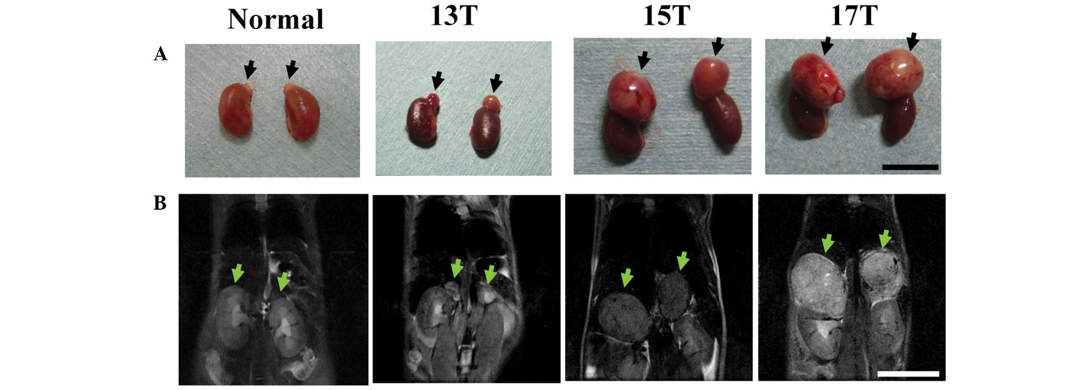
bilateral large adrenal tumors in 12–13 week-old mice (9). Transgenic mice, beginning at 13 weeks
of age, developed carcinoma of the adrenal gland (Fig. 1A), and by 15 weeks of age, most
adrenal tumors were between 5 and 10 mm in diameter. At 17 weeks of
age, tumors of the adrenal glands had enlarged to 10–15 mm in
diameter. Next, we evaluated the adrenal tumor progression of
transgenic mice by MRI as a non-invasive monitoring method. The
tumor was hyperintense on T2-weighted images relative to
T1-weighted ones (data not shown); therefore, we
followed the growth of adrenal tumors by coronal
T2-weighted images without exogenous contrast agents
(Fig. 1B). The adrenal glands of
normal mice were barely evident in this procedure due to their
small size. In contrast, the adrenal tumors in the transgenic mice
after 13 weeks of age were detected above the kidneys and were
easily delineated from adjacent organs.
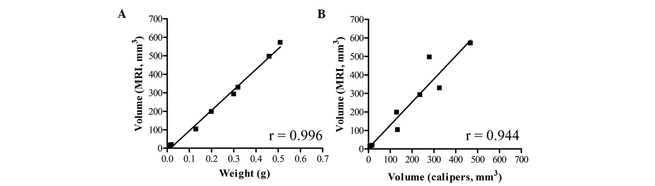
To assess the accuracy of the adrenal tumor
volumetric determination by MRI, we calculated the tumor volumes
from MRI data and compared these with tumor wet weights and tumor
volumes calculated by caliper measurement (Fig. 2). Linear regression analyses
demonstrated that tumor volume determined by MRI correlated well
with tumor wet weight (r=0.996) and caliper measurement (r=0.944).
These findings indicate that tumor volume in transgenic mice can be
quantitatively measured by MRI as well as by weighing.
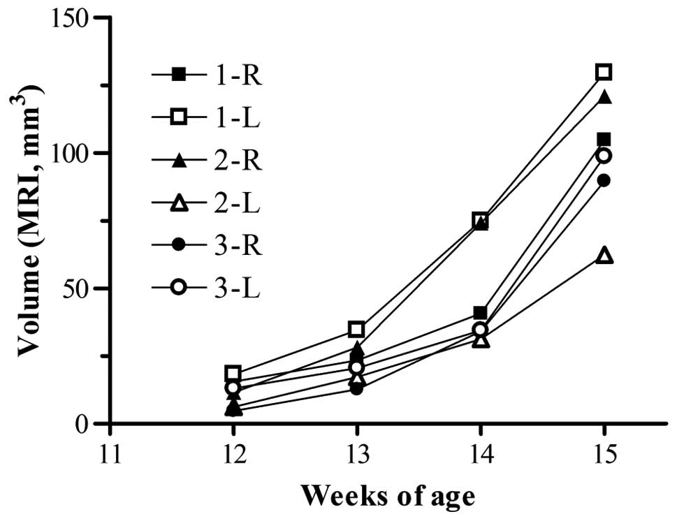
Adrenal tumors in transgenic mice at 13 weeks of age
were detected by MRI (Fig. 1).
Therefore, we assessed the growth of individual tumors by
sequential MRI in mice from 12 to 15 weeks of age (Fig. 3). The sizes of all the tumors in
the transgenic mice were detectable at 12 weeks of age, and
increased over time. Although the total tumor growth curves for
each of the three groups of mice were similar, there was
considerable variation in the volume of individual tumors for each
mouse. In subsequent evaluation of tumor growth by treatment with
antitumor drug, the sum of right and left adrenal tumor volumes was
used to represent the tumor volume of each mouse.
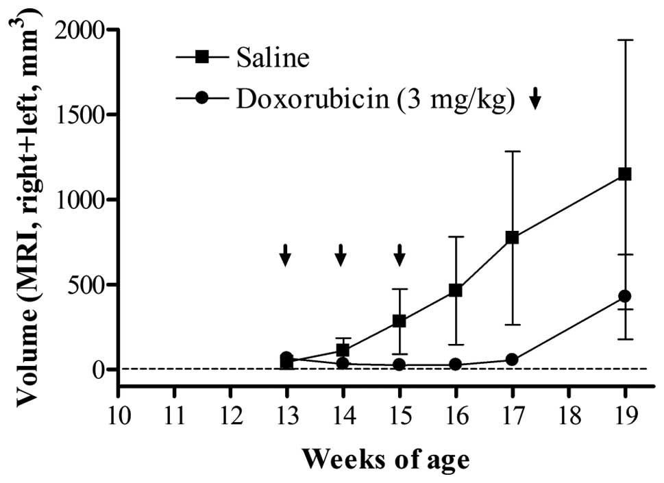
Tumor progression and regression by
doxorubicin treatment
Finally, we examined the effects of a therapeutic
drug on transgenic mice with the primary objective of determining
whether adrenal tumor progression would be delayed or if tumors
would regress in treated mice compared with those of the controls
(saline injection). In a previous study, we found that the
expression of DNA topoisomerase IIα (Topo IIα) mRNA in the adrenal
tumors of transgenic mice was strongly increased compared with that
in non-transgenic mice (10);
therefore, we evaluated the therapeutic effect of doxorubicin,
which is an inhibitor of Topo IIα (Fig. 4). MRI with a fast spin echo
sequence minimized the scan time, without loss of image resolution
necessary for size determination. As a result, tumor volume
regression was observed in mice treated with doxorubicin and the
size of a number of tumors was under the detection limit (∼1.5 mm).
In tumor sections following injection of doxorubicin, large areas
of necrosis were observed (data not shown). These findings
corresponded with the prognostic results from the DNA array.
However, tumor regrowth was observed at approximately 19 weeks of
age. We could evaluate the continuous change in tumor size
following drug treatment using MRI.
Discussion
Transgenic mice exhibiting spontaneous tumors are a
useful model for evaluation of the efficacy of anticancer drug
treatment. Since spontaneous tumors are embedded in a
micro-environment that closely mimics that of human disease,
reliable methods for measurement of tumor size are required. Tumor
volume determination by autopsy is not suited for sequential
observation. MRI permits a wide variety of longitudinal studies not
possible with other destructive analytical methods. Recently, MRI
for tumors has become more readily available in cancer research and
has been used for several transgenic models including lung
(11) and ovarian tumors (6). Transgenic tumor models have been
evaluated by MRI not only to assess tumor size but also to evaluate
therapeutic response (6,12). However, to the best of our
knowledge, there is little information regarding quantitative
measurement of tumor volume by MRI. In this study, we monitored the
adrenal tumor progression of transgenic mice by MRI. To establish a
non-invasive MRI scanning protocol that has high resolution for
tumors, we characterized the intrinsic MR contrast parameter
(T2) of the adrenal tumors. Adrenal tumors were easily
identifiable without using exogenous contrast agents on
T2-weighted spin echo images. We established a
correlation between tumors detected by MRI and those observed ex
vivo following harvesting of the adrenal tumors. Tumor volume
determined by MRI was better correlated with that by weighing
(r=0.996) than that by caliper measurements (r=0.944), indicating
that MRI is able to measure the tumor volume three-dimensionally
and, therefore, more accurately than two-dimensional measurement.
Although we used MRI with a high resolution with a 9.4 T
instrument, there was limited detection of small tumors (1.5 mm in
diameter and less than 2 mm3 in volume). An MR
instrument with a stronger magnetic field or the use of exogenous
contrast agents might provide higher resolution of much smaller
tumors in a small animal model (3).
Researchers are studying chemotherapy drugs in order
to find an effective therapy for neuroblastoma. In chemotherapy for
high-risk neuroblastoma, the following drugs are often used:
Cyclophosphamide, ifosfamide, cisplatin, carboplatin, vincristine,
doxorubicin, melphalan, etoposide, teniposide, and topotecan. In
previous study, we found that the expression of DNA Topo IIα mRNA
in adrenal tumors of transgenic mice increased 120- to 150-fold
compared with that in non-transgenic mice (10); therefore, we evaluated the
therapeutic effect of doxorubicin. As expected from the gene
expression profile (10), delay of
adrenal tumor progression by doxorubicin treatment was observed by
serial MRI. Furthermore, therapeutic response, including tumor
regression and regrowth, was also measured using longitudinal MRI.
MRI provides important insights into the factors that control the
onset and development of tumors and serves as an important platform
for the preclinical development and evaluation of novel
chemotherapeutic agents having a high likelihood of efficacy in
human clinical trials.
In conclusion, MRI is a powerful imaging modality
for the in vivo characterization of adrenal tumors in mice.
The use of repeated scans by MRI in the same animal reduces sample
size, and this method enables efficient screening of novel
therapeutic approaches for neuroblastoma.
Acknowledgements
We thank Ms. Yuko Iwase, Ms. Kimiko
Koga, Mr. Yuki Shimojo, Ms. Yukimi Taniguchi, Mr. Takuya Minowa,
Mr. Tomohiro Izumisawa, and Mr. Haruya Ubukata for their assistance
with the experimental work. This research was supported in part by
grants from the Ministry of Education, Culture, Sports, Science and
Technology, Japan, the Ministry of Health, Labour and Welfare,
Japan, the Science Research Promotion Fund from the Promotion and
Mutual Aid Corporation for Private Schools of Japan, and the Open
Research Center Project.
References
|
1
|
Talmadge JE, Singh RK, Fidler IJ and Raz
A: Murine models to evaluate novel and conventional therapeutic
strategies for cancer. Am J Pathol. 170:793–804. 2007. View Article : Google Scholar : PubMed/NCBI
|
|
2
|
Zhang L, Smith KM, Chong AL, Stempak D,
Yeger H, Marrano P, Thorner PS, Irwin MS, Kaplan DR and Baruchel S:
In vivo antitumor and antimetastatic activity of sunitinib in
preclinical neuroblastoma mouse model. Neoplasia. 11:426–435.
2009.PubMed/NCBI
|
|
3
|
Moats R, Ma LQ, Wajed R, Sugiura Y,
Lazaryev A, Tyszka M, Jacobs R, Fraser S, Nelson MD Jr and DeClerck
YA: Magnetic resonance imaging for the evaluation of a novel
metastatic orthotopic model of human neuroblastoma in
immunodeficient mice. Clin Exp Metastasis. 18:455–461. 2000.
View Article : Google Scholar : PubMed/NCBI
|
|
4
|
Connolly DC, Bao R, Nikitin AY, Stephens
KC, Poole TW, Hua X, Harris SS, Vanderhyden BC and Hamilton TC:
Female mice chimeric for expression of the simian virus 40 TAg
under control of the MISIIR promoter develop epithelial ovarian
cancer. Cancer Res. 63:1389–1397. 2003.PubMed/NCBI
|
|
5
|
Bock NA, Zadeh G, Davidson LM, Qian B,
Sled JG, Guha A and Henkelman RM: High-resolution longitudinal
screening with magnetic resonance imaging in a murine brain cancer
model. Neoplasia. 5:546–554. 2003. View Article : Google Scholar : PubMed/NCBI
|
|
6
|
Hensley H, Quinn BA, Wolf RL, Litwin SL,
Mabuchi S, Williams SJ, Williams C, Hamilton TC and Connolly DC:
Magnetic resonance imaging for detection and determination of tumor
volume in a genetically engineered mouse model of ovarian cancer.
Cancer Biol Ther. 6:1717–1725. 2007.PubMed/NCBI
|
|
7
|
Maris JM, Hogarty MD, Bagatell R and Cohn
SL: Neuroblastoma. Lancet. 369:2106–2120. 2007. View Article : Google Scholar : PubMed/NCBI
|
|
8
|
Brodeur GM: Neuroblastoma: biological
insights into a clinical enigma. Nat Rev Cancer. 3:203–216. 2003.
View Article : Google Scholar : PubMed/NCBI
|
|
9
|
Iwakura H, Ariyasu H, Kanamoto N, Hosoda
K, Nakao K, Kangawa K and Akamizu T: Establishment of a novel
neuroblastoma mouse model. Int J Oncol. 33:1195–1199.
2008.PubMed/NCBI
|
|
10
|
Hattori Y, Kanamoto N, Kawano K, Iwakura
H, Sone M, Miura M, Yasoda A, Tamura N, Arai H, Akamizu T, Nakao K
and Maitani Y: Molecular characterization of tumors from a
transgenic mouse adrenal tumor model: Comparison with human
pheochromocytoma. Int J Oncol. 37:695–705. 2010. View Article : Google Scholar
|
|
11
|
Degrassi A, Russo M, Nanni C, Patton V,
Alzani R, Giusti AM, Fanti S, Ciomei M, Pesenti E and Texido G:
Efficacy of PHA-848125, a cyclin-dependent kinase inhibitor, on the
K-Ras(G12D)LA2 lung adenocarcinoma transgenic mouse model:
evaluation by multimodality imaging. Mol Cancer Ther. 9:673–681.
2010. View Article : Google Scholar
|
|
12
|
He Z, Evelhoch JL, Mohammad RM, Adsay NV,
Pettit GR, Vaitkevicius VK and Sarkar FH: Magnetic resonance
imaging to measure therapeutic response using an orthotopic model
of human pancreatic cancer. Pancreas. 21:69–76. 2000. View Article : Google Scholar : PubMed/NCBI
|