Introduction
Obstructive jaundice (OJ) refers to a type of
jaundice caused by mechanical obstruction of the extrahepatic and
intrahepatic bile ducts due to tumor, stones, inflammation and
other causes. Gallbladder tissue damage is a major pathological
manifestation of OJ (1). According
to the literature, obstruction of the biliary tract can cause
reduced cholate in the intestinal tract and decreased ability to
clear endotoxin, which can directly induce apoptosis of liver cells
(2). Obstruction of the biliary
tract also can impair electron transport chain function of liver
mitochondria and promote oxygen free radical generation.
Simultaneously, obstruction of the biliary tract reduces the
activity of free radical scavenging enzymes in the liver, causing
decreased free radical scavenging capacity and further induction of
liver cell apoptosis.
Traditional Chinese medicine describes capillary
artemisia as bitter, acrid and slightly cold, able to remove damp
heat from the spleen, stomach, liver and gall bladder. It is
considered an important drug for treating jaundice. Capillary
artemisia impacts primary Qi of Shaoyang and affects the ascending
vitality of liver. Its acridness can dispel stagnation of the liver
and gallbladder (3). Studies show
that capillary artemisia can protect the liver, relax the
gallbladder and remove jaundice; this provides a pharmacological
basis for treating liver disease with capillary artemisia (3–5).
This study investigated the protective effects of capillary
artemisia polysaccharide on oxidative injury to the liver using the
common bile duct ligation method in growing rats.
Materials and methods
Experimental animals and grouping
Healthy male Wistar rats (n=40, age 3–4 weeks old,
weight 85–95 g) were randomized into four groups (n=10 each): a
normal control group, a sham operation group, a OJ group and a OJ +
capillaris polysaccharide group or ‘study group’. The rats were
kept in a quiet and ventilated environment with a room temperature
of 22–26°C and relative humidity of 40–70%. All animals had free
access to a normal chow diet and water during the experiment.
Methods
Extraction of capillary artemisia
polysaccharide
Dry powder of crushed capillary artemisia was
weighed and added to water at a ratio of 1:20, then placed in a
water bath at 80°C for 3 h. The sample was centrifuged to remove
filter residues and harvest the supernatant. The supernatant was
concentrated followed by addition of a 3-fold volume of anhydrous
ethanol. The sample was centrifuged to obtain the sediment. Saline
was added to prepare a 1% solution, and the sample was
deproteinized with 3% trichloroacetic acid and freeze-dried to
obtain crude polysaccharides (6).
The phenol-sulfuric acid method was used to determine
polysaccharide with glucose as the standard substance and a 721
spectrophotometer was used for colorimetric analysis (7). The extracted capillary artemisia
polysaccharide was prepared as a 0.5 g/ml solution.
Preparation of a rat pup model of
OJ
The rats were fasted for 12 and 4 h for food and
drink, respectively. Rats were anesthetized with ketamine (20
mg/kg) by intraperitoneal (i.p.) injection, then cut along the
mid-line of the upper abdomen to the hepatoduodenal ligament. The
common bile duct was dissociated and ligated near the porta
hepatis. Rats in the sham operation group underwent common bile
duct dissociation but no ligation. The standard of a successful
model preparation was that the OJ rat pup model had total bilirubin
and direct bilirubin values (measured by blood sampling) that were
5-times higher than the normal control group two days after the
procedure (8). Rat pups in the
study group were administered 5 ml/kg capillary artemisia
polysaccharide solution (0.5 g/ml) via once-daily i.p. injection;
the other groups were given 5 ml/kg saline via once-daily i.p.
injection. After 4 weeks, the animals were sacrificed and their
livers were rapidly removed, washed with ice-cold saline, blotted
dry and weighed. Liver weight was also expressed relative to grams
body weight, as the ‘liver coefficient’. A sample (0.2 g) was taken
from each liver, mixed with 0.2 mol/l phosphate buffer into 10%
tissue homogenate and centrifuged at 3000 rpm/min. The supernatant
was obtained, and xanthine oxidase superoxide was used to determine
the activity level of superoxide dismutase (SOD). The NADPH
coupling method was used to determine the activity of glutathione
peroxidase (GSH-Px). Visible light was used to determine the
activity of catalase (CAT) and thiobarbituric acid (TBA) was used
to determine the content of malondialdehyde (MDA) (9).
Statistical analysis
SPSS 13.0 software was used for statistical
analysis. The data are expressed as the means ± standard deviation
(SD). Single factor variance analysis was used to compare the test
results of each index across groups, followed with pairwise
comparison between groups using the Student-Newman-Keuls test. The
above analysis was performed as a two-sided test; the test level α
was 0.05 with a P-value of <0.05 considered statistically
significant.
Results
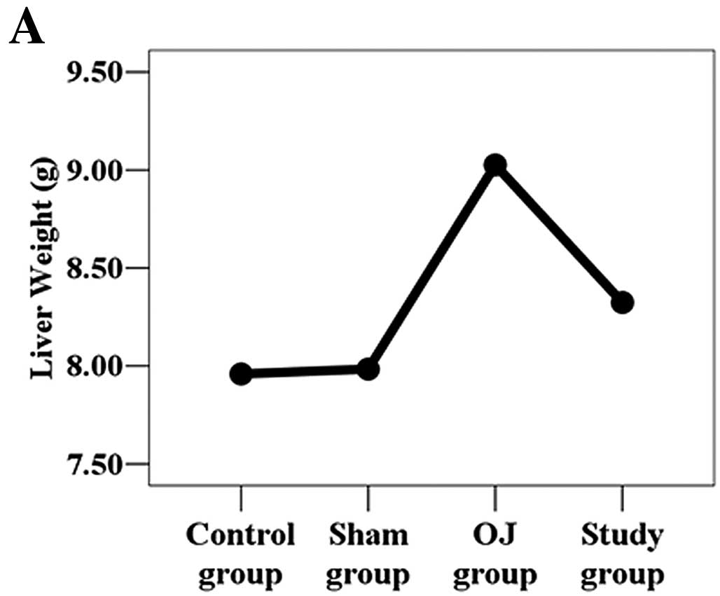
Liver weight and liver coefficient
The liver weight and liver coefficient in the normal
control, sham operation, OJ and study groups were: 7.96±0.26 g,
8.93±0.40%; 7.98±0.27 g, 8.92±0.46%; 9.03±0.26 g, 10.06±0.38%; and
8.32±0.44 g, 9.34±0.57%, respectively. The liver weight and liver
coefficient in the OJ model group were the highest followed by the
study group, the normal control group and the sham operation group
(Fig. 1). Liver weight and liver
coefficient were statistically significant among the groups
(P<0.05). Liver weight and liver coefficient for the study group
were significantly reduced compared to the OJ model group, but
still higher than the normal control group and the sham operation
group (P<0.05).
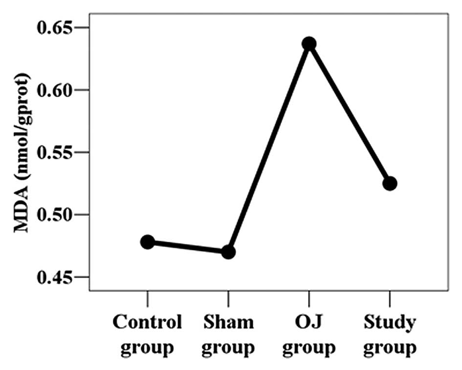
MDA content in the liver homogenate
MDA content in the liver homogenate was 0.48±0.01,
0.47±0.01, 0.64±0.05 and 0.53±0.04 nmol/g prot in the normal
control group, sham operation group, OJ group and study group,
respectively. MDA content in the liver homogenate in the OJ model
group was the highest followed by the study group, normal control
group and sham operation group (Fig.
2). The difference in MDA content was statistically significant
among the groups (P<0.05). MDA content in the study group was
significantly reduced compared to that in the OJ model group, but
was still higher than the content in the normal control group and
sham operation group (P<0.05).
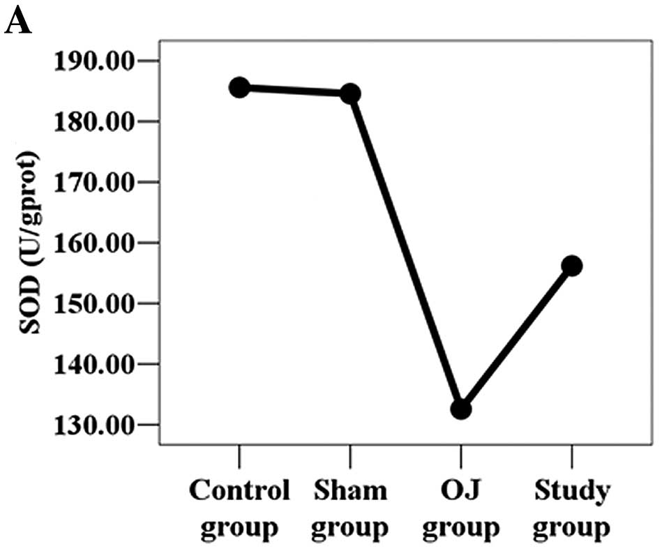
Activity levels of SOD, GSH-Px and CAT in
the liver homogenate
The activity of SOD, GSH-Px and CAT in the liver
homogenate in the normal control, sham operation, OJ and study
groups, respectively, were 185.61±2.82, 184.59±2.75, 132.56±11.51,
156.18±14.79 U/g prot (SOD); 577.12±9.21, 572.25±7.05,
370.74±18.37, 528.76±18.76 U/g prot (GSH-Px); 1031.80±64.08,
1014.07±64.65, 681.81±125.02, 861.66±32.48 U/g prot (CAT). Among
the groups, the difference in activity of SOD, GSH-Px and CAT in
the liver homogenate was statistically significant (P<0.05). The
activity levels of SOD, GSH-Px and CAT were lowest in the OJ model
group. The study group had higher activities that the OJ group
(P<0.05), and the normal control group and sham operation group
had the highest activities (Fig.
3).
Discussion
Obstructive jaundice (OJ) is a jaundice caused by
mechanical obstruction of the bile duct caused by congenital
malformations, inflammation, stones, tumors, parasitic disease and
cholestasis. Its main syndromes include tissue damage to the liver
and gallbladder, and a series of pathological and physiological
changes in various body systems. Furthermore, it can cause
dysfunction of intestinal mucous membrane, sepsis and multiple
organ failure (10).
In recent years, numerous studies concerning the
mechanisms of liver injury in OJ have been carried out and reveal
that reactive oxygen species (ROS) play an important role. Oxygen
radicals can peroxidize unsaturated fatty acids in biomembranes to
form lipid hydroxide (LPO). LPO can reduce membrane fluidity,
destroy membrane integrity, increase membrane permeability, and
cause cell swelling and increased liver volume (11). MDA is one of the end-products of
lipid peroxidation and can be used as an index of lipid
peroxidation (12). Normal cells
have a complex anti-ROS defense system that includes
enzyme-catalyzed and non-enzymatic methods to reduce active oxygen
radicals. Antioxidant enzymes include SOD, GSH-Px and CAT.
Non-enzymatic antioxidants include cellulose, amino acids and
metalloproteins. Cells produce small amounts of active oxygen
radicals under normal physiological states that can be removed by
intracellular antioxidant enzymes. SOD acts upon the superoxide
anion to generate hydrogen peroxide which can be subsequently
broken down into H2O and O2 by CAT and GSH-Px
(13,14). This sequence reduces potential
damage from hydrogen peroxide and prevents hydrogen peroxide from
generating more harmful radicals (such as the hydroxide radical and
alkoxy) through its interactions with O2 (9).
Capillary artemisia is the dry, ground part of
Artemisia scoparia Waldst and Kit or Artemisia
capillaris or feverfew. It is harvested when its spring
seedlings are 6–10 cm high or when the autumn buds are growing. The
stems are removed before drying. The products harvested during
spring are usually called capillary wormwood, and those harvested
in autumn are called Artemisia capillaris (15). Capillary artemisia is bitter, acrid
and slightly cold, and moves across channels of the spleen,
stomach, liver and gallbladder. Modern pharmacological studies have
found that capillary artemisia has the following functions: it
relaxes the gallbladder, protects the liver, reduces blood sugar,
has antioxidant action and removes heat, dampness and jaundice. It
is widely used in the clinical treatment of jaundice, hepatitis and
other diseases (16).
In the present study, caapillary artemisia
polysaccharide reduced liver weight, liver coefficient and MDA
content as well as increased the activity levels of SOD, GSH-Px and
CAT. These results indicate outstanding antioxidant and
liver-protective effects, and suggest that these antioxidant
effects of capillary artemisia may be a principal mechanism of
effect. The results were consistent with our hypothesis. Our
findings provide a theoretical basis for the clinical treatment of
OJ using capillary artemisia polysaccharide.
References
|
1
|
Weng MZ, Zhou XP, Jia JG, et al: The
hepatic protective mechanism of Ginkgo biloba extract in
rats with obstructive jaundice. Bosn J Basic Med Sci. 11:209–213.
2011.PubMed/NCBI
|
|
2
|
Li D, Sun J, Sun H, et al: Bile salt
induces apoptosis of hepatocytes: the mechanism of hepatic function
injury during obstructive jaundice. Zhonghua Wai Ke Za Zhi.
36:624–626. 1998.(In Chinese).
|
|
3
|
Lee TY, Chang HH, Kuo JJ, et al: Changes
of hepatic proteome in bile duct ligated rats with hepatic fibrosis
following treatment with Yin-Chen-Hao-Tang. Int J Mol Med.
23:277–484. 2009.PubMed/NCBI
|
|
4
|
Lee TY, Chang HH, Lo WC, et al:
Alleviation of hepatic oxidative stress by Chinese herbal medicine
Yin-Chen-Hao-Tang in obese mice with steatosis. Int J Mol Med.
25:837–844. 2010.PubMed/NCBI
|
|
5
|
Huang JX and Zhang BH: Clinical study of
the effect of Yin Chen Dan Dao Tang on the solid contents of the
bile. Zhong Xi Yi Jie He Za Zhi. 6:154–156. 1986.(In Chinese).
|
|
6
|
Chen XQ: Antimicrobial activities of the
polysaccharide extracts from Houttuynia cordata thunb and
Artemisia capillaris thunb. Stud Trace Elem Health.
24:17–18. 2007.
|
|
7
|
Xu XL: Experimental and Guidance of
Biochemistry. Chinese Medicine Science and Technology Press;
Beijing: 1994
|
|
8
|
Zhang L, Liu YS, Fu TL, et al: Effects of
nutrison on liver endothelin mRNA expression in growing rats with
obstructive jaundice. J Chin General Practice. 8:2–4. 2010.
|
|
9
|
Huang MQ, Wang XM, Wang SX, et al:
Protective effect on ethanol extract of Kandelia candel to
oxidative damage of liver in type-II diabetic rat models. Lishizhen
Med Mater Med Res. 22:2237–2238. 2011.
|
|
10
|
Grau GE, Mili N, Lou JN, et al: Phenotypic
and functional analysis of pulmonary microvascular endothelial
cells from patients with acute respiratory distress syndrome. Lab
Invest. 74:761–770. 1996.
|
|
11
|
Moran M, Oruc MT, Ozmen MM, et al: Effect
of erythropoietin on oxidative stress and liver injury in
experimental obstructive jaundice. Eur Surg Res. 43:228–234. 2009.
View Article : Google Scholar : PubMed/NCBI
|
|
12
|
Tuma DJ: Role of
malondialdehyde-acetaldehyde adducts in liver injury. Free Radic
Biol Med. 32:303–308. 2002.PubMed/NCBI
|
|
13
|
Liu JQ, Chen ZD, Liao ZX, et al: A
comparison of the its sequences of the Tibetan medicine ‘zang yin
chen’ – Swertia mussotti and its adulterant species. Yao Xue
Xue Bao. 36:60–70. 2001.(In Chinese).
|
|
14
|
Lee TY, Chang HH, Chen JH, et al: Herb
medicine Yin-Chen-Hao-Tang ameliorates hepatic fibrosis in bile
duct ligation rats. J Ethnopharmacol. 109:318–324. 2007. View Article : Google Scholar : PubMed/NCBI
|
|
15
|
Kim YS, Bahn KN, Hah CK, Gang HI and Ha
YL: Inhibition of 7,12-dimethylbenz[a]anthracene induced mouse skin
carcinogenesis by Artemisia capillaris. J Food Sci.
73:T16–T20. 2008.
|
|
16
|
Chen FP, Kung YY, Chen YC, et al:
Frequency and pattern of Chinese herbal medicine prescriptions for
chronic hepatitis in Taiwan. J Ethnopharmacol. 117:84–91. 2008.
View Article : Google Scholar : PubMed/NCBI
|