Introduction
Olecranon fractures are among the most common
traumas of the elbow joint, representing approximately 10% of all
fractures in the proximity the elbow (1). In addition to direct trauma,
overloading of the triceps muscle may also cause a fracture
(1,2). Fractures due to direct trauma are
usually comminuted fractures which impact into the interior of the
distal humerus (2). With the
exception of certain avulsion fractures, the majority of olecranon
fractures involve the articular surface. When an olecranon fracture
occurs, its normal anatomical association is damaged and the
biomechanical balance is disrupted. Due to the proximal traction of
the triceps brachii and counteraction of the trochlea of the
humerus, pressure is generated on the olecranon and tension occurs
in the cortex. Thus, the fractured end has a tendency to separate.
An olecranon fracture is a type of internal joint fracture which,
if not properly treated, may result in fracture nonunion,
synarthrophysis, myositis ossificans, articular instability,
traumatic arthritis and delayed paralysis of the cubital nerve
(3). Therefore, three criteria
should be met in the treatment of olecranon fractures (4): i) anatomical reduction and the
restoration of smooth articular surfaces; ii) firm fixation to
allow positive and non-intensive functional training to begin prior
to the confirmation of complete healing by X-ray; and iii)
early-stage functional training to restore the function of the
elbow joint. Olecranon fractures are usually transverse or oblique.
However, with the intensification of external forces, the articular
surface of the olecranon may be comminuted and crushed in the
center or an avulsion fracture of the coronoid process may occur
(5). Open reduction internal
fixation is the basic method for treating olecranon fractures.
There are a number of internal fixation methods and
types of equipment for treating olecranon fractures. Halling et
al (6) advocated the tension
band technique while Assom et al (7) applied the thread fixation technique.
However, each fixation technique has its advantages and
disadvantages. For example, although tension-band wiring with
Kirschner wires is widely used in the surgical treatment of
olecranon fractures, particularly in non-comminuted fractures, and
may provide a stable construct to allow early joint motion
(1,8,9), it
may result in tenderness at pin sites at later stages (10). In comminuted fractures,
particularly in cases with bone loss, initiating early movements
following tension-band wiring may cause problems (11). The use of tension-band wiring in
comminuted fractures may also cause contractions in the sigmoid
notch (12). In the biomechanical
study by Fyfe et al (13),
adequate rigidity was ensured by using tension-band wiring in
models with transverse osteotomies but a significantly more stable
fixation was achieved using plate fixation in comminuted
osteotomies. Therefore, it is essential to identify a convenient,
reliable, less invasive, less costly and practical internal
fixation method.
Since 1987, the Department of Orthopedics (the
Fuzhou No. 2 Hospital, Fuzhou, China) have practiced improved
internal fixation by wiring, i.e., using figure of eight plus
circular wiring fixation to treat olecranon fractures, with
satisfactory clinical results. This method has an additional
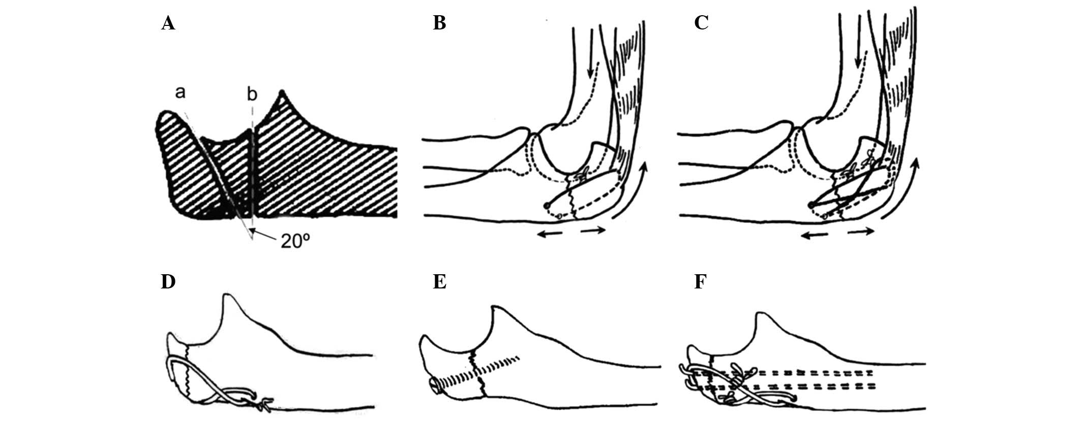
circular wire to the figure of eight wire (Fig. 1C), thus the fracture stability and
the tension band effect of the wire are reinforced. This conforms
to the concept and principles of the Association for the Study of
Internal Fixation (ASIF), as well as the tension band and flexible
fixation principles. It may be applied to all types of fractures,
providing firm fixation and permitting early-stage functional
training. It appears that by using this method, fracture healing is
faster and has a greater therapeutic effect. However, this
technique lacks quantitative indices and experimental data to
verify its suitability for treating olecranon fractures. In the
present study, an experiment was designed for the biomechanical
comparison of this method with four others: circular wiring, figure
of eight wiring, screw fixation and Kirschner wire tension band
fixation. The fixation stability of this method for treating
olecranon fractures was studied, clinical cases were collected and
the efficacy of the treatment was observed. The results of the
present study demonstrated that figure of eight plus circular
wiring fixation is a convenient, reliable, less invasive, less
costly and practical internal fixation method.
Materials and methods
Model establishment
A total of 20 fresh cadaveric elbow joints obtained
from adult males were selected (provided by the Teaching and
Researching Section of Anatomy, Fuzhou Medical University, Fuzhou,
China). The joints contained the upper and lower 2/3 of the
diaphysis. The possibility of pathological changes of the sclerotin
was ruled out by visual observation and X-ray scanning. The
attachment of the triceps brachii tendon to the olecranon and joint
capsule was preserved while the remaining soft tissues were
removed. The samples were sealed in two-layer plastic bags and
preserved in a refrigerator at −40°C. Prior to use, the samples
were thawed at room temperature. A fretsaw with a diameter of 0.6
mm was used to amputate the bones which were made into three
models: a transverse fracture model, an oblique fracture model and
a comminuted fracture model which combined the two models (Fig. 1A). All treatment of cadavers was
strictly in accordance with the international ethical guidelines
and the National Institute of Health guide concerning the care and
use of the human body. The experiments were approved by the
Institutional human Care and Use Committee of the Fujian University
of Traditional Chinese Medicine (Fuzhou, China).
Load and measurement
The samples were divided randomly into two groups:
transverse fracture and oblique fracture models. Each group
contained 10 samples. Five internal fixation techniques were
applied to each fracture model (Fig.
1B–F). The samples were then made into comminuted fracture
models and 10 samples were selected to test the five internal
fixation techniques. The proximal end of the humerus was vertically
fixed on the base of a WWL-100B electronic universal testing
machine. A special clamping apparatus was used to hold and fix the
aponeurosis of the triceps brachii and the elbow joint was flexed
at 90°. The ulnar side was fixed to the clamping apparatus of the
testing machine. A high-precision cantilever displacement sensor
was placed at the bilateral sides of the olecranon fracture line
where the tension was exerted. The sensor was connected to an
SY-III digital strain meter. The load was applied level-by-level at
a rate of 2 mm/min. The association between the load and
displacement and the value of the tensile force when the fractured
end was separated by 2 mm was recorded. Finally, two samples of the
comminuted fracture which were fixed by each internal fixation
technique were selected and a destructive load was applied. The
relevant data were recorded.
Fixation materials
The materials used were 1.0-mm diameter wires,
2.0-mm diameter Kirschner needles and 3.5-mm diameter screws. An
SY-III digital strain meter was used to apply the load at a
constant speed and in a level-by-level manner. The association
between the load and displacement and the tensile force value when
the fractured end was separated by 2 mm was recorded. A destructive
load was applied to the comminuted fracture model to test each
internal fixation technique. The relevant data were recorded.
Surgical method
Brachiplex blocking anesthesia was administered and
a pneumatic tourniquet was used to arrest bleeding. The affected
limb was placed in front of the chest. A vertical incision was made
at the center of the posterior cubital region, around the fracture
site. The incision extended upwards and downwards for 3 and 4 cm,
respectively, to expose the layers of tissues. The ulnar nerve was
protected. The periosteum was cut open along the bony ridge and
stripped to the bilateral sides to expose the fractured end and
joint cavity. Hematoma and scar tissue were then removed. The
fascia at the back of the olecranon was preserved as much as
possible. The two ends of the fracture were repositioned and fixed
with towel forceps. For old fractures, the triceps brachii was
appropriately separated to reduce the tension so that the fracture
was simple to reposition. For open fractures, thorough cleaning was
necessary prior to the internal fixation. Next, a channel was
drilled transversally at 1.5 to 2.0 cm away from the fracture line
(equivalent to 1/2 of the olecranon thickness). Two wires 0.8 and
1.0 mm in diameter were passed through the channel. A wire with the
ulnar side attached to the olecranon, was guided by a bone awl
through the aponeurosis of the triceps branchii to the radial side.
The wire thus formed a circle. The ulnar side of the other wire was
guided from the back of the fracture to the radial side in an
oblique manner. It also passed through the aponeurosis of the
triceps brachii and reached the radial side from behind the
fracture by attaching to the olecranon in an oblique manner. This
wire formed a figure of eight configuration. When the fracture was
repositioned, the circular wiring fixation was performed, followed
by the figure of eight wiring. The free ends of the two wires were
placed at the radial side of the distal end of the fracture and
buried under the aponeurosis. The affected elbow was moved to
examine its function and the condition of the articular surface.
The wound was washed and stitched layer-by-layer. A rubber sheet
was placed for drainage and removed the following day. Following
the surgery, there was no need for external plaster fixation.
Functional training began 2–3 days later.
Statistical analysis
Load-displacement curves were plotted for the three
fracture models. A destructive load was applied to samples fixed by
different internal fixation techniques to record the limit load.
The SPSS 16.0 software was used for the statistical analyses.
Variance analysis was performed on the means of multiple groups and
if the variance analysis indicated significant differences,
multiple comparisons were further employed. P<0.05 was
considered to indicate statistically significant differences.
Results
Load and displacement
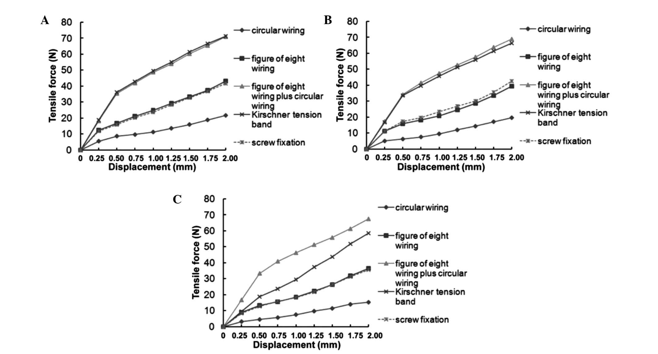
The association between the load and displacement is
shown in Fig. 2. The average
tensile force values required for the separation of the fractured
end by 2 mm in the three types of model using the five internal
fixation techniques are shown in Table
I.
 | Table ITensile force required for the
separation of the fracture end by 2 mm. |
Table I
Tensile force required for the
separation of the fracture end by 2 mm.
| | Tensile force, mean ±
SD (N)
|
|---|
| Group | n | Circular wiring | Figure of eight
wiring | Figure of eight plus
circular wiring | Kirschner tension
band | Screw |
|---|
| Transverse | 6 | 21.72±1.54 | 43.21±1.82a |
70.92±2.34b,c |
71.40±2.16b,d | 41.75±2.37a |
| Oblique | 6 | 19.92±2.29 | 40.99±1.97a |
70.26±2.16b,c |
69.18±1.99b,d | 42.63±1.80a |
| Comminuted | 6 | 15.23±1.30 | 36.42±2.34a |
67.42±2.17b,c |
58.52±2.17b,d | 35.73±3.23a |
Table I and
Fig. 2 show that when elbow
flexion reached 90°, the strain was essentially proportional to the
load for all five fixation techniques. The circular wiring had the
lowest fixation strength, while those of the figure of eight wiring
and screw fixation were higher and similar to each other
(P>0.05). The figure of eight plus circular wiring had a higher
fixation strength than the circular wiring, figure of eight wiring
or screw fixation (P<0.05). In the transverse and oblique
fracture models, the figure of eight plus circular wiring exhibited
no significant difference from the Kirschner tension band in terms
of fixation strength (P>0.05). However, in the comminuted
fracture model, the fixation strength of the figure of eight plus
circular wiring was higher than that of the Kirschner tension band
and the difference was significant (P<0.05).
Limit load
The values of tensile force required to damage the
samples fixed using the five internal fixation techniques are shown
in Table II and Figs. 3A and B. Table II shows that the Kirschner tension
band had the largest limit load while circular wiring had the
smallest. The figure of eight wiring and screw fixation had similar
fixation strengths. The figure of eight plus circular wiring
exhibited no significant differences from the Kirschner tension
band (P>0.05). The absolute value of the limit load of the
Kirschner tension band reached 84.5 N, higher than that of the
figure of eight plus circular wiring.
| Table IILimit loads of the five internal
fixation techniques. |
Table II
Limit loads of the five internal
fixation techniques.
| Internal fixation
technique | n | Limit load, mean ± SD
(N) |
|---|
| Circular wiring | 4 | 35.14±2.46 |
| Figure of eight
wiring | 4 | 56.61±1.97a |
| Figure of eight plus
circular wiring | 4 |
81.43±2.61b,c |
| Kirschner tension
band | 4 |
84.45±2.52b,c |
| Screw fixation | 4 | 57.62±2.91a |
Clinical application
General data
A total of 152 olecranon fracture patients have been
treated using figure of eight plus circular wiring internal
fixation since 2001. The follow-up information was complete for 108
patients. The follow-up visits had an average duration of 12 months
(Table III).
| Table IIIGeneral olecranon fracture patient
information. |
Table III
General olecranon fracture patient
information.
| Fracture type | Number of
patients | Gender
(male/female) | Affected limb
(left/right) | Age, years, mean ±
SD | Time interval between
injury and surgery, days, mean ± SD | Duration of follow-up
visit, months,mean ± SD |
|---|
| Transverse | 35 | 18/17 | 15/20 | 38±7.6 | 3.4±1.2 | 12.3±1.94 |
| Oblique | 26 | 15/11 | 9/17 | 32±12.7 | 2.8±1.5 | 12.20±2.28 |
| Comminuted | 47 | 26/21 | 20/27 | 33±12.5 | 5.5±1.3 | 12.17±1.51 |
Clinical assessment of therapeutic
effectX-ray images of the 108 patients revealed good
positioning of the wires and the anatomical restoration of the
fracture (Fig. 3). Follow-up
visits, lasting for 12 months (range, 9–16 months), revealed the
complete recovery of the fractures in all cases and the average
recovery time was eight weeks. According to the Broberg scoring
system (14), the recovery of 89
patients (82.4%) was excellent, 17 (15.7%)were fairly good and 2
(1.9%)were ordinary.
Discussion
The olecranon is the endpoint of the triceps brachii
muscle which is attached to the rear upper section of the
olecranon. Following fracture, the proximal end becomes subject to
the traction of the triceps brachii muscle and counteraction of the
trochlea of the humerus, which generates pressure at the front of
the olecranon and tension at the rear part. Therefore, the
displacement of the fractured end was set as the experimental
index. By referring to the method by Murphy et al (15), poor fixation was defined as the
separation of the fractured end by 2 mm under load. According to
the actual fixation position, load measurement was performed when
the elbow was flexed at 90°.
Tables I and
II and Fig. 2 show that the circular wiring had
the lowest fixation strength, while figure of eight wiring and
screw fixation had similar fixation strengths (P>0.05) and
figure of eight plus circular wiring had a higher fixation strength
than circular wiring, figure of eight wiring and screw fixation.
The differences were statistically significant (P<0.05). In the
transverse and oblique fracture models, the figure of eight plus
circular wiring and the Kirschner tension band wiring exhibited no
significant differences (P>0.05). However, in the comminuted
fracture model, the fixation strength of eight plus circular wiring
was significantly higher than that of the Kirschner tension band
wiring (P<0.05). For the same fixation technique, the
differences between the transverse and oblique fracture models were
not significant (P>0.05). However, the difference was
significant when compared with the comminuted fracture model
(P<0.05). This observation indicated that the fixation stability
of the transverse and oblique fracture models were not
significantly different and the comminuted fracture model was less
stable. The load curve of the circular wiring was the most flat in
the three fracture models, without an apparent inflexion point. The
load was linearly correlated with displacement. This finding
suggested that this technique was unstable. In the transverse and
oblique fracture models, both the screw fixation and figure of
eight wiring had apparent inflexion points (S= 0.25 mm),
corresponding to a load of approximately 18 N. The Kirschner
tension band and figure of eight plus circular wiring both had
inflexion points (S= 0.5 mm) in the transverse and oblique fracture
models, corresponding to a load of 36 N. Beyond this point, the
load was linearly correlated with displacement. This revealed that
in the transverse and oblique fracture models, the four internal
fixation techniques exhibited no separation of the fracture line
before the load was increased to 18 or 36 N (S<0.25 or 0.5 mm,
respectively). The fixation strength was similar. In the comminuted
fracture model, only the Kirschner tension band and figure of eight
plus circular wiring had apparent inflexion points. The difference
was not significant before the displacement reached 0.5 mm. As the
load increased, the figure of eight plus circular wiring had a
higher fixation strength than the Kirschner tension band
(P<0.05).
In the analysis of the experimental methods, there
were three considerations: i) a 3-dimensional finite element model
does not reflect the non-continuity of the fracture and the load of
internal fixation; ii) a photoelastic test model does not fully
simulate fracture models and the load of the internal fixation; and
iii) the resistor disc used for measurement may not be easily fixed
and is not suitable for the measurement of the displacement of the
fracture end. Therefore, in the present study, we used a
high-precision displacement sensor for a micro-study of the
association between the load of an olecranon fracture and its
stress. Simultaneously, a mechanical measurement method was
performed to apply a destructive load. The new internal fixation
technique was compared with four common internal fixation
techniques: circular wiring, figure of eight wiring, Kirschner
tension band and screw fixation. The purpose was to stimulate
actual clinical models and to provide an experimental basis for the
selection of appropriate internal fixation techniques clinically.
Although the experimental data differed due to the variation in
experimental conditions and methods, they all reflected the
coherent mechanical characteristics of the commonly used internal
fixation techniques. The circular wiring internal fixation had the
lowest fixation strength, the figure of eight wiring and screw
fixation were superior in terms of fixation strength and the figure
of eight plus circular wiring and Kirschner tension band had the
highest fixation strength. In the comminuted fracture model, the
figure of eight plus circular wiring was the most stable.
Our long-term clinical observations also suggest
that the figure of eight plus circular wiring internal fixation
method has the following advantages for treating olecranon
fractures: i) this technique conforms to the tension band
principles and overcomes the shortcomings of screw fixation, such
as a low bearing capacity, easy opening and separation at the rear;
ii) a fixation wire is added to form a semi-spherical fixation
structure, which significantly enhances the stability and
resistance to tensile stress compared with figure of eight wiring;
iii) this technique is easy to practice, overcoming the
shortcomings of Kirschner tension band internal fixation of not
being suitable for comminuted fractures and being difficult to
adjust after fixation. It also prevents the tenderness around the
pin site which may occur with the Kirschner tension band (16); iv) the technique requires less
stripping and bleeding. It maintains the stability of the joint,
and is less invasive, costly and time-consuming. Compared with hook
plate internal fixation, it has a simplified procedure for internal
fixation, thereby reducing the pain and economic burden for the
patient; v) the technique makes use of the continuous force exerted
by the aponeurosis of the triceps branchii, which is in accordance
with the flexible fixation principle. It does not require drilling
at the proximal end of the fracture and requires a smaller incision
in the fascia of the olecranon for comminuted fractures. The
technique makes use of the integrity of the aponeurosis of the
triceps branchii for repositioning, making the surgery simpler to
perform for comminuted fractures; and vi) during the removal, only
a small cut under local anaesthesia is required to loosen and
remove the wires. This process is simple to perform, thus
hospitalization is not necessary for the removal of this internal
fixation.
In conclusion, figure of eight plus circular wiring
internal fixation may be applied to transverse, oblique and
comminuted olecranon fractures. It appears to be a particularly
good treatment for comminuted fractures. Characterized by low
invasiveness, simple surgery, firm fixation and lower cost, this
technique is safe, reliable and practical for internal fixation and
should be popularized in clinical practice.
Acknowledgements
This study was supported by the Youth
Foundation of the Fujian Provincial Health Department (No.
2006-2-57).
References
|
1
|
Veillette CJ and Steinmann SP: Olecranon
fractures. Orthop Clin North Am. 39:229–236. 2008. View Article : Google Scholar
|
|
2
|
Newman SD, Mauffrey C and Krikler S:
Olecranon fractures. Injury. 40:575–581. 2009. View Article : Google Scholar : PubMed/NCBI
|
|
3
|
Ishigaki N, Uchiyama S, Nakagawa H,
Kamimura M and Miyasaka T: Ulnar nerve palsy at the elbow after
surgical treatment for fractures of the olecranon. J Shoulder Elbow
Surg. 13:60–65. 2004. View Article : Google Scholar : PubMed/NCBI
|
|
4
|
McKay PL and Katarincic JA: Fractures of
the proximal ulna olecranon and coronoid fractures. Hand Clin.
18:43–53. 2002. View Article : Google Scholar : PubMed/NCBI
|
|
5
|
Heim U: Combined fractures of the radius
and the ulna at the elbow level in the adult. Analysis of 120 cases
after more than 1 year. Rev Chir Orthop Reparatrice Appar Mot.
84:142–153. 1998.(In French).
|
|
6
|
Halling KB, Lewis DD, Cross AR, Kerwin SC,
Smith BA and Kubilis PS: Complication rate and factors affecting
outcome of olecranon osteotomies repaired with pin and tension-band
wiring fixation in dogs. Can Vet J. 43:528–534. 2002.PubMed/NCBI
|
|
7
|
Assom M, Lollino N, Caranzano F, Rossi R
and Castoldi F: Polyester tension-band wiring of olecranon
fractures of elderly people: a simple technique. Injury.
39:1474–1476. 2008. View Article : Google Scholar : PubMed/NCBI
|
|
8
|
Gordon MJ, Budoff JE, Yeh ML, Luo ZP and
Noble PC: Comminuted olecranon fractures: a comparison of plating
methods. J Shoulder Elbow Surg. 15:94–99. 2006. View Article : Google Scholar : PubMed/NCBI
|
|
9
|
Rommens PM, Küchle R, Schneider RU and
Reuter M: Olecranon fractures in adults: factors influencing
outcome. Injury. 35:1149–1157. 2004. View Article : Google Scholar : PubMed/NCBI
|
|
10
|
Huang TW, Wu CC, Fan KF, Tseng IC, Lee PC
and Chou YC: Tension band wiring for olecranon fractures: relative
stability of Kirschner wires in various configurations. J Trauma.
68:173–176. 2010. View Article : Google Scholar : PubMed/NCBI
|
|
11
|
Anderson ML, Larson AN, Merten SM and
Steinmann SP: Congruent elbow plate fixation of olecranon
fractures. J Orthop Trauma. 21:386–393. 2007. View Article : Google Scholar : PubMed/NCBI
|
|
12
|
Boyer MI, Galatz LM, Borrelli J Jr,
Axelrod TS and Ricci WM: Intra-articular fractures of the upper
extremity: new concepts in surgical treatment. Instr Course Lect.
52:591–605. 2003.PubMed/NCBI
|
|
13
|
Fyfe IS, Mossad MM and Holdsworth BJ:
Methods of fixation of olecranon fractures. An experimental
mechanical study. J Bone Joint Surg Br. 67:367–372. 1985.PubMed/NCBI
|
|
14
|
Broberg MA and Morrey BF: Results of
delayed excision of the radial head after fracture. J Bone Joint
Surg Am. 68:669–674. 1986.PubMed/NCBI
|
|
15
|
Murphy DF, Greene WB and Dameron TB Jr:
Displaced olecranon fractures in adults. Clinical evaluation. Clin
Orthop Relat Res. 224:215–223. 1987.PubMed/NCBI
|
|
16
|
Rommens PM, Schneider RU and Reuter M:
Functional results after operative treatment of olecranon
fractures. Acta Chir Belg. 104:191–197. 2004.PubMed/NCBI
|