Introduction
Head and neck squamous cell carcinoma (HNSCC) is
known for its rapid clinical progression and poor prognosis. The
survival rates for many types of HNSCC have improved little over
the past 40 years. Anti-monoclonal antibody is often used as a
first-line and primary treatment of malignancies, such as malignant
lymphomas and breast cancer. Cetuximab, an anti-epidermal growth
factor receptor (EGFR) antibody, is the only EGFR inhibitor
approved in HNSCC. Several combined methods with cetuximab, such as
irradiation or many types of anticancer drugs, have been applied to
enhance the effect of treatment. However, the efficacy of such
treatment is inadequate. Therefore, new therapeutic methods should
be developed to improve the survival rate.
In non-surgical treatment, induction of apoptosis is
a preferred mode of killing cancer cells with fewer side effects
and immune reactions (1).
Apoptosis is a physiologic process that contributes to the
homeostasis of multi-cellular organisms and maintains the balance
between cell proliferation and cell death (2), as a cell response to stress stimuli,
such as exposure to chemotherapeutic drugs, oxidative stress, free
radicals, X-rays, ultraviolet radiation, shear stress and
ultra-sound (US) irradiation.
Since the biological effect of US was first reported
in 1927, numerous reports concerning the application of US have
been published. The mechanisms of US-induced apoptosis on physical
and biochemical levels have been discussed in several studies
(3–7). In addition, US irradiation can be
divided into low-intensity ultrasound (LIU) and high-intensity
focused ultrasound (HIFU) depending on the intensity. LIU has a
great potential in apoptosis therapy for cancer (8), while HIFU can thermally ablate
tissues via hyperthermia in various types of cancer (9,10).
LIU has been subjected to numerous studies to evaluate its
biological effects and their possible application in therapeutic
strategies in cancer treatment. Thes]e outcomes include the
enhancement of cellular uptake of drugs (11) and the induction of apoptosis and
cell killing (12,13). Besides, it is likely that the
selectivity of LIU for cancer cells is higher than that for normal
cells (14).
The combined effects of US and several anticancer
drugs, such as (DOX) and Cisplatin (CDDP) have often been reported
(15,16). Thus, sonication by US may also be
considered as a candidate for the enhancement of anticancer drugs.
However, few reports associated with anti-monoclonal antibody are
available. We hypothesized that US has the potential to become an
enhancer of cetuximab owing to its effect of changing cell membrane
structure and making cancer cells more sensitive to cetuximab as
well as rituximab, as previously reported (17).
In the present study, we assessed the therapeutic
potential of the combination of cetuximab and LIU in vitro
and demonstrated that this combination induces the effect of tumor
cell killing and increases the therapeutic efficacy in head and
neck cancer HSC-3 and HSC-4 cells. In addition, we focused on EGFR
signaling and apoptosis signaling through the mitochondria-caspase
pathway to confirm cetuximab enhancement by US.
Materials and methods
Cell culture and drugs
Human HNSCC cells, HSC-3 and HSC-4, were employed in
this study. The HSC-3 and HSC-4 cells, obtained from the Japanese
Cancer Research Resource Bank (Tokyo, Japan), were cultured in DMEM
(Invitrogen, Carlsbad, CA, USA) supplemented with 10%
heat-inactivated fetal bovine serum (Invitrogen), 100 U/ml
penicillin, and 100 mg/ml streptomycin (Gibco, Grand Island, NY,
USA) at 37°C in 5% CO2. Cetuximab (Merck KgaA,
Darmstadt, Germany) was dissolved in phosphate-buffered saline
(PBS) at a concentration of 100 nM and then stored at 4°C until
use.
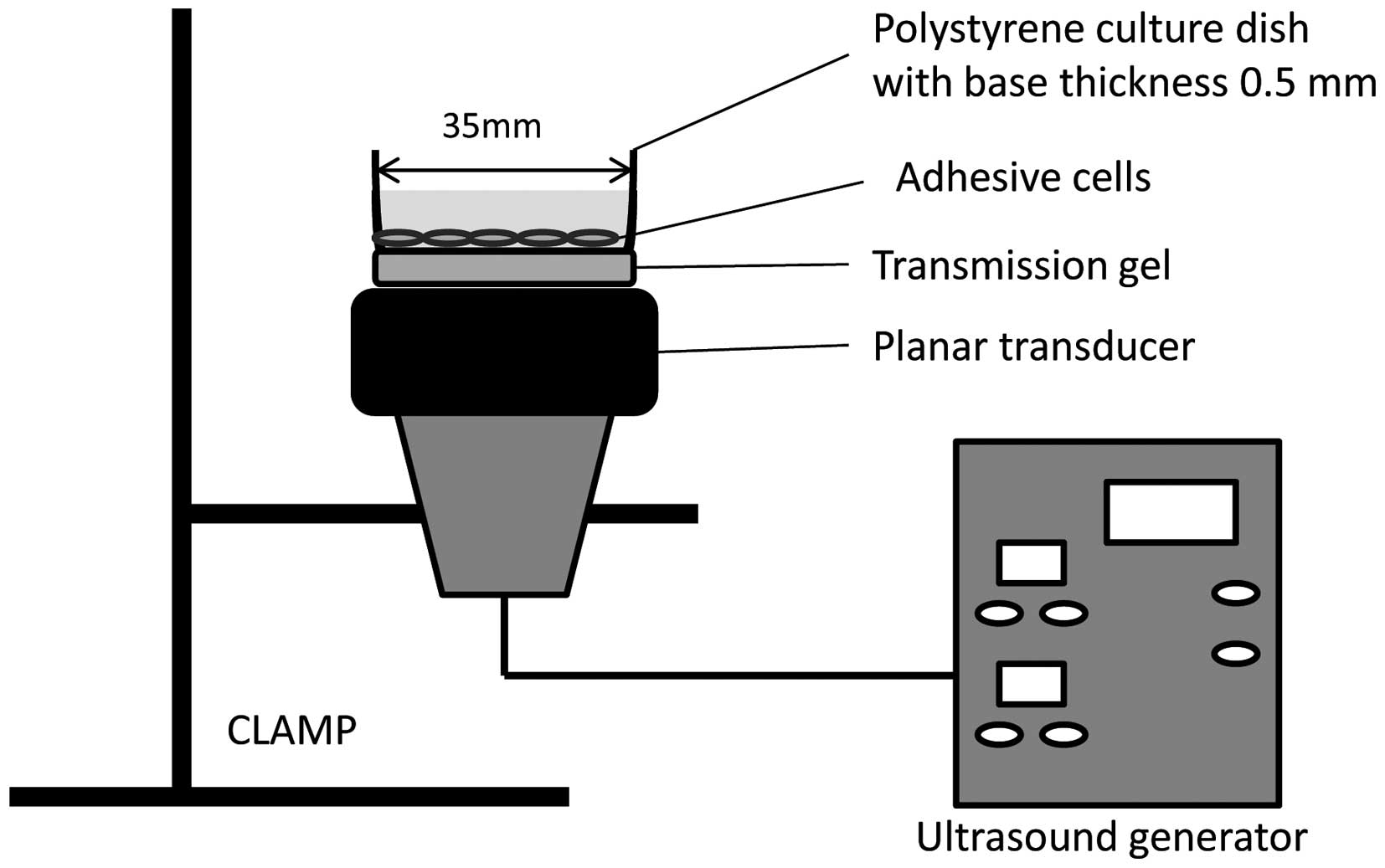
US apparatus and intensity
A 1.0 MHz ultrasonic generator (KUS-2S; ITO
Ultrasonic Co., Ltd., Tokyo, Japan) with a fixed duty factor of 50%
and with 1 Hz pulse repetition frequency (PRF) was used. The
sonication was conducted at an intensity of 0.5 W/cm2 (=
output intensity of 0.38 W/cm2) and exposure time of 1
min. The intensity and time were used in all of the sonication
experiments.
To keep the transducer-facing directory upward for
the sonication procedure, the transducer, with a diameter of 2.7
cm, was fixed with a clamp attached to a metal stand. A 3.5-cm
culture dish was placed on the center of the transducer with
intermediate gel (Fig. 1).
Experimental protocol
The experimental groups for this study were: i)
non-treated (CNTRL), ii) cetuximab-treated (CETU), iii) US-treated
(UST) and iv) the combination of cetuximab and US-treated (COMB).
Cells (1×10⁶) in 1.5 ml medium were seeded in a 3.5-cm
dish and incubated at 37°C for 12 h. At 30 min prior to sonication
another 1.5 ml of fresh medium, with or without cetuximab at the
final concentration of 100 nM, was added to each dish to avoid
cavitation attenuation due to the high concentration of carbon
dioxide accumulated in the dish, as previously described (18,19).
Following the treatment, the cells were subjected to different
analyses.
Measurement of cell viability
Cell viability was assessed by trypan blue staining
assay at 24 h after cetuximab and US, as previously described
(6). The number of cells was
counted using a hemocytometer to estimate the viability. The cell
viability was calculated as the number of viable cells in the
treated group/the number of viable cells in the non-treated group.
Each measurement was repeated three times independently.
Detection of apoptosis
Apoptosis was assessed by fluorescein isothiocyanate
(FITC)-Annexin V and propidium iodide (PI) staining assay according
to the manufacturer’s instructions (Roche Diagnostic GmbH,
Penzberg, Germany). Following the treatment of cetuximab and US,
the cells were incubated at 37.0°C for 24 h. The cells were then
washed with cold PBS, stained with FITC-Annexin V/PI for 10 min and
observed under a fluorescent microscope for typical morphological
changes characteristic of apoptosis.
FITC-Annexin V/PI staining assay was performed to
detect the phosphatidylserine expression using FITC-Annexin V
labeling as an endpoint indicator of early apoptosis, and the PI
uptake as an indicator of necrosis. Annexin V-positive and
PI-negative cells were considered to be early apoptosis, while
Annexin V-positive and PI-positive cells were considered to be
secondary necrosis (6,15). Apoptosis was calculated as the
number of apoptotic cells/the number of total cells in each group.
Each measurement was repeated three times independently.
Antibodies and immunoblot analysis
EGFR, phospho-EGFR, and activation of caspase-3
signaling on HSC-3 cells after the treatment were also evaluated
with western blotting. Cells were cultured in 3.5-cm dishes for 6 h
after the treatment, respectively. The cells were collected and
frozen in 100 μl RIPA buffer, and stored in a −30°C ultra
low temperature freezer. Briefly, total protein extracts were
prepared according to the freeze-thaw lysis method (20) and protein concentrations were
measured with the bovine serum albumin (BSA) protein assay. Extract
samples containing 20 μg of protein were then separated by
sodium dodecylsulfate-polyacrylamide gel electrophoresis (SDS-PAGE)
and transferred to polyvinylidene difluoride membranes. The
membranes were incubated with anti-EGFR, anti-phospho-EGFR, and
anti-caspase-3 antibody [Cell Signaling Technology, Danvers, MA,
USA; diluted 1:2000, 1:1000 and 1:1000 in PBS with Tween-20 (PBST)
respectively] at 4°C overnight, and then with peroxidase-conjugated
secondary anti-rabbit or goat immunoglobulin G (IgG) (Cell
Signaling Technology; diluted 1:1000 in PBST) for 1 h. After
rinsing in PBST (4 times, 5 min each), immuno detection was
performed using an enhanced chemiluminescence (ECL) western blot
analysis detection reagants and analysis system. The membranes were
subsequently exposed to X-ray film, as previously described
(21).
Statistical analysis
Data were presented as the mean ± standard deviation
(SD). Significant differences between groups were assessed using
two-way factorial analysis of variance (ANOVA). P<0.01 was
considered to be a statistically significant difference.
Results
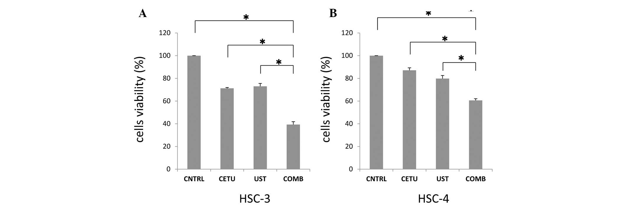
Enhancement of cetuximab-induced cell
killing by US
Cell viability was assessed by trypan blue staining
assay at 24 h after cetuximab and US. The cell viabilities of HSC-3
cells (Fig. 2A) and HSC-4 cells
(Fig. 2B) in the CETU group were
71.2 and 87.1% compared to those of the CNTRL group, respectively.
The cell viabilities in the UST group were 73.0 and 79.8%, while
those in the COMB group decreased to 39.3 and 60.6%, respectively,
thus showing a synergistic enhancement of cetuximab-induced cell
killing by US.
Enhancement of cetuximab-induced
apoptosis by US
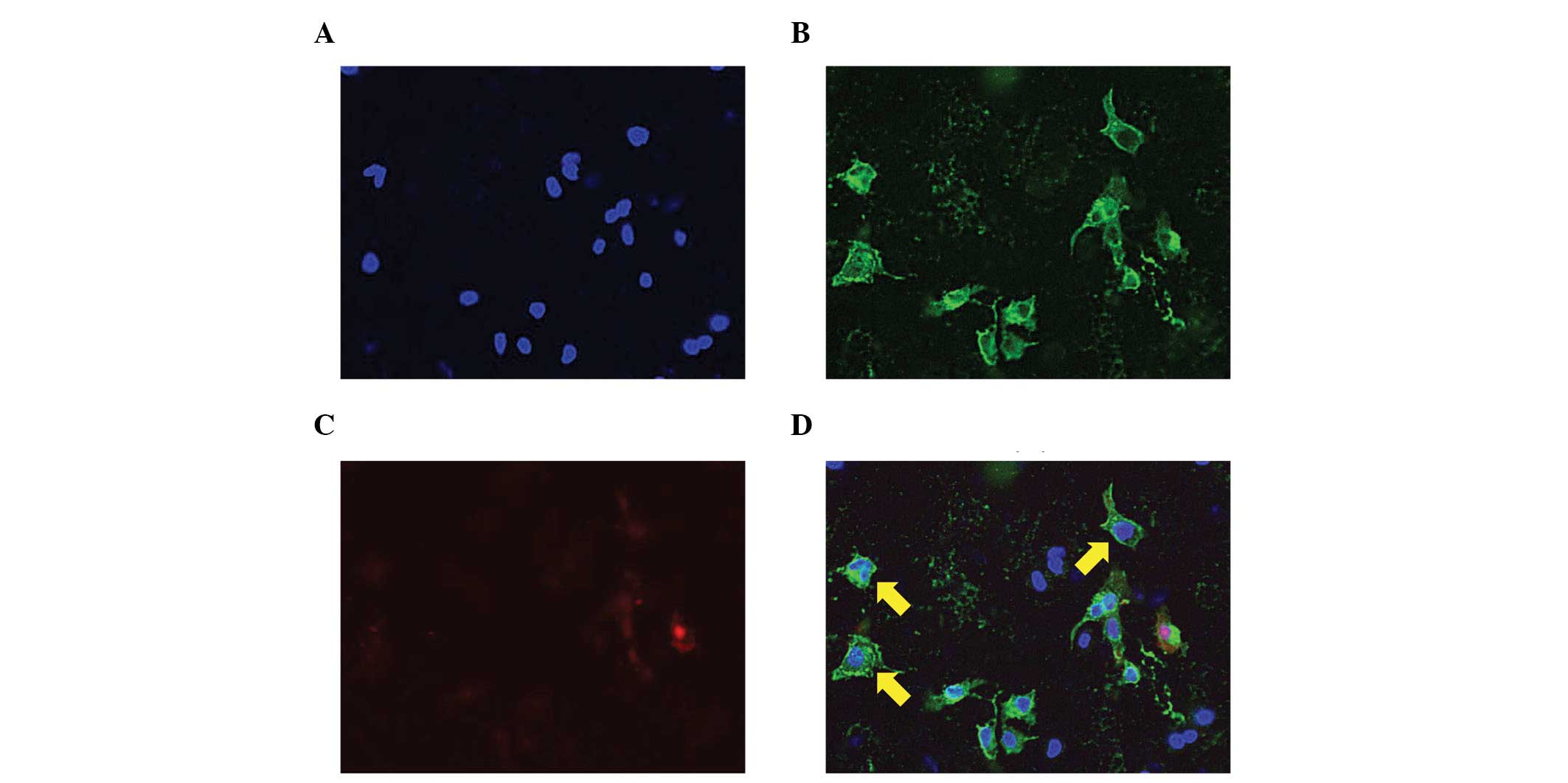
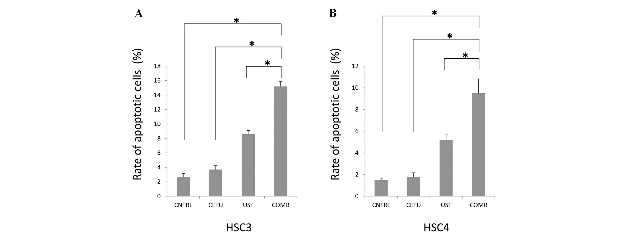
In order to estimate the enhancement of cell
killing, the induction of apoptosis was assessed by FITC-Annexin
V/PI staining assay (Figs. 3 and
4). FITC-Annexin V/PI staining
assay was performed to detect the phosphatidylserine expression on
the cell membrane as an endpoint of early apoptosis, showing the
exact percentage of cells in viable cells (Annexin V−/PI−), early
apoptotic cells (Annexin V+/PI−) and secondary necrosis (Annexin
V+/PI+) under a fluorescent microscope. Fig. 3 shows (A) DAPI, (B) Annexin V, (C)
PI and (D) merged images of the combined treatment on HSC-3 cells,
respectively. The rates of apoptotic features of HSC-3 cells
(Fig. 4A) and HSC-4 cells
(Fig. 4B) in the CNTRL group were
2.7 and 1.5%, respectively, while in the CETU group, they were 3.7
and 1.8%, respectively. No statistically significant difference was
observed between these two groups. In the UST group, the apoptotic
features were 8.6 and 5.2%, respectively, while those in the COMB
group were 15.2 and 9.5%, respectively. These data suggest that US
induced apoptosis in these cells and that the combination of US and
cetuximab enhanced apoptosis synergistically.
Effects of EGFR signaling and apoptosis
following the treatments
To elucidate the effects of EGFR signaling and
apoptosis on HSC-3 cells after the treatments were administered,
the expression of EGFR, phospho-EGFR, and the activation of
caspase-3 were evaluated using western blotting (Fig. 5). No statistically significant
differences in EGFR expression among the CETU, UST and COMB groups
was observed, while the expression of phospho-EGFR was much more
downregulated in the COMB group compared with the remaining groups,
although phospho-EGFR was downregulated in the CETU group. These
data suggest that US induced the inhibition of EGFR-signaling
pathway by cetuximab. To elucidate the induction of apoptosis, the
activation of caspase-3 was estimated. The activation, the cleavage
of caspase-3, in the UST group was upregulated compared with that
in the CNTRL group. Additionally, caspase-3 activation was much
more upregulated in the COMB group compared with that in the
remaining groups, suggesting that US induced apoptosis through the
activation of caspase-3 by cetuximab.
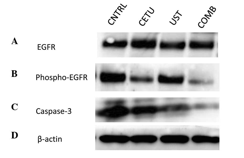 | Figure 5The expression of EGFR, phospho-EGFR,
and activation of caspase-3 signaling on HSC-3 cells after the
treatment with western blot analysis. (A), EGFR; (B),
phosphor-EGFR; (C), caspase-3; (D), β-actin. There were no
differences of the expression of EGFR among the CETU, UST and COMB
groups, while the expression of phospho-EGFR was down-regulated and
moreover the cleavage of caspase-3 was upregulated in the COMB
group compared with the other groups. CNTRL, non-treated; CETU,
cetuximab-treated; UST, US-treated; COMB, combination of cetuximab
and US-treated |
Discussion
In recent years, there has been a focus on the
potential of LIU as well as HIFU in clinical applications. These
outcomes include the enhancement of cellular uptake of drugs and
the induction of cell killing or apoptosis (15,16,22).
LIU primarily inhibits cell proliferation through its heating,
cavitation and mechanical effect. Such investigations indicate that
the biological effectiveness of US is linked to the acoustic
conditions (23). In general, the
impulsive pressures generated by the collapse of cavitation bubbles
associated with US may permeabilize the plasma membrane of
neighboring cells (24–27). This process allows exogenous
molecules to enter the cells resulting in a biological response
(27–32). Following the application of US, the
cell membrane surface becomes rough and reversible within 24 h
after US exposure (33,34). Tumor cells are more sensitive to US
compared with normal cells and there is a threshold dose that
effectively treats tumor cells with little effect on normal cells
(4). In addition, the duration
time and dose of LIU depend on tumor cell types. Generally, to
avoid unexpected immune responses in vivo resulting from
cell lysis induced by focused US at higher intensity, the dose of
LIU is should be 0.5 W/cm2 below. In a previous report,
there was a significant effect of US-induced apoptosis at more than
0.3 W/cm2 for 1 min, but not at less than 0.2
W/cm2 compared with non-treatment (15), suggesting that the threshold of the
US effect is between 0.2 and 0.3 W/cm2. It has been
shown that LIU, such as 1 MHz pulsed US at an intensity of 0.3 or
0.5 W/m2 for 1 min, is effective enough to induce
apoptosis for cancer cells (15).
Therefore, we utilized this dose of 0.5 W/cm2 for 1 min
to obtain more effective apoptosis with minimal lysis for the HSC-3
and HSC-4 cells used in this study.
Furthermore, the possible mechanisms of US-induced
apoptosis reportedly involve its cavitations (6,7,12,29,35).
Apoptosis is known to require the activation of caspases, a group
of enzymes involved in apoptotic cascade events (36). Apoptotic stimulus is capable of
activating apoptosis-related proteins to enter mitochondria
inducing the mitochondrial membrane to form pores to release
molecules into the cytosol, such as cytochrome c. The released
molecules activate caspase-9, which cleaves procaspase-3 to
caspase-3, inducing apoptosis (37,38).
Among these apoptosis-related proteins, caspase-3 plays a crucial
role in apoptosis through the mitochondria-caspase pathway, and its
activation is often used as a marker of apoptosis. Previous studies
have shown that on a molecular level, several proteins, including
p53, Bid and Bcl-2, were identified as responding to US
irradiation, suggesting that mitochondrial membrane
permeabilization, or pore formation, was involved in LIU-induced
apoptosis (22,39). Additionally, disruption of the
mitochondrial transmembrane potential, cytochrome c release, and
caspase activation were also observed after US treatment (30). Thus, evidence suggests that US
exposure is associated with cell apoptosis, which is
mitochondria-caspase-dependent. This theory has provided a
foundation for the clinical application of US. Consequently,
numerous reports concerning the enhancement of anticancer drug by
LIU are available. However, there have been few reports associated
with anti-monoclonal antibody for cancer cells.
Cetuximab, a chimeric human:murine IgG1 monoclonal
antibody against EGFR, is the most studied targeted therapy in
HNSCC. It can be either inhibitory for cell growth or cytotoxic and
can enhance the cytotoxic effect of chemotherapeutic drugs or
ionizing radiation, resulting in cell apoptosis (40). Cetuximab is currently approved in
combination with radiation therapy in locally advanced disease, as
a single agent in platinum-refractory recurrent/metastatic disease,
and in combination with platinum (carboplatin or CDDP) and
5-fluorouracil, as first-line therapy in recurrent/metastatic
disease. However, response rates as a single agent have been less
than satisfactory (13%) and of limited duration (2–3 months)
(41). Thus, we suggest that US
has the potential to become an enhancer of cetuximab owing to its
effect of changing cell membrane structure and making cancer cells
more sensitive to cetuximab as well as rituximab, as previously
reported (17).
In the present study, we demonstrated that there
were more tumor cell killing features in the COMB group than in the
other groups and that the phosphorylation of EGFR was
down-regulated, while the activation of caspase-3 was upregulated
in the COMB group compared with that in the remaining groups. These
data suggest that LIU was able to increase the induction rate of
cetuximab in head and neck cancer cells, leading to enhancement of
the effect of cetuximab in the COMB group. In addition, as
previously reported, the activation of caspase-3 also increased
when apoptosis was induced by US alone in cancer cells (22). These results suggest that an
increase of the induction rate of cetuximab by US could
subsequently activate caspase-cascade reaction more than US
alone.
In summary, the present study has shown the
synergistic enhancement of apoptosis induction by the combination
of LIU and cetuximab in HNSCC cells. The mitochondria-caspase
pathway is considered to be crucial in this process. These data
suggest that LIU enhanced the anticancer effect of cetuximab in
HSC-3 and HSC-4 cells of HNSCC. Although the precise involvement of
the EGFR and caspase signaling modifications by cetuximab in the
enhancement remains to be elucidated, they may be involved in the
latent effect. The critical mechanisms remain to be further
investigated. The majority of studies on US effects have been
performed in vitro to evaluate clinical cancer therapies.
However, it should be noted that the results of in vitro
studies on the biological effects of therapeutic US cannot be
translated to the same cells in vivo, even if the same US
exposure is used. Although it is still too early to discuss its
clinical efficiency, LIU may prove to be a valid treatment option
for HNSCC.
Acknowledgements
We thank Dr Takashi Kondo (University
of Toyama, Japan) for his critical input. This study was supported
in part by Grants-in Aids for Scientific Research from the Ministry
of Education, Culture, Sports, Science and Technology of Japan.
References
|
1
|
Fulda S and Debatin KM: Apoptosis pathways
in neuroblastoma therapy. Cancer Lett. 197:131–135. 2003.
View Article : Google Scholar
|
|
2
|
Mandal D, Mazumder A, Das P, Kundu M and
Basu J: Fas-, caspase 8-, and caspase 3-dependent signaling
regulates the activity of the aminophospholipid translocase and
phosphatidylserine externalization in human erythrocytes. J Biol
Chem. 280:39460–39467. 2005. View Article : Google Scholar
|
|
3
|
Ashush H, Rozenszajn LA, Blass M, et al:
Apoptosis induction of human myeloid leukemic cells by ultrasound
exposure. Cancer Res. 60:1014–1020. 2000.PubMed/NCBI
|
|
4
|
Lagneaux L, de Meulenaer EC, Delforge A,
et al: Ultrasonic low-energy treatment: a novel approach to induce
apoptosis in human leukemic cells. Exp Hematol. 30:1293–1301. 2002.
View Article : Google Scholar : PubMed/NCBI
|
|
5
|
Honda H, Zhao QL and Kondo T: Effects of
dissolved gases and an echo contrast agent on apoptosis induced by
ultrasound and its mechanism via the mitochondria-caspase pathway.
Ultrasound Med Biol. 28:673–682. 2002. View Article : Google Scholar : PubMed/NCBI
|
|
6
|
Feril LB Jr, Kondo T, Zhao QL and Ogawa R:
Enhancement of hyperthermia-induced apoptosis by non-thermal
effects of ultrasound. Cancer Lett. 178:63–70. 2002. View Article : Google Scholar : PubMed/NCBI
|
|
7
|
Feril LB Jr, Kondo T, Zhao QL, et al:
Enhancement of ultrasound-induced apoptosis and cell lysis by
echo-contrast agents. Ultrasound Med Biol. 29:331–337. 2003.
View Article : Google Scholar : PubMed/NCBI
|
|
8
|
Abdollahi A, Domhan S, Jenne JW, et al:
Apoptosis signals in lymphoblasts induced by focused ultrasound.
Faseb J. 18:1413–1414. 2004.PubMed/NCBI
|
|
9
|
Huber PE, Jenne JW, Rastert R, et al: A
new noninvasive approach in breast cancer therapy using magnetic
resonance imaging-guided focused ultrasound surgery. Cancer Res.
61:8441–8447. 2001.PubMed/NCBI
|
|
10
|
Blana A, Walter B, Rogenhofer S and
Wieland WF: High-intensity focused ultrasound for the treatment of
localized prostate cancer: 5-year experience. Urology. 63:297–300.
2004.PubMed/NCBI
|
|
11
|
Larkin JO, Casey GD, Tangney M, et al:
Effective tumor treatment using optimized ultrasound-mediated
delivery of bleomycin. Ultrasound Med Biol. 34:406–413. 2008.
View Article : Google Scholar : PubMed/NCBI
|
|
12
|
Feril LB Jr, Kondo T, Cui ZG, et al:
Apoptosis induced by the sonomechanical effects of low intensity
pulsed ultrasound in a human leukemia cell line. Cancer Lett.
221:145–152. 2005. View Article : Google Scholar : PubMed/NCBI
|
|
13
|
Feng Y, Tian ZM, Wan MX and Zheng ZB: Low
intensity ultrasound-induced apoptosis in human gastric carcinoma
cells. World J Gastroenterol. 14:4873–4879. 2008. View Article : Google Scholar : PubMed/NCBI
|
|
14
|
Lejbkowicz F and Salzberg S: Distinct
sensitivity of normal and malignant cells to ultrasound in vitro.
Environ Health Perspect. 105(Suppl 6): 1575–1578. 1997. View Article : Google Scholar : PubMed/NCBI
|
|
15
|
Yoshida T, Kondo T, Ogawa R, et al:
Combination of doxorubicin and low-intensity ultrasound causes a
synergistic enhancement in cell killing and an additive enhancement
in apoptosis induction in human lymphoma U937 cells. Cancer
Chemother Pharmacol. 61:559–567. 2008. View Article : Google Scholar : PubMed/NCBI
|
|
16
|
Watanabe Y, Aoi A, Horie S, et al:
Low-intensity ultrasound and microbubbles enhance the antitumor
effect of cisplatin. Cancer Sci. 99:2525–2531. 2008. View Article : Google Scholar : PubMed/NCBI
|
|
17
|
Danno D, Kanno M, Fujimoto S, Feril LB Jr,
Kondo T and Nakamura S: Effects of ultrasound on apoptosis induced
by anti-CD20 antibody in CD20-positive B lymphoma cells. Ultrason
Sonochem. 15:463–471. 2008. View Article : Google Scholar : PubMed/NCBI
|
|
18
|
Mendelsohn J: Blockade of receptors for
growth factors: an anticancer therapy - the fourth annual Joseph H
Burchenal American Association of Cancer Research Clinical Research
Award Lecture. Clin Cancer Res. 6:747–753. 2000.
|
|
19
|
Feril LB Jr and Kondo T: Major factors
involved in the inhibition of ultrasound-induced free radical
production and cell killing by pre-sonication incubation or by high
cell density. Ultrason Sonochem. 12:353–357. 2005. View Article : Google Scholar : PubMed/NCBI
|
|
20
|
Bollag DM and Edelstein SJ: Protein
methods. Wiley-Liss; New York: 1991
|
|
21
|
Hayashi K, Motoyama S, Koyota S, et al:
REG I enhances chemo- and radiosensitivity in squamous cell
esophageal cancer cells. Cancer Sci. 99:2491–2495. 2008. View Article : Google Scholar : PubMed/NCBI
|
|
22
|
Feng Y, Tian Z and Wan M: Bioeffects of
low-intensity ultrasound in vitro: apoptosis, protein profile
alteration, and potential molecular mechanism. J Ultrasound Med.
29:963–974. 2010.PubMed/NCBI
|
|
23
|
Tezel A, Sens A, Tuchscherer J and
Mitragotri S: Frequency dependence of sonophoresis. Pharm Res.
18:1694–1700. 2001. View Article : Google Scholar : PubMed/NCBI
|
|
24
|
Chen WS, Matula TJ and Crum LA: The
disappearance of ultrasound contrast bubbles: observations of
bubble dissolution and cavitation nucleation. Ultrasound Med Biol.
28:793–803. 2002. View Article : Google Scholar : PubMed/NCBI
|
|
25
|
Miller DL and Dou C: Membrane damage
thresholds for 1- to 10-MHz pulsed ultrasound exposure of
phagocytic cells loaded with contrast agent gas bodies in vitro.
Ultrasound Med Biol. 30:973–977. 2004. View Article : Google Scholar : PubMed/NCBI
|
|
26
|
Hallow DM, Mahajan AD, McCutchen TE and
Prausnitz MR: Measurement and correlation of acoustic cavitation
with cellular bioeffects. Ultrasound Med Biol. 32:1111–1122. 2006.
View Article : Google Scholar : PubMed/NCBI
|
|
27
|
Kodama T, Tomita Y, Koshiyama K and
Blomley MJ: Transfection effect of microbubbles on cells in
superposed ultrasound waves and behavior of cavitation bubble.
Ultrasound Med Biol. 32:905–914. 2006. View Article : Google Scholar : PubMed/NCBI
|
|
28
|
Larina IV, Evers BM and Esenaliev RO:
Optimal drug and gene delivery in cancer cells by
ultrasound-induced cavitation. Anticancer Res. 25:149–156.
2005.PubMed/NCBI
|
|
29
|
Honda H, Kondo T, Zhao QL, Feril LB Jr and
Kitagawa H: Role of intracellular calcium ions and reactive oxygen
species in apoptosis induced by ultrasound. Ultrasound Med Biol.
30:683–692. 2004. View Article : Google Scholar : PubMed/NCBI
|
|
30
|
Firestein F, Rozenszajn LA,
Shemesh-Darvish L, Elimelech R, Radnay J and Rosenschein U:
Induction of apoptosis by ultrasound application in human malignant
lymphoid cells: role of mitochondria-caspase pathway activation.
Ann N Y Acad Sci. 1010:163–166. 2003. View Article : Google Scholar : PubMed/NCBI
|
|
31
|
Feril LB Jr, Tsuda Y, Kondo T, et al:
Ultrasound-induced killing of monocytic U937 cells enhanced by
2,2′-azobis(2-amidino-propane) dihydrochloride. Cancer Sci.
95:181–185. 2004.
|
|
32
|
Koshiyama K, Kodama T, Yano T and Fujikawa
S: Structural change in lipid bilayers and water penetration
induced by shock waves: molecular dynamics simulations. Biophys J.
91:2198–2205. 2006. View Article : Google Scholar : PubMed/NCBI
|
|
33
|
Duvshani-Eshet M, Adam D and Machluf M:
The effects of albumin-coated microbubbles in DNA delivery mediated
by therapeutic ultrasound. J Control Release. 112:156–166. 2006.
View Article : Google Scholar : PubMed/NCBI
|
|
34
|
Taniyama Y, Tachibana K, Hiraoka K, et al:
Local delivery of plasmid DNA into rat carotid artery using
ultrasound. Circulation. 105:1233–1239. 2002. View Article : Google Scholar : PubMed/NCBI
|
|
35
|
Kagiya G, Ogawa R, Tabuchi Y, et al:
Expression of heme oxygenase-1 due to intracellular reactive oxygen
species induced by ultrasound. Ultrason Sonochem. 13:388–396. 2006.
View Article : Google Scholar : PubMed/NCBI
|
|
36
|
Yuan J: Molecular control of life and
death. Curr Opin Cell Biol. 7:211–214. 1995. View Article : Google Scholar
|
|
37
|
Majno G and Joris I: Apoptosis, oncosis,
and necrosis. An overview of cell death. Am J Pathol. 146:3–15.
1995.PubMed/NCBI
|
|
38
|
Wyllie AH, Kerr JF and Currie AR: Cell
death: the significance of apoptosis. Int Rev Cytol. 68:251–306.
1980. View Article : Google Scholar
|
|
39
|
Kroemer G, Galluzzi L and Brenner C:
Mitochondrial membrane permeabilization in cell death. Physiol Rev.
87:99–163. 2007. View Article : Google Scholar : PubMed/NCBI
|
|
40
|
Ciardiello F and Tortora G: A novel
approach in the treatment of cancer: targeting the epidermal growth
factor receptor. Clin Cancer Res. 7:2958–2970. 2001.PubMed/NCBI
|
|
41
|
Vermorken JB, Trigo J, Hitt R, et al:
Open-label, uncontrolled, multicenter phase II study to evaluate
the efficacy and toxicity of cetuximab as a single agent in
patients with recurrent and/or metastatic squamous cell carcinoma
of the head and neck who failed to respond to platinum-based
therapy. J Clin Oncol. 25:2171–2177. 2007.
|