Introduction
Left atrial (LA) function plays a critical role in
the cardiac cycle, particularly in the left ventricular (LV)
filling and stroke volume during LV diastolic dysfunction,
including the ventricular systole right ventricle (RV) function
(reservoir), early ventricular diastolic conduit function (conduit)
and ventricular diastolic ejection fraction (EF) function (booster
pump) (1). Thus, the importance of
LA is clear. Previously, the methods used to assess LA included
motion-mode ultrasonography of LV size, ultrasonography area-length
measurement of LV volume, spectral Doppler echocardiography of
pressure of LA, LAEF pressure left atria (PLA), LAEF, pulmonary
vein flow parameters (PVFP), acoustic quantification technique (AQ)
of LA volume, quantitative tissue velocity imaging, 3D ultrasound
and strain rate imaging methods. These methods have limitations in
showing a complete view of the LA function, which imposes great
challenge in performing LA function measurement.
Two-dimensional speckle tracking imaging (2DSTI)
provides the velocity, strain and strain rate of the myocardium
with 2D echocardiography.
Studies exist with regard to the STI of LA function
(2,3), but LA studies on different age groups
are lacking. The current study concentrates on 2D LA strain and
time-to-peak (TP) measurements of healthy subjects of various age
groups. The present study analyzes the regular correlation of 2D LA
strain and TP of healthy adults of different ages and the potential
of STI in clinical applications.
Subjects and methods
Subjects
A total of 142 healthy volunteers (80 male and 62
female) were selected between July 2010 and October 2011. The 142
volunteers were divided into age groups. The volunteers aged
between 18 and 45 years accounted for 52 cases and belonged to the
young adult group. The middle-aged group consisted of 49 cases,
aged between 46 and 64 years. The elderly group consisted of 41
cases, whose ages were ≥65 years and averaged 70.40±4.76.
Conventional echocardiography
During examination, the volunteers were asked to lie
down in the left lateral position and to breathe normally. A
synchronous Electrocardiogram (ECG) was connected to each volunteer
to assess the left ventricular end systole (LVES) and left
ventricular end diastole (LVED) from the parasternal longitudinal
LV view. Biplane Simpson’s method was used to measure LVEF and
apical four-chamber (Ap4C) for mitral forward flow and ratio of
peak early to peak atrial (E/A ratio).
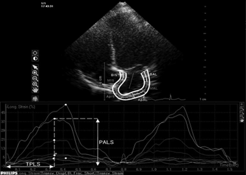
2DSTI tracking
Three sets of 2D echocardiography images of Ap4C,
apical two-chamber (Ap2C) and LV longitudinal view were obtained
and stored for offline analysis. During the QLAB software (version
8.1; Philips Ultasound, Bothell, WA, USA) analysis, samples were
obtained at anterior, posterior, lateral and inferior walls of LA,
atrial septa superior, central and inferior segments [where the
superior segment was taken close to pulmonary vein (PV), the
inferior segment was extracted at the mitral valve annulus, and the
central was taken in between], and the myocardium inferior
membrane. Samples were then measured to obtain the peak strain (PS)
and TP of each segment as well as the average peak value (4).
Statistical analysis
SPSS 13.0 (SPSS, Chicago, IL, USA) was used to
analyze the data (expressed as mean ± standard deviation). One-way
ANOVA was used to obtain P-values. P<0.05 was considered to
indicate a statistically significant result.
Results
Volunteers
Variances of height, weight, cardiac rate, LVED and
LVES diameters and LVEF of the healthy volunteers from the three
groups did not show statistical significance
(Pave>0.05). Compared with the young adult group, the
mitral E/A of the middle-aged and elderly groups decreased. By
contrast, Epeak deceleration increased, demonstrating a significant
variance (P<0.05; Table I).
 | Table IComparison of the three age
groups. |
Table I
Comparison of the three age
groups.
| Variable | Young adult | Middle-aged | Elderly |
|---|
| Age (years), mean ±
SD | 32.46±8.46 | 54.01±5.63a |
70.40±4.76a,b |
| Male, n (%) | 30 (57.7) | 29 (59.2) | 21 (51.2) |
| Height (cm), mean ±
SD | 169.13±3.92 | 168.73±4.19 | 167.20±3.21 |
| Weight (kg), mean ±
SD | 60.53±4.94 | 60.60±3.15 | 61.46±4.29 |
| Cardiac rate (bpm),
mean ± SD | 69.33±1.54 | 71.01±3.02 | 70.11±2.43 |
| LVEDD (mm), mean ±
SD | 44.86±2.82 | 44.93±2.68 | 44.93±2.78 |
| LVESD (mm), mean ±
SD | 27.06±1.43 | 26.33±1.34 | 26.88±1.74 |
| LVEF (%), mean ±
SD | 61.01±3.61 | 60.93±4.39 | 59.93±4.35 |
| Mitral E/A, mean ±
SD | 1.56±0.06 | 1.21±0.11a |
0.77±0.04a,b |
| Epeak
deceleration (msec), mean ± SD | 171.93±5.39 | 206.73±8.15a |
229.26±5.16a,b |
LA strain and TP changes in healthy
volunteer segments
The LA strain changes in the inferior segments of
each group were higher than the results of the central segments.
The changes in the central segments were higher than the results of
the superior segments (P<0.05). The strains of the inferior
segments of the elderly group decreased as age increased compared
with the young adult and middle-aged groups. The central segments
of the elderly group were lower than the results of the young adult
and middle-aged groups. The young adult and middle-aged groups
showed no significant variance. No statistical significance was
observed in the superior segment strains of the elderly group as
with the other two groups (Table
II, Figs. 1 and 2).
| Table IIComparisons of strain and TP of the
three age groups. |
Table II
Comparisons of strain and TP of the
three age groups.
| Segment | Young adult
| Middle-aged
| Elderly
|
|---|
| Strain (%) | Time to peak
(msec) | Strain (%) | Time to peak
(msec) | Strain (%) | Time to peak
(msec) |
|---|
| Inferior | 42.2±7.4 | 389.0±20.0 | 27.1±4.5a | 422.6±14.0 | 25.5±5.5a,b |
482.8±84.1a,b |
| Central | 27.8±7.1 | 368.5±20.5 | 25.3±3.8 | 422.1±37.1a | 16.3±0.4a,b | 436.8±31.7a |
| Superior | 9.1±3.5 | 518.0±99.1 | 8.2±2.9 | 522.0±56.4 | 9.6±3.4 | 526.1±11.8 |
As age increased, LA TP of the inferior segment of
each wall had a longer duration, which was observed in the elderly
group but not with the other two groups (P<0.05). The central
segments of the elderly and middle-aged groups were longer than the
young adult group (P<0.05). No statistical significance was
observed between the middle-aged and elderly groups (P>0.05) as
well as in the changes in the superior segment (P>0.05; Table II, Figs. 1 and 2).
Discussion
LA augments the ventricular reservoir function
during atrial contraction, conduit function of early diastole and
booster pump function at the end diastole (1). Regardless of physical conditions, LA
modulates the LV filling and maintains the normal stroke volume
during diastolic dysfunction (5).
The conventional LA function measurement has several
approaches (6–8). The most common approaches are tissue
Doppler imaging (TDI) and strain rate imaging, which have gained
acceptance in the medical field (9,10).
These techniques measure the velocity of myocardial motion with
accuracy in myocardial diastolic and systolic activities within the
cardiac cycle and time and spatial resolution. The surrounding
segment and cardiac motions do not affect the results (11). However, these techniques are
sensitive to angle-Doppler offset, and require small angle attacks
between the acoustic beam and the direction of myocardial motion
during measurement. 2DSTI is the latest technology based on the
strain and strain rate imaging. The image generated by STI
comprises thousands of pixels or acoustic speckles that cover the
myocardium uniformly, and form synchronously with myocardial
mechanics with no distortion to adjacent images. The STI technique
tracks every speckle, calculates the kinetic trajectory in
successive frames and provides measurements of velocity, strain and
strain rate of the heart tissue. This technique does not use the
Doppler principle, therefore, is not sensitive to the angles of
attack. 2DSTI provides the longitudinal abnormal motion and the
radial and annular activities of the heart, and offers more
advantages than the TDI.
LV relaxation and compliance prior to the atrial
systole decrease as age increases (12,13),
i.e., early peak velocity of the E wave of the mitral valve
diastole will drop and the end diastole A wave will increase as
measured by a pulse Doppler. In this study, the strain value
decreased and TP increased as age increased. These conditions may
lead to the possible correlation with the decrease of myocardial
elasticity caused by the myocardial stiffness induced by the
interstitial fibrosis that resulted from the increase of myocardial
interstitial collagen synthesis. In addition, the decrease of LA
inferior segment strain and the longer TP were more obvious in the
elderly group than in the young adult and middle-aged groups. This
condition may be associated with the close location to the mitral
annulus, in which the superior segment did not show significant
variance for the three groups. This case may be associated with its
close location to the PV entrance and far location from the mitral
annulus, which was affected less by the LV compliance.
Telagh et al discovered that being closer to
the mitral annulus leads to a higher peak velocity of the LA
(14). The top of the LA is fixed
and not involved in the atrial movement and active contraction. In
all three groups of this study, the strain of the inferior segment
was higher than the central segment, while the central segment was
higher than the superior segment. These findings confirm the
results of Telagh et al (14). The
strain of the inferior segment of the elderly group was markedly
lower than the results of the other groups, whereas the central and
superior segments did not demonstrate significance. This condition
might be associated with the decrease of the LV relaxation and
compliance of the elderly volunteers. LA function changes caused by
age increase require further study since this study only confirmed
that the LA function examination must consider the age factor.
LA function measurements provided novel insight and
has prognostic significance for several cardiovascular diseases.
STI technique provides an improved sensitivity and conventional
measurement of LA function. The proposed technique supported a
regular and comprehensive method of examining LA function, assisted
early monitoring of sub-clinical diseases and provided useful
auxiliary information to clinicians for heart examination.
References
|
1
|
Stefanadis C, Dernellis J and Toutouzas P:
A clinical appraisal of left atrial function. Eur Heart J.
22:22–36. 2001. View Article : Google Scholar : PubMed/NCBI
|
|
2
|
Di Salvo G, Drago M, Pacileo G, et al:
Atrial function after surgical and percutaneous closure closure of
atrial septal defect: a strain rate imaging study. J Am Soc
Echocardiogr. 18:930–933. 2005.PubMed/NCBI
|
|
3
|
D’Andrea A, Caso P, Romano S, et al:
Association between left atrial myocardial function and exercise
capacity in patients with either idiopathic or ischemic dilated
cardiomyopathy: a two-dimensional speckle strain study. Int J
Cardiol. 132:354–363
|
|
4
|
Cameli M, Caputo M, Mondillo S, et al:
Feasibility and reference values of left atrial longitudinal strain
imaging by two-dimensioal speckle tracking. Cardiovascular
ultrasound. 7:62009. View Article : Google Scholar : PubMed/NCBI
|
|
5
|
Kono T, Sabbah HN, Rosman H, Alam M, Stein
PD and Goldstein S: Left atrial contribution to ventricular filling
during the course of evolving heart failure. Circulation.
86:1317–1322. 1992. View Article : Google Scholar : PubMed/NCBI
|
|
6
|
Matsuda Y, Toma Y, Ogawa H, et al:
Importance of left atrial function in patients with myocardial
infarction. Circulation. 67:566–571. 1983. View Article : Google Scholar : PubMed/NCBI
|
|
7
|
Terzi S, Dayi SU, Akbulut T, et al: Value
of left atrial function in predicting exercise capacity in heart
failure with moderate to severe left ventricular systolic
dysfunction. Int Heart J. 46:123–131. 2005. View Article : Google Scholar : PubMed/NCBI
|
|
8
|
Vaturi M, Levine RA, Yosefy C, O’Neil MJ,
Picard MH and Hung J: Usefulness of left atrial emptying fraction
to predict exercise capacity in patients with normal systolic left
ventricular function and without myocardial ischemia. Am J Cardiol.
95:1014–1017. 2005. View Article : Google Scholar : PubMed/NCBI
|
|
9
|
Yip G, Abraham T, Belohlavek M and
Khandheria BK: Clinical applications of strain rate imaging. J Am
Soc Echocardiogr. 16:1334–1342. 2003. View Article : Google Scholar : PubMed/NCBI
|
|
10
|
Leitman M, Lysyansky P, Sidenko S, et al:
Two-dimensional strain-a novel software for real-time quantitative
echocardiographic assessment of myocardial function. J Am Soc
Echocardiogr. 17:1021–1029. 2004. View Article : Google Scholar : PubMed/NCBI
|
|
11
|
Inaba Y, Yuda S, Kobayashi N, et al:
Strain rate imaging for noninvasive functional quantification of
the left atrium: comparative studies in controls and patients with
atrial fibrillation. J Am Soc Echocardiogr. 18:729–736. 2005.
View Article : Google Scholar : PubMed/NCBI
|
|
12
|
Castro PL, Greenberg NL, Drinko J, Garcia
MJ and Thomas JD: Potential pitfalls of strain rate imaging: angle
dependency. Biomed Sci Instrum. 36:197–202. 2000.PubMed/NCBI
|
|
13
|
Sirbu C, Herbots L, D’Hooge J, et al:
Feasibility of strain and strain rate imaging for the assessment of
regional left atrial deformation: a study in normal subjects. Eur J
Echocardiogr. 7:199–208. 2006. View Article : Google Scholar : PubMed/NCBI
|
|
14
|
Telagh R, Hui W, Abd El Rahman M, Berger
F, Lange PE and Abdul-Khaliq H: Assessment of regional atrial
function in patients with hypertrophic cardiomyopathies using
tissue Doppler imaging. Pediatr Cardiol. 29:301–308. 2008.
View Article : Google Scholar : PubMed/NCBI
|