Introduction
Hawk tea is a beverage with a long history of use in
southwest China, particularly in Chongqing, Sichuan and Guizhou
(1). The utilization of Hawk tea
has surpassed that of green tea for a long time. Hawk tea is made
from a plant that belongs to the Litsea coreana Levl. var.,
its full scientific name being Litsea coreana Levl. var.
lanuginose(2). Litsea
coreana leve is a traditional Chinese medicine which has been
recorded in ‘Ben Cao Gang Mu’ (the Compendium of Materia Medica, a
classical traditional Chinese medicine book) that has traditionally
been used as a hypolipidemic drug in southern China for hundreds of
years (3). Certain studies have
demonstrated that Hawk tea possesses antioxidant, hyperglycemic,
hypolipemic and anti-inflammatory effects (1).
Numerous lines of evidence have suggested oxidative
stress and inflammation in the etiology of liver disease,
cardiovascular disease and cancer (4,5). As
a result, carbon tetrachloride (CCl4), which produces
reactive-free radicals when metabolized, has been widely used as a
solvent to develop hepatic damage in animal models (6). CCl4 is able to increase
lipid peroxidation on the cell membrane and alter enzyme activity,
thereby inducing hepatic injury and necrosis (7).
The release of aspartate aminotransferase (AST) and
alanine aminotransferase (ALT) into the blood has been shown to
increase as cell membranes of injured or dying hepatocytes lose
their integrity (8). AST is
released into the serum in proportion to cell damage and is most
elevated in the acute phase of cell necrosis. ALT is released early
in liver damage and remains elevated (9). Lactate dehydrogenase (LDH) is of
greatest use in monitoring liver injury disorders (10). Cytokines, including IL-6, IFN-γ and
TNF-α, are small proteins that are produced and released from
numerous cells under certain physiological and pathological
conditions. Cytokines may be centralized around this organ as it
hosts cells that are highly susceptible to the action of the
proteins (11). Unlike sustained
hepatocellular damage, acute hepatic damage is temporary and ends
with the return of normal liver histology and function. A stress
situation may be induced in hepatocytes by hypatotoxins with a
subsequent release of chemokines followed by the accumulation of
inflammatory cells and hepatocellular damage (12). Blood assays of ALT, AST, LDH and
cytokines are an important standard measure of the hepatic damage
levels.
The iNOS, COX-2, IL-lβ and TNF-α genes are
responsible for the hepatic damage and deleterious effects in the
liver caused as by a response to inflammatory stimuli (13,14).
In the present study, the in vivo
anti-inflammatory effects of Hawk tea were investigated. Serum
assays of AST, ALT, LDH, TG (triglyceride), TC (total cholesterol)
levels and inflammation-related cytokines and examinations of liver
tissue histology and inflammation-related gene expression were used
to determine the preventative effects of Hawk tea against
CCl4-induced hepatic damage in Sprague-Dawley (SD)
rats.
Materials and methods
Preparations of Hawk tea
Hawk tea was purchased in a local market (Yuzhong
District, Chongqing City, China) and stored at −80°C prior to being
freeze-dried to produce a powder. A 20-fold volume of methanol was
added to the powdered samples and then extracted twice by stirring
overnight. The methanol extract was evaporated using a rotary
evaporator (N-1100; Eywla, Tokyo, Japan), concentrated and then
dissolved in dimethyl sulfoxide (DMSO; Amresco, Solon, OH, USA) to
adjust to the stock concentration (20%, w/v).
Induction of hepatic damage
Male SD rats (n=60, 7-weeks-old) were purchased from
the Experimental Animal Center of Chongqing Medical University
(Chongqing, China). The animals were maintained in a temperature
controlled facility (temperature 25±2°C, relative humidity 50±5%)
with a 12-h light/dark cycle and free access to a standard rat diet
and water.
To investigate the preventive effects of Hawk tea
against CCl4-induced hepatic damage, the animals were
divided into six groups consisting of 10 rats each. The
experimental design was as follows: the normal control group were
administered with distilled water for 14 days and a single dose of
vehicle [2 ml/kg body weight (bw) olive oil, p.o.], while the
CCl4 control group received a 14-day repeated oral
administration of distilled water followed by a single
administration of CCl4 (2 ml/kg bw dissolved in olive
oil, 1:1, v/v) on the last day to induce liver damage. The three
Hawk tea + CCl4 groups received 100, 200 or 400 mg/kg bw
of Hawk tea and the positive control group received 2 weeks of 100
mg/kg bw silymarin dissolved in water, with hepatic damage induced
in the same manner as that mentioned above. The rats were
anesthetized 24 h after the administration of CCl4 and
sacrificed using CO2(15). Blood and livers were collected and
preserved at −70°C until biological assays were performed. These
experiments followed a protocol approved by the Animal Ethics
Committee of Chongqing Medical University (Chongqing, China).
AST, ALT and LDH levels in serum
The AST and ALT levels of the serum were determined
using commercially available kits (Shanghai Institute of Biological
Products Co., Ltd., Shanghai, China). LDH levels of the serum were
determined using specific commercially available kits (Cayman
Chemical Company, Ann Arbor, MI, USA).
ELISA analysis of inflammation-related
cytokines in serum
For the serum cytokine assays, blood from the
inferior vena cava was collected into tubes and centrifuged (3,000
rpm, 10 min, 4°C). The serum was then aspirated. The serum
concentrations of the inflammatory-related cytokines IL-6, IFN-γ
and TNF-α (Biolegend, San Diego, CA, USA) were measured by ELISA
according to the manufacturer’s protocol. Briefly, following the
addition of the biotinylated antibody reagent to the 96-well
plates, the supernatants of the homogenized serum were incubated at
37°C in CO2 for 2 h. Subsequent to washing with PBS,
streptavidin-peroxidase (HRP) solution was added and the plate was
incubated for 30 min at room temperature. The absorbance was
measured at 450 nm using a microplate reader (16).
Histological examination of the liver
tissue
At the end of the experimental period, the liver
tissues were removed and cleaned in saline to remove any residue.
For the histological investigations, the liver tissues were fixed
in 10% (v/v) buffered formalin for 24 h, cut into 2 longitudinal
halves and then embedded into paraffin. Paraffin sections 4
μm-thick were stained with hematoxylin and eosin (H&E)
prior to microscopic observation (BX41, Olympus, Tokyo, Japan)
(17).
Reverse transcription-polymerase chain
reaction (RT-PCR) of inflammation-related gene expressions in the
liver tissue
Total RNA was isolated from the liver tissue using
TRIzol reagent (Invitrogen, Carlsbad, CA, USA) according to the
manufacturer’s recommendations. The RNA was digested with
RNase-free DNase (Roche, Basel, Switzerland) for 15 min at 37°C and
purified using a RNeasy kit (Qiagen, Hilden, Germany) according to
the manufacturer’s instructions. cDNA was synthesized from 2
μg total RNA through incubation at 37°C for l h with AMV
reverse transcriptase (GE Healthcare, Little Chalfont, UK) and
random hexanucleotides, according to the manufacturer’s
instructions.
The primers used to specifically amplify the genes
of interest were: iNOS forward: 5′-AGA GAG ATC GGG TTC ACA-3′ and
reverse: 5′-CAC AGA ACT GAG GGT ACA-3′; COX-2 forward: 5′-TTA AAA
TGA GAT TGT CCG AA-3′ and reverse: 5′-AGA TCA CCT CTG CCT GAG
TA-3′; TNF-α forward: 5′-GAC CCT CAG ACT CAG ATC ATC CTT CT-3′ and
reverse: 5′-ACG CTG GCT CAG CCA CTC-3′; IL-1β forward: 5′-CTC CAT
GAG CTT TGT ACA AGG-3′ and reverse: 5′-TGC TGA TGT ACC AGT TGG
GG-3′. The internal control gene of GAPDH was amplified using the
primers: forward: 5′-CGG AGT CAA CGG ATT TGG TC-3′ and reverse:
5′-AGC CTT CTC CAT GGT CGT GA-3′. Amplification was performed in a
thermal cycler (Eppendorf, Hamburg, Germany) with cycles of
denaturation. The amplified PCR products were run in 1.0% agarose
gels and visualized by ethidium bromide (EtBr) staining (18).
Protein extraction and western blot
analysis in the liver tissue
Total liver tissue protein was obtained with RIPA
buffer as described by Kim et al(19). Protein concentrations were
determined with a Bio-Rad protein assay kit (Hercules, CA, USA).
For the western blot analysis, aliquots of the lysate containing
30–50 μg protein were separated by sodium dodecyl
sulfate-polyacrylamide gel electrophoresis (SDS-PAGE) and then
electrotransferred onto a nitrocellulose membrane (Schleicher and
Schuell, Keene, NH, USA). The membranes were subjected to
immunoblot analysis and the proteins were visualized by an enhanced
chemiluminescence (ECL) method (GE Healthcare). The cell lysates
were separated by 12% SDS-PAGE, transferred onto a polyvinylidene
fluoride membrane (GE Healthcare), blocked with 5% skimmed milk and
hybridized with primary antibodies (diluted 1:1,000). The
antibodies against iNOS, COX-2, TNF-α and IL-1β were obtained from
Santa Cruz Biotechnology Inc. (Santa Cruz, CA, USA), then incubated
with the horseradish peroxidase-conjugated secondary antibody
(Santa Cruz Biotechnology Inc.) for 1 h at room temperature. The
blots were washed three times with PBS-T and then developed by
enhanced chemiluminescence (Amersham Life Science, Arlington
Heights, IL, USA).
Statistical analysis
Data were presented as the mean ± SD. Differences
between the mean values for individual groups were assessed using a
one-way ANOVA with Duncan’s multiple range test. P<0.05 was
considered to indicate a statistically significant difference. SAS
version 9.1 (SAS Institute Inc., Cary, NC, USA) was used for
statistical analyses.
Results
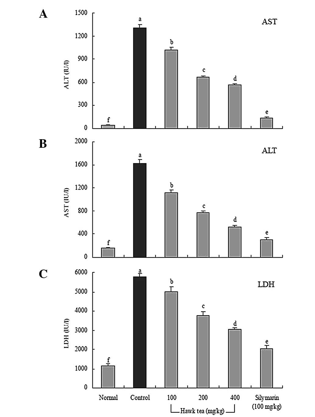
Effect of Hawk tea on serum levels of
AST, ALT and LDH
The AST level of normal rats was 155.5±15.6 IU/l,
however, the level in the CCl4 control rats was
significantly increased to 1623.7±67.6 IU/l. The levels of AST in
the 100, 200 or 400 mg/kg Hawk tea groups were 1121.3±44.2,
765.6±32.3 and 524.6±31.2 IU/l, respectively. The level of AST in
rats treated with silymarin (100 mg/kg) decreased to 303.0±37.3
IU/l, which was lower than the level in rats treated with Hawk tea
(Fig. 1A).
The ALT level in the normal group was 43.5±6.5 IU/l,
whereas that of the control group was 1308.8±44.2 IU/l, reflecting
a marked increase. The ALT levels in the 100 and 200 mg/kg Hawk tea
groups decreased to 1024.5±31.2 and 667.8±18.5 IU/l, respectively.
A high concentration (400 mg/kg) of Hawk tea treatment resulted in
a further decreased ALT level (570.6±14.5; Fig. 1B). Thus, from the results of serum
levels of AST and ALT, it was confirmed that Hawk tea generally
inhibited hepatic damage induced by CCl4, although the
400 mg/kg Hawk tea group demonstrated this to the best effect.
The levels of LDH in the 100 and 200 mg/kg Hawk tea
groups were 5000.1±258.6 and 3785.2±179.5 IU/l, respectively, which
were slightly lower than the level of the control group
(5784.2±145.6 IU/l). However, LDH levels in the 400 mg/kg and
silymarin groups were 3033.6±88.8 and 2023.6±194.5 IU/l,
respectively, whereas the normal group showed the lowest level at
1155.8±98.6 IU/l (Fig. 1C). The
LDH levels in the Hawk tea groups were lower than those of the
control group, similar to the observations of the AST and ALT
levels.
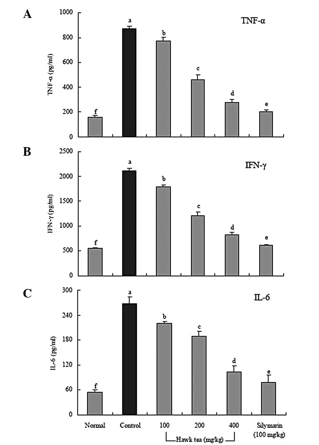
Effect of Hawk tea on serum levels of
IL-6, IFN-γ and TNF-α
Serum IL-6, IFN-γ and TNF-α levels in rats in the
100 and 200 mg/kg Hawk tea-treated groups were significantly lower
than those of the control group (Fig.
2). The reductions observed in the IL-6 and IFN-γ levels of the
400 mg/kg Hawk tea group were 78.0% and 82.4%, respectively,
compared with the control group. The TNF-α level in rats treated
with 400 mg/kg Hawk tea also decreased by 83.8%. However, the
levels of these proinflammatory cytokines in rats treated with
silymarin were similar to those of rats in the normal group.
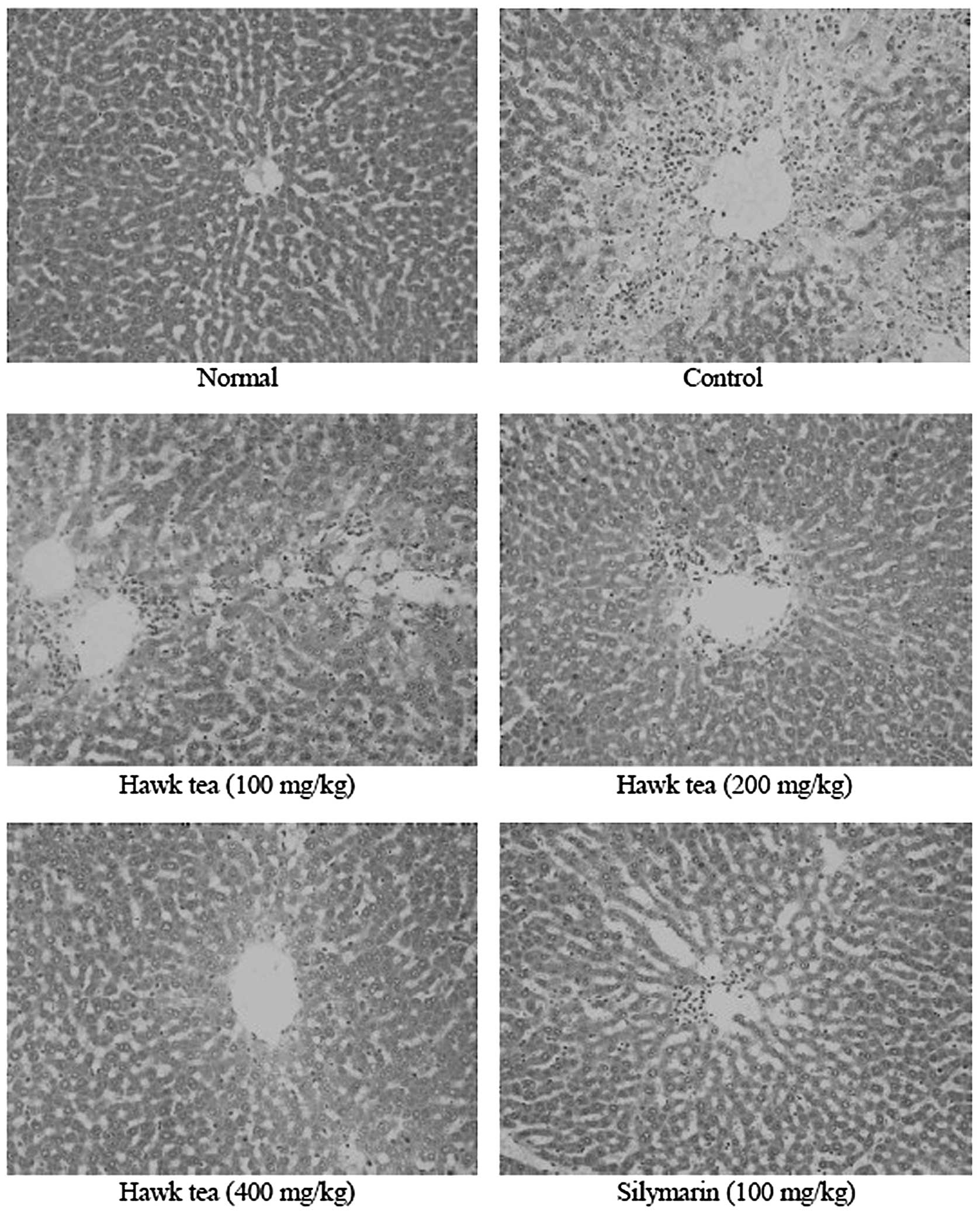
Effect on the liver tissues in rats with
CCl4-induced hepatic damage
The sections from the mice in the control group
showed widespread areas of congestion and hemorrhages in the
centrilobular zone, as well as necrosis involving all the
hepatocytes in the centrilobular zone (moderate grade 3). There was
also bridging of the areas of necrosis (Fig. 3). The sections from the silymarin
(100 mg/kg)-treated group did not show any evident hepatic damage,
but were also different from normal (moderate grade 0). The 100
mg/kg Hawk tea group showed moderate congestion and hemorrhages in
the area around the centrilobular vein and extending into the
midzonal cells (mild grade 2), with the majority of lobules being
affected. Areas of confluent necrosis were limited to the liver
cells surrounding the centrilobular vein. The tissue sections of
the 200 and 400 mg/kg Hawk tea groups appeared less like normal
tissue (grade 1). The livers showed minimal congestion and necrosis
of single hepatocytes limited to the area immediately around the
centrilobular vein, with many of the lobules not being affected.
The 200 mg/kg Hawk tea group showed mild inflammatory infiltration,
whereas the 400 mg/kg Hawk tea group did not show any inflammatory
infiltration. These results demonstrate that a higher concentration
of Hawk tea decreased the degree of inflammation.
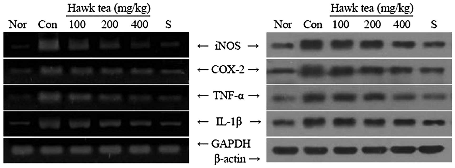
Effects of Hawk tea on mRNA and protein
expression levels of iNOS, COX-2, TNF-α and IL-1β in the liver
tissues of rats treated with CCl4
RT-PCR and western blot analyses were conducted to
investigate whether the inhibitory effect of the Hawk tea on
inflammation was due to transcriptional regulation of inflammatory
mediators in the liver, including iNOS, COX-2, TNF-α and IL-1β.
CCl4 treatment significantly increased the mRNA and
protein levels of these inflammatory mediators (Fig. 4). However, 400 mg/kg Hawk tea
significantly decreased the mRNA and protein expression levels of
iNOS, COX-2, TNF-α and IL-1β, similar to the silymarin group
(P<0.05). Liver tissues from the 100 and 200 mg/kg Hawk
tea-treated rats showed a decreased expression of these genes
compared with the control group.
Discussion
AST and ALT are enzymes located in liver cells that
leak out into the general circulation when the cells are injured.
AST is located in many body tissues, including the heart, muscle,
kidney, brain and lung tissues. ALT is located predominately in the
liver, with lesser quantities located in the kidneys, heart and
skeletal muscles (20). LDH is an
enzyme located in numerous body tissues, including the liver where
elevated levels of LDH may indicate liver damage. AST and ALT
levels have also been used to evaluate the hepatoprotective
activities of medicinal plants in CCl4-induced hepatic
damage in animal models (21,22).
Raju et al(23) reported
that the serum AST and ALT levels in CCl4-treated rats
was markedly increased compared with those of rats in the normal
group, which indicated that liver damage was significantly induced
by CCl4. Tea is a beverage consumed worldwide and
regarded as a healthy source of pleasure. Green tea was originally
recommended in traditional Chinese medicine and has gained
considerable attention due to its preventive effects against
CCl4-induced hepatic damage (24). Hawk tea and green tea were
important beverages in southwest China for hundreds of years. Hawk
tea contains a higher content of catechins, minerals and flavonoid
compounds (25). Drinking Hawk tea
for a long period of time is believed to keep a person mentally and
physically healthy. Certain studies have demonstrated that Hawk tea
has anti-inflammatory effects (1).
Silymarin is a unique flavonoid complex, with both in vitro
and animal studies suggesting that silymarin possesses
hepatoprotective properties that protect liver cells against toxins
(26). In the present study, Hawk
tea and silymarin had an in vivo preventive effect on
CCl4-induced hepatic damage.
The serum levels of cytokines, including IL-6,
TNF-α, IL-1β and IFN-γ, in patients with inflammatory diseases are
higher than those in healthy people (27). Thus, lower levels of IL-6, IFN-γ
and TNF-α are indicative of improved anti-inflammatory effects.
Hawk tea has consequently demonstrated a good protective effect
against hepatic damage in studies thus far. Hepatocytes bear a
variety of cytokine receptors and the inflammatory cytokines IL-6,
IFN-γ and TNF-α play pathogenic roles in liver disease (28). Although systemic IL-6 levels are
elevated following traumatic hemorrhage, hepatocellular function is
impaired and liver injury occurs (29). IFN-γ increases such injury by
stimulating hepatic inflammation and aggravating liver damage
(30). TNF-α is also a key
mediator in a number of experimental liver injury models (31).
Histopathological analysis is an important clinical
standard used to diagnose hepatic damage (32). In addition, the histopathological
examination of rat liver sections is reported as an effective
method to check the hepatoprotective activity against the
CCl4-induced hepatic damage in the rat model (33). From the sections examined in the
present study, Hawk tea was observed to exert a preventive effect
against CCl4-induced hepatic damage. Hawk tea exhibits
antioxidative effects (1).
Compounds with antioxidant properties may also have
anti-inflammatory effects and actually prevent the activation of
inflammatory signals (34).
The present study demonstrated that Hawk tea was
effective in the prevention of CCl4-induced hepatic
damage in SD rats. The results show that the hepatoprotective
effects of Hawk tea may be due to decreased serum levels of AST,
ALT, LDH and proinflammatory cytokines, including TNF-α, IL-6 and
IFN-γ. Histopathological studies also showed that Hawk tea was able
to prevent CCl4-induced inflammation in the liver.
Furthermore, mRNA and protein expression levels of
inflammation-related genes in the liver, including iNOS, COX-2,
TNF-α and IL-1β were significantly reduced in the Hawk tea-treated
rats (P<0.05). These results suggest that Hawk tea is
potentially useful in the treatment or prevention of
chemical-induced hepatic damage in vivo. The present study
demonstrated that Hawk tea was effective in the prevention of
CCl4-induced hepatic damage in SD rats.
References
|
1.
|
Ji HF, Nan HJ, Zhang LW, Yang MD and Zhang
H: Extraction and antioxidant properties of total flavonoids from
Litsea Coreana L. China Food Addit. 2:121–125. 2011.
|
|
2.
|
Shen JZ, Ye M, Shu AQ, Yin KQ and Yang L:
Extraction and purification of polyphenolic compounds and
determination of EGCG from eagle tea (Litsea coreana Lévl
var. lanuginosa). J Zhejiang Univ (Agric and Life Sci).
36:329–334. 2010.
|
|
3.
|
Liu B: Simultaneous quantification of
seven flavonoids in eagle tea using pressurized liquid extraction
and HPLC. Chin J Pharm Anal. 30:1424–1427. 2010.
|
|
4.
|
Seitz HK and Stickel F: Risk factors and
mechanisms of hepatocarcinogenesis with special emphasis on alcohol
and oxidative stress. Biol Chem. 387:349–360. 2006. View Article : Google Scholar : PubMed/NCBI
|
|
5.
|
Cauwels A and Brouckaert P: Survival of
TNF toxicity: dependence on caspases and NO. Arch Biochem Biophys.
462:132–139. 2007. View Article : Google Scholar : PubMed/NCBI
|
|
6.
|
Rechnagel RO and Glende EA Jr: Carbon
tetrachloride hepatotoxicity: an example of lethal cleavage. CRC
Crit Rev Toxicol. 2:263–297. 1973. View Article : Google Scholar : PubMed/NCBI
|
|
7.
|
Weber LW, Boll M and Stampfl A:
Hepatotoxicity and mechanism of action of haloalkanes: carbon
tetrachloride as a toxicological model. Crit Rev Toxicol.
33:105–136. 2003. View Article : Google Scholar : PubMed/NCBI
|
|
8.
|
Raoul JL, Bruix J, Greten TF, Sherman M,
Mazzaferro V, Hilgard P, Scherubl H, Scheulen ME, Germanidis G,
Dominguez S, Ricci S, Nadel A, Moscovici M, Voliotis D and Llovet
JM: Relationship between baseline hepatic status and outcome, and
effect of sorafenib on liver function: SHARP trial subanalyses. J
Hepatol. 56:1080–1088. 2012. View Article : Google Scholar : PubMed/NCBI
|
|
9.
|
Pal M and Ghosh M: Studies on comparative
efficacy of α-linolenic acid and α-eleostearic acid on prevention
of organic mercury-induced oxidative stress in kidney and liver of
rat. Food Chem Toxicol. 50:1066–1072. 2012.
|
|
10.
|
Small DM and Gobe GC: Cytochrome c:
potential as a noninvasive biomarker of drug-induced acute kidney
injury. Expert Opin Drug Metab Toxicol. 8:655–664. 2012. View Article : Google Scholar : PubMed/NCBI
|
|
11.
|
Ramadori G and Armbrust T: Cytokines in
the liver. Eur J Gastroenterol Hepatol. 13:777–784. 2001.
View Article : Google Scholar : PubMed/NCBI
|
|
12.
|
Diehl AM: Cytokine regulation of liver
injury and repair. Immunol Rev. 174:160–171. 2000. View Article : Google Scholar : PubMed/NCBI
|
|
13.
|
Suliburk JW, Helmer KS, Gonzalez EA,
Robinson EK and Mercer DW: Ketamine attenuates liver injury
attributed to endotoxemia: role of cyclooxygenase-2. Surgery.
138:134–140. 2005. View Article : Google Scholar : PubMed/NCBI
|
|
14.
|
de Waal RM: The anti-inflammatory activity
of glucocorticoids. Mol Biol Rep. 19:81–88. 1994.PubMed/NCBI
|
|
15.
|
Park CM, Youn HJ, Chang HK and Song YS:
TOP1 and 2, polysaccharides from Taraxacum officinale,
attenuate CCl(4)-induced hepatic damage through the modulation of
NF-κB and its regulatory mediators. Food Chem Toxicol.
48:1255–1261. 2010.PubMed/NCBI
|
|
16.
|
Park HS, Park JY and Yu R: Relationship of
obesity and visceral adiposity with serum concentrations of CRP,
TNF-α and IL-6. Diabetes Res Clin Pract. 69:29–35. 2005.
|
|
17.
|
Melgar S, Karlsson L, Rehnström E,
Karlsson A, Utkovic H, Jansson L and Michaëlsson E: Validation of
murine dextran sulfate sodium-induced colitis using four
therapeutic agents for human inflammatory bowel disease. Int
Immunopharmacol. 8:836–844. 2008. View Article : Google Scholar : PubMed/NCBI
|
|
18.
|
Bak SS, Kong CS, Rhee SH, Rho CW, Kim NK,
Choi KL and Park KY: Effect of sulfur enriched young radish kimchi
on the induction of apoptosis in AGS human gastric adenocarcinoma
cells. J Food Sci Nutr. 12:79–83. 2007. View Article : Google Scholar
|
|
19.
|
Kim YA, Rhee SH, Park KY and Choi YH:
Antiproliferative effect of resveratrol in human prostate carcinoma
cells. J Med Food. 6:273–280. 2003. View Article : Google Scholar : PubMed/NCBI
|
|
20.
|
Delić R and Štefanović M: Optimal
laboratory panel for predicting preeclampsia. J Matern Fetal
Neonatal Med. 23:96–102. 2010.PubMed/NCBI
|
|
21.
|
Ranawat L, Bhatt J and Patel J:
Hepatoprotective activity of ethanolic extracts of bark of
Zanthoxylum armatum DC in CCl4 induced hepatic damage in
rats. J Ethnopharmacol. 127:777–780. 2010. View Article : Google Scholar : PubMed/NCBI
|
|
22.
|
Surendran S, Eswaran MB, Vijayakumar M and
Rao CV: In vitro and in vivo hepatoprotective activity of
Cissampelos pareira against carbon-tetrachloride induced
hepatic damage. Indian J Exper Biol. 49:939–945. 2011.PubMed/NCBI
|
|
23.
|
Raju K, Anbuganapathi G, Gokulakrishnan V,
Rajkapoor B, Jayakar B and Manian S: Effect of dried fruits of
Solanum nigrum LINN against CCl4-induced hepatic
damage in rats. Biol Pharm Bull. 26:1618–1619. 2003.PubMed/NCBI
|
|
24.
|
Noori S, Rehman N, Qureshi M and Mahboob
T: Reduction of carbon tetrachloride-induced rat liver injury by
coffee and green tea. Pakistan J Nutr. 8:452–458. 2009. View Article : Google Scholar
|
|
25.
|
Yang XF, Han M and Hu L: Determination of
total flavonoids in Litsea coreana by spectrophotometric
method. Southwest China J Agri Sci. 24:1234–1235. 2011.
|
|
26.
|
Berger J and Kowdley KV: Is silymarin
hepatoprotective in alcoholic liver disease? J Clin Gastroenterol.
37:278–279. 2003. View Article : Google Scholar : PubMed/NCBI
|
|
27.
|
Gratacós J, Collado A, Filella X, Sanmartí
R, Cañete J, Llena J, Molina R, Ballesta A and Muñoz-Gómez J: Serum
cytokines (IL-6, TNF-α, IL-1β and IFN-γ) in ankylosing spondylitis:
a close correlation between serum IL-6 and disease activity and
severity. Br J Rheumatol. 33:927–931. 1994.
|
|
28.
|
Wang JY, Wang XL and Liu P: Detection of
serum TNF-α, IFN-β, IL-6 and IL-8 in patients with hepatitis B.
World J Gastroenterol. 5:38–40. 1999.
|
|
29.
|
Toth B, Yokoyama Y, Schwacha MG, George
RL, Rue LW III, Bland KI and Chaudry IH: Insights into the role of
interleukin-6 in the induction of hepatic injury after
trauma-hemorrhagic shock. J Appl Physiol. 97:2184–2189. 2004.
View Article : Google Scholar : PubMed/NCBI
|
|
30.
|
Knight B, Lim R, Yeoh GC and Olynyk JK:
Interferon-γ exacerbates liver damage, the hepatic progenitor cell
response and fibrosis in a mouse model of chronic liver injury. J
Hepatol. 47:826–833. 2007.
|
|
31.
|
Segner WP, Schmidt CF and Boltz JK: Effect
of sodium chloride and pH on the outgrowth of spores of type E
Clostridium botulinum at optimal and suboptimal
temperatures. Appl Microbiol. 14:49–54. 1966.PubMed/NCBI
|
|
32.
|
Girish C, Koner BC, Jayanthi S, Rao KR,
Rajesh B and Pradhan SC: Hepatoprotective activity of six
polyherbal formulations in CCl4-induced liver toxicity
in mice. Indian J Exp Biol. 47:257–263. 2009.PubMed/NCBI
|
|
33.
|
Manjrekar AP, Jisha V, Bag PP, Adhikary B,
Pai MM, Hegde A and Nandini M: Effect of Phyllanthus niruri
Linn. treatment on liver, kidney and testes in CCl4
induced hepatotoxic rats. Indian J Exp Biol. 46:514–520. 2008.
|
|
34.
|
Karlsen M, Hovden AO, Vogelsang P, Tysnes
BB and Appel S: Bromelain treatment leads to maturation of
monocyte-derived dendritic cells but cannot replace PGE2 in a
cocktail of IL-1β, IL-6, TNF-α and PGE2. Scand J Immunol.
74:135–143. 2011.PubMed/NCBI
|