Angular vessels as a new vascular pedicle of an island nasal chondromucosal flap: Anatomical study and clinical application

  • Authors:
    • Dianju Hou
    • Lin Fang
    • Zhenmin Zhao
    • Chuande Zhou
    • Mingyong Yang
  • View Affiliations

  • Published online on: December 18, 2012     https://doi.org/10.3892/etm.2012.860
  • Pages: 751-756
Metrics: Total Views: 0 (Spandidos Publications: | PMC Statistics: )
Total PDF Downloads: 0 (Spandidos Publications: | PMC Statistics: )


Abstract

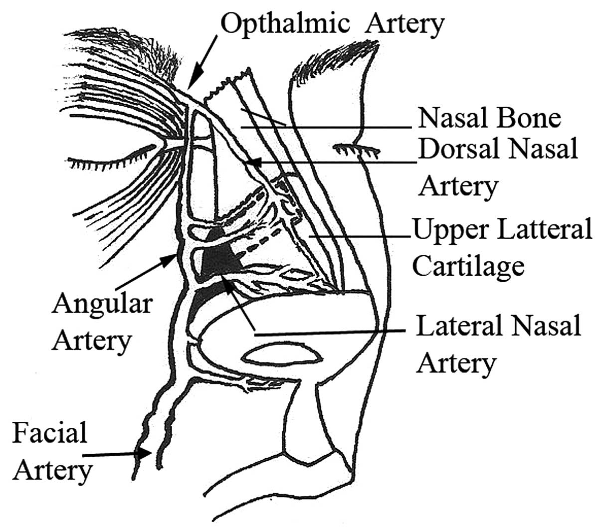
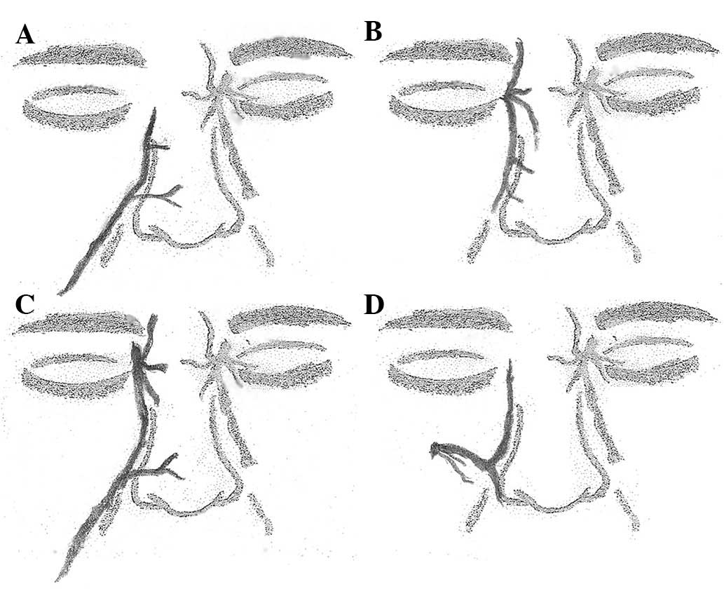
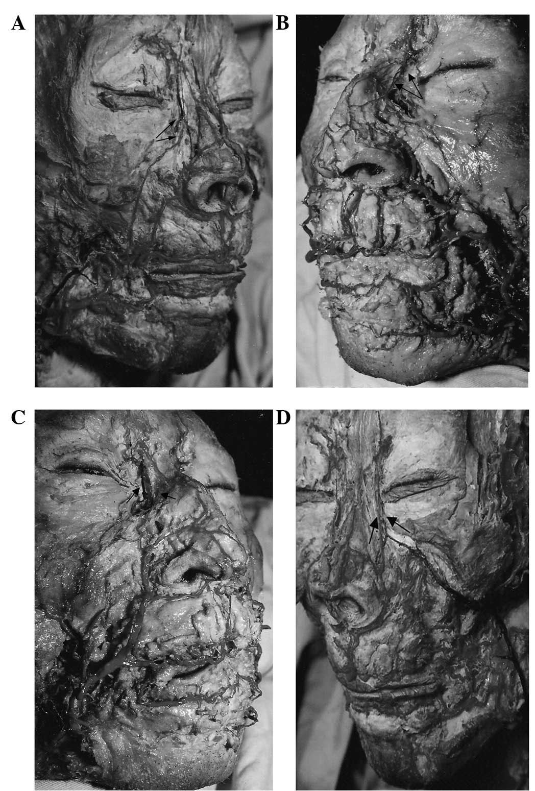
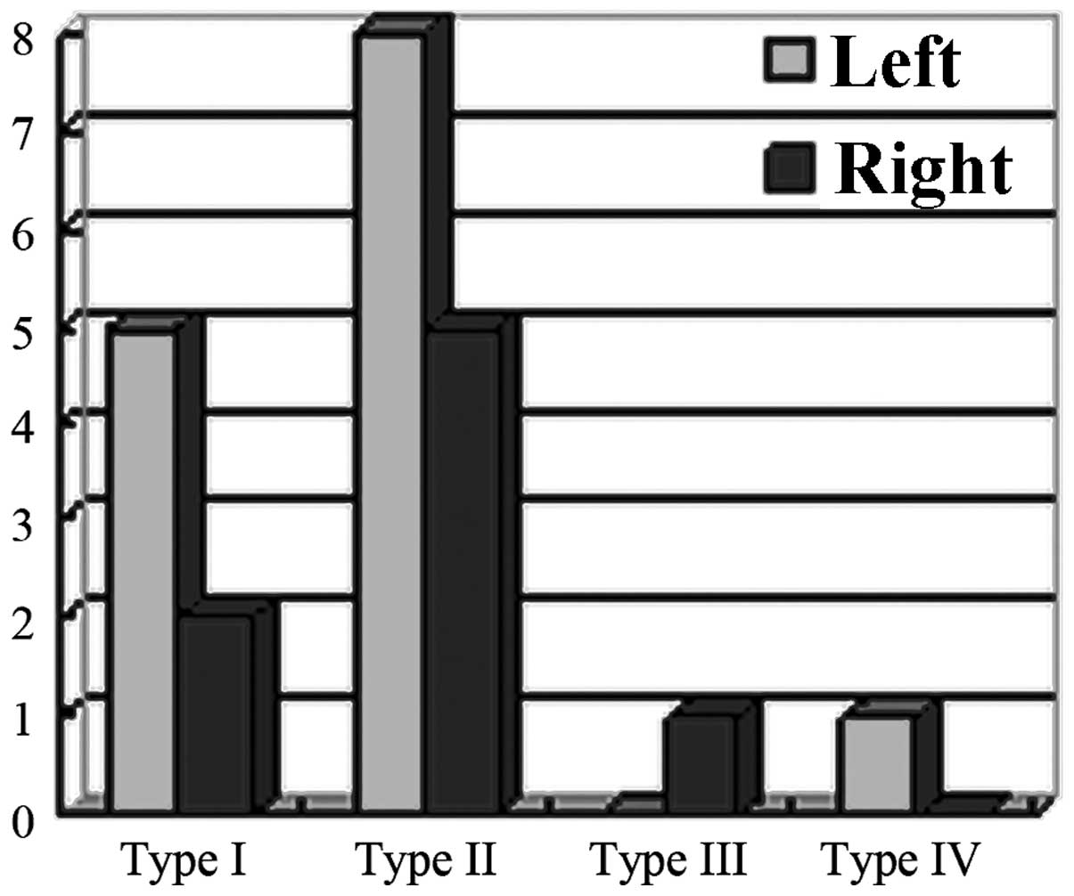
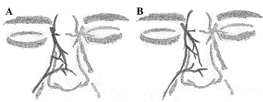
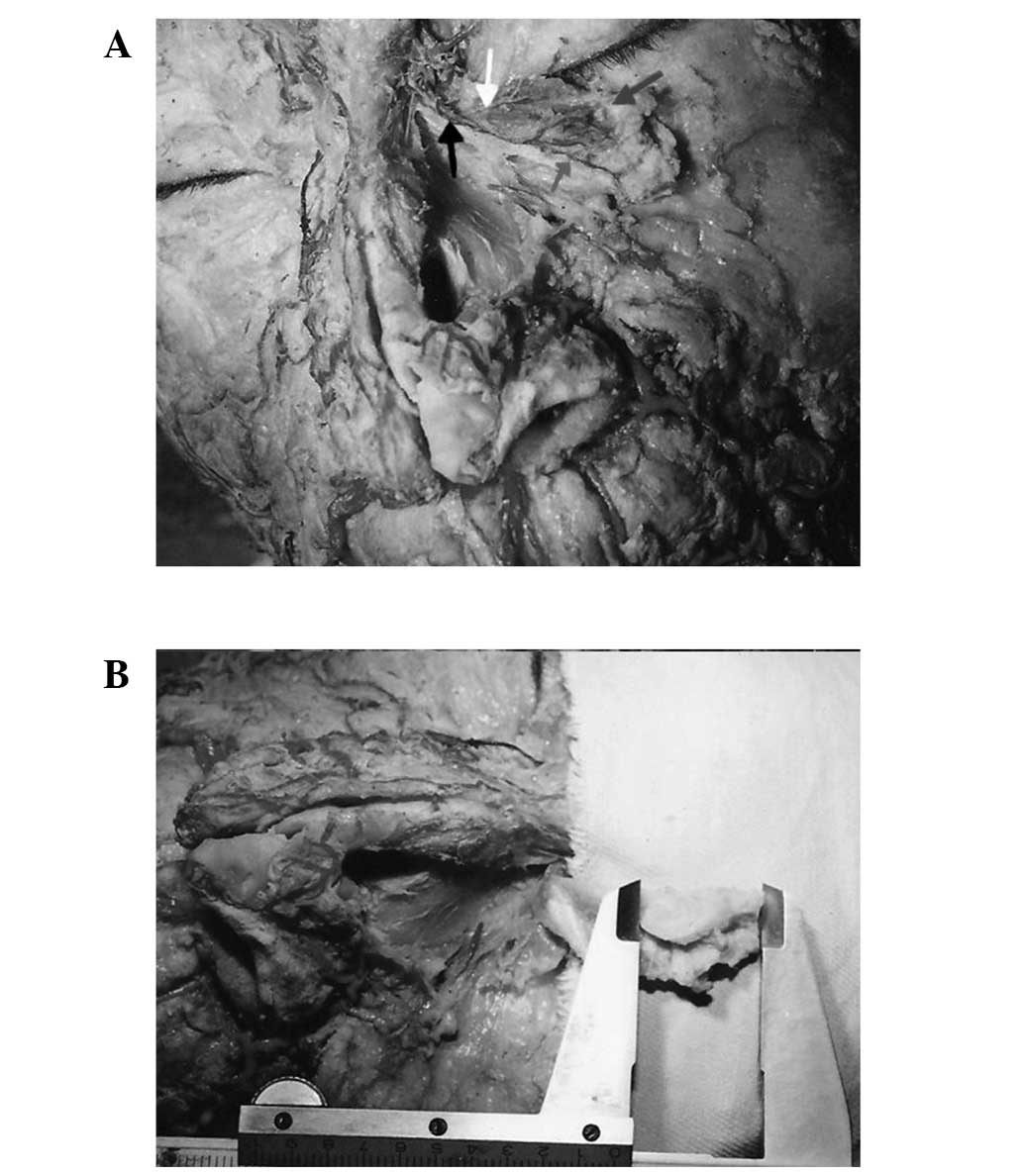
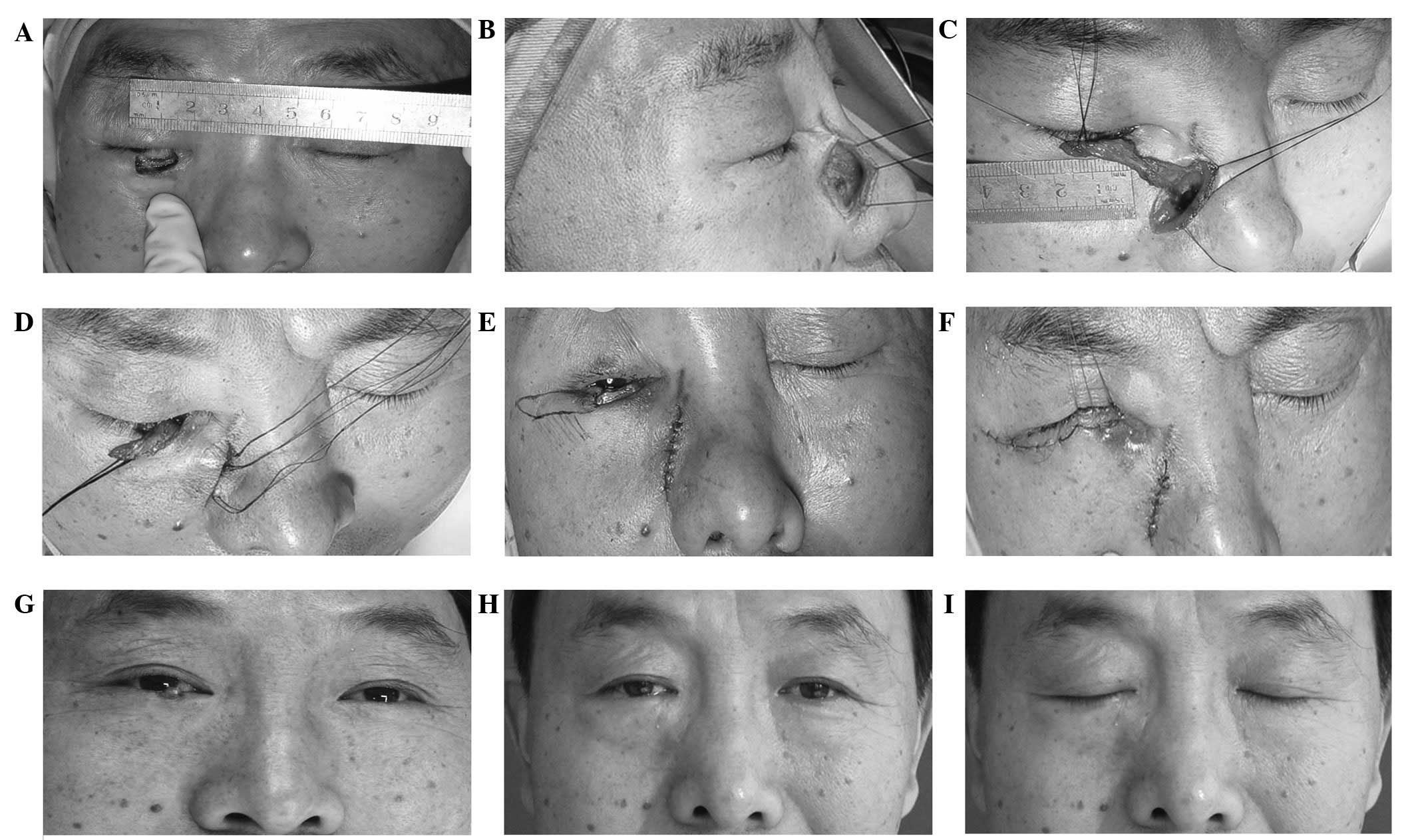
Successful eyelid reconstructions have been reported when using an axial nasal chondromucosal flap based on the dorsal nasal artery. The present study aimed to present a detailed anatomical description of the blood supply of the lateral nasal region and the angular artery, in order to propose the angular vessels as a new vascular pedicle for the island nasal chondromucosal flap. A total of 11 cadavers (22 hemi‑faces) were examined. Observations with regard to the origin, course and distribution patterns of the angular artery were recorded. Based on the anatomical study findings, the angular vessels were proposed as a vascular source for the island nasal chondromucosal flap. Observations with regard to the varying origins of the angular artery were categorized into four types. The course of the angular artery along the nasojugal fold was constant. The angular artery branched off into the upper two‑thirds of the lateral nasal region and anastomosed with the other vascular branches on the nasal dorsum. Clinically, reconstruction of a full‑thickness defect of the lower eyelid was successfully performed by using this composite flap based on the angular vessels and an adjacent orbicularis oculi myocutaneous flap. Satisfactory esthetic outcomes were obtained for the donor and recipient sites. The angular artery is a good vascular source for an island nasal chondromucosal flap. The flap may be created safely and successfully in clinic. Island nasal chondromucosal flaps and nasolabial groove skin flaps based on the angular vessels may be designed simultaneously for use on full‑thickness defects of the eyelid.
View Figures
View References

Related Articles

Journal Cover

March 2013
Volume 5 Issue 3

Print ISSN: 1792-0981
Online ISSN:1792-1015

Sign up for eToc alerts

Recommend to Library

Copy and paste a formatted citation
x
Spandidos Publications style
Hou D, Fang L, Zhao Z, Zhou C and Yang M: Angular vessels as a new vascular pedicle of an island nasal chondromucosal flap: Anatomical study and clinical application. Exp Ther Med 5: 751-756, 2013
APA
Hou, D., Fang, L., Zhao, Z., Zhou, C., & Yang, M. (2013). Angular vessels as a new vascular pedicle of an island nasal chondromucosal flap: Anatomical study and clinical application. Experimental and Therapeutic Medicine, 5, 751-756. https://doi.org/10.3892/etm.2012.860
MLA
Hou, D., Fang, L., Zhao, Z., Zhou, C., Yang, M."Angular vessels as a new vascular pedicle of an island nasal chondromucosal flap: Anatomical study and clinical application". Experimental and Therapeutic Medicine 5.3 (2013): 751-756.
Chicago
Hou, D., Fang, L., Zhao, Z., Zhou, C., Yang, M."Angular vessels as a new vascular pedicle of an island nasal chondromucosal flap: Anatomical study and clinical application". Experimental and Therapeutic Medicine 5, no. 3 (2013): 751-756. https://doi.org/10.3892/etm.2012.860