Introduction
Cervical radiculopathy is a common disease
manifesting as radiated pain, weakness or numbness of the upper
extremities and stiffness or limited motion of the neck (1). However, the syndrome of cervical
radiculopathy often affects areas far beyond the innervation area
of the affected nerves and is not consistent with the magnetic
resonance imaging (MRI) findings, which makes identification of the
location of the disease and treatment difficult and may even lead
to mismanagement (1). The
explanation of this phenomenon remains unclear.
c-Fos, the protein product of immediate-early gene
(IEG) c-fos, has been widely used as a tool for the study of
neural correlates of nociception (2–5) and
as a marker for neuronal activation following noxious stimulation.
c-Jun, the protein product of another IEG c-jun, is also
reported to be a marker for neuronal activation and noxious
stimulation (6,7).
We hypothesize that there are neural pathways
between adjacent segments of the cervical spinal cord. This
hypothesis may explain the mismatch between the symptoms and the
affected spinal cord segment. In order to test our hypothesis, the
present study was designed to investigate the expression of c-Fos
and c-Jun in ipsilateral C5–7 segments following
unilateral C7 nerve root rhizotomy in rats. The findings
may offer a possible explanation for this clinical phenomenon.
Materials and methods
Animals
All experiments were performed according to the
Guidelines on Ethical Standards for Investigation of Experimental
Pain in Animals (Zimmermann, 1983). Experiments were performed on
adult male Wistar rats (220–250 g). All animals were provided by
the Experimental Animal Center of Tianjin Medical University
(Tianjin, China) and housed in groups of 2 or 3 in clear plastic
cages. Food and water were freely available during this study.
Animals were randomly divided into two groups: i) the C7
rhizotomy group (rhizotomy group, n=24), for which animals received
right C7 nerve root rhizotomy and ii) the sham-operated
group (sham group, n=24), for which animals underwent the same
surgery without right C7 nerve root rhizotomy. Each
group was subdivided into two subgroups (2 and 4 h after
surgery).
Surgery
All the animals were intraperitoneally anesthetized
with chloral hydrate (300 mg/kg). An incision was made in the
middle of the back and the skin and superficial muscle were
retracted. A laminectomy was performed from C6 to
T1. During surgery, the surgeon was extremely careful to
avoid any damage to the cervical spinal cord. The right
C7 nerve root was exposed by the operating microscope
and then broken completely. The incision was carefully closed.
c-Fos and c-Jun immunohistochemistry
At 2 and 4 h after surgery, the animals were deeply
anesthetized with an overdose of chloral hydrate and underwent
transcardial perfusion with 150 ml normal saline (4°C), followed by
4% paraformaldehyde in 0.1 M phosphate-buffered saline (PBS; pH
7.4). The C5–7 segments of the spinal cord were removed
and post-fixed in 4% paraformaldehyde containing sucrose (10%) at
4°C for 1 h and then transferred to PBS containing sucrose (30%)
overnight. Then, the spinal cord segments were serially sectioned
at 10 μm thickness in a transverse plane with a freezing
microtome, and one from every eight serial sections was collected
and processed for c-Fos and c-Jun immunohistochemistry.
For c-Fos or c-Jun immunostaining, a standard
avidin-biotin-peroxidase complex (ABC) technique was performed
using a Histostain SP kit (Boshide Biological Technology Co.,
Wuhan, China) according to the manufacturer’s instructions.
Briefly, free-floating sections were incubated overnight in a
rabbit monoclonal anti-c-Fos or c-Jun antibody diluted in PBS
(1:300). After several washes in PBS, the sections were incubated
in biotinylated goat anti-rabbit IgG diluted to 1:200 in PBS for 20
min, rinsed in PBS and incubated with avidin-biotin reagents
(1:100) for 20 min at room temperature. After three washes in
Tris-buffered saline (TBS), sections were developed in
3,3′-diaminobenzidine tetrahydrochloride solution containing 0.05%
H2O2 in TBS for 30 min. Sections were then
washed in distilled water, mounted on slides, air-dried, dehydrated
through graded ethanol solutions followed by xylene and then
coverslipped with Permount for cell counting under a light
microscope.
Image analysis and quantification
Analysis of c-Fos or c-Jun immunoreactivity was
quantified by determining the number of c-Fos- or c-Jun-positive
neurons in the right spinal gray matter. The regions of the gray
matter corresponded to Rexed’s superficial laminae I–III and deeper
laminae IV–VI. The neurons were considered as c-Fos- or
c-Jun-positive if the immunostaining of their nuclei was brown and
were considered negative if the staining within the nuclei was at
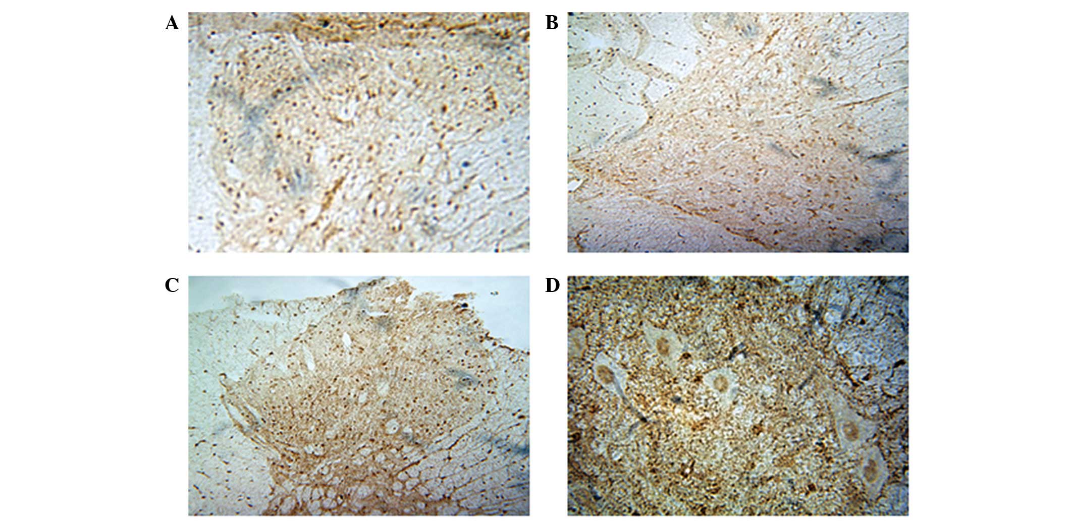
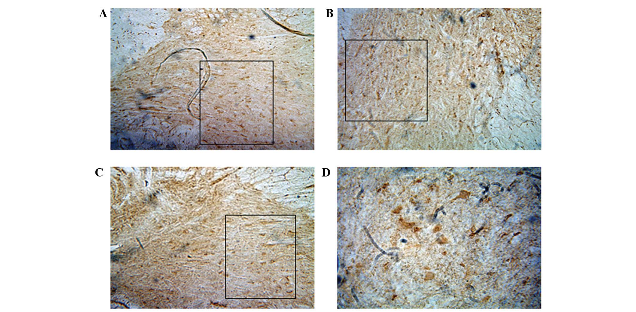
background levels (Figs. 1 and ).
Tissue sections were examined using a CAS Immunohistochemistry
Image Analysis System (CAS, East Rutherford, NJ, USA). Four
sections in one spinal cord segment were randomly selected for
counting of c-Fos- or c-Jun-positive neurons, respectively, in a
high magnification field. The c-Fos- or c-Jun-positive neurons were
recorded in three groups at the different time-points from
C5–7 right spinal gray matter, respectively.
Statistical analysis
All data are expressed as mean ± SEM. Statistical
analysis was performed using SPSS 13.0 (SPSS, Inc., Chicago, IL,
USA) for Windows. Statistical significance was calculated by the
t-test. P<0.05 was considered to indicate a statistically
significant difference.
Results
In the present study, we identified that the
expression of c-Fos and c-Jun in control animals was extremely
weak, with only small numbers of c-Fos- and c-Jun-positive neurons
scattered in the grey matter (data not shown).
In the rhizotomy and sham-operated groups, c-Fos-
and c-Jun-positive neurons were visible not only in right
C7, but also in ipsilateral C5–6 segments of
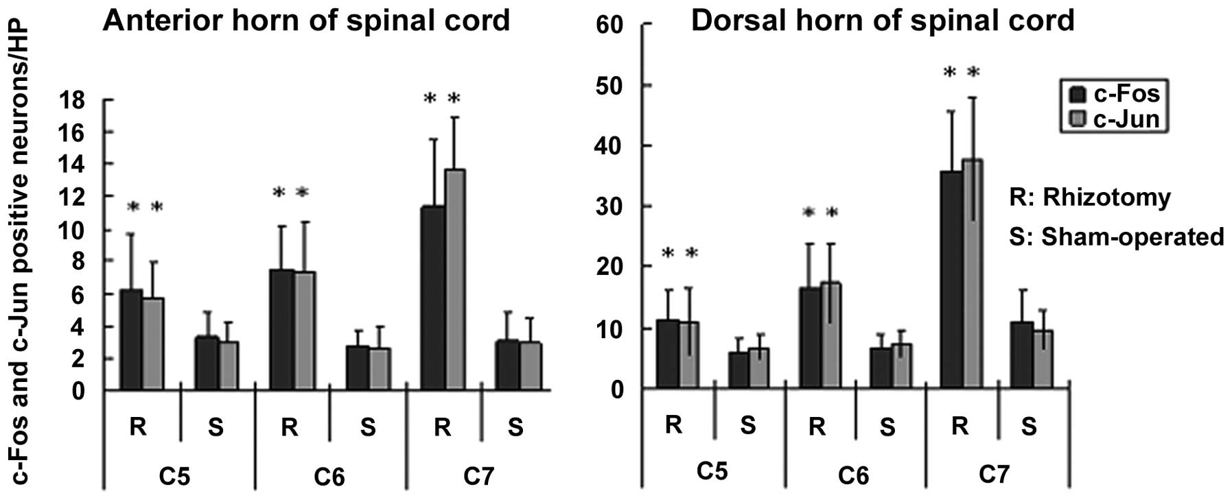
the spinal cord at 2 and 4 h after surgery (Figs. 1 and 2). The numbers of c-Fos- and
c-Jun-positive neurons in the rhizotomy group were markedly
increased compared with those in the sham group, at the same
time-point and in the same segment (P<0.05; Figs. 3 and 4). The location of these neurons was
similar in the rhizotomy and sham groups, which was mainly in
Rexed’s lamina IX (anterior horn of the grey matter) and in Rexed’s
laminae I–II (dorsal horn of the grey matter) of the cervical cord.
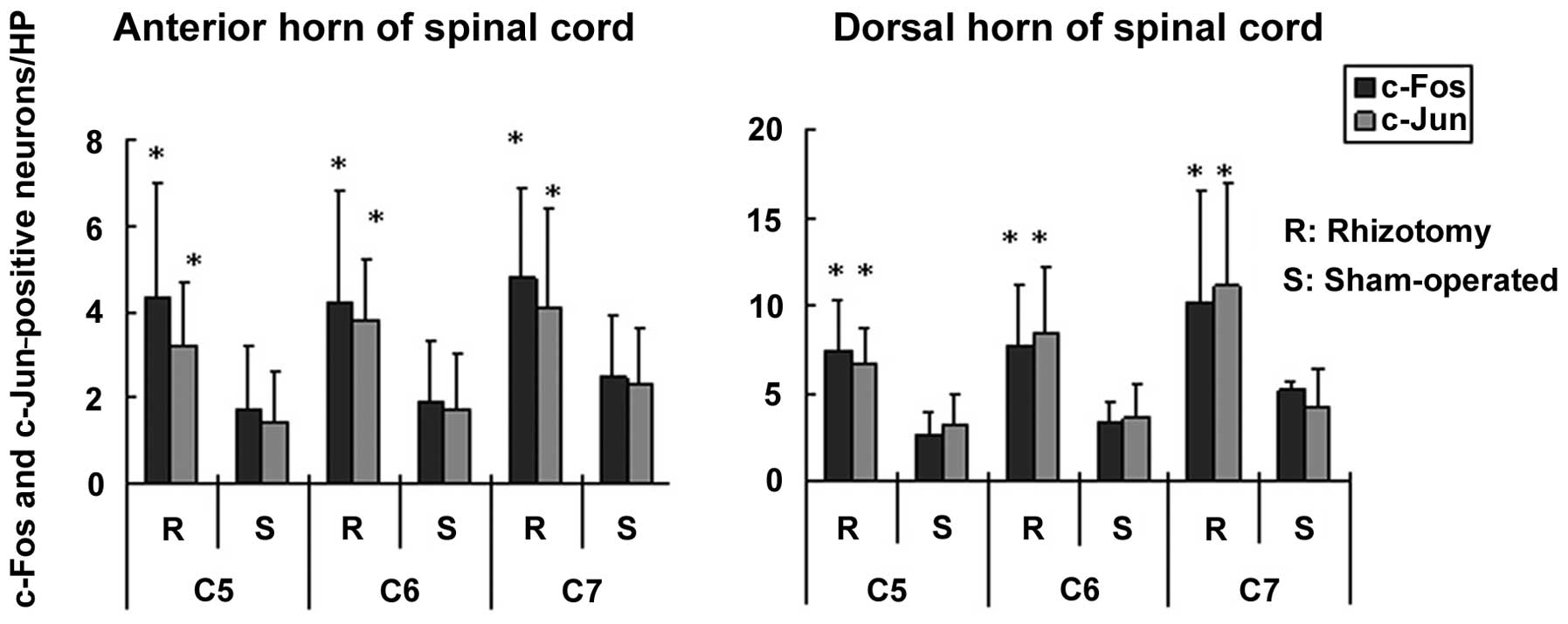
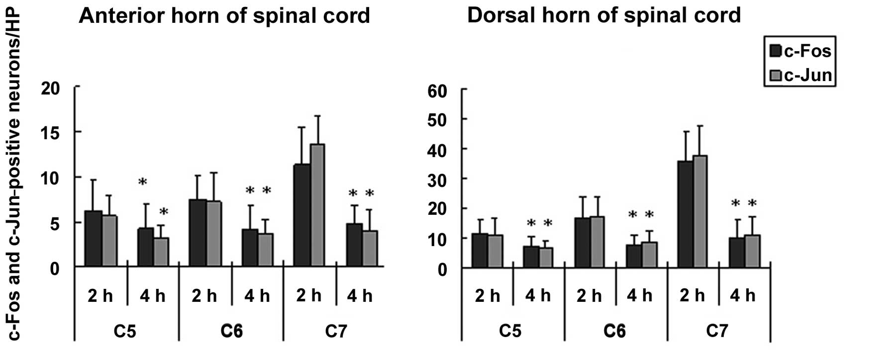
Moreover, in the rhizotomy group, the numbers of c-Fos- and
c-Jun-positive neurons in the anterior horn and posterior horn of
the grey matter were significantly lower at 4 h after surgery than
at the 2 h time-point (P<0.05; Fig.
5). The location of c-Fos- and c-Jun-positive neurons was
similar at 2 and 4 h after surgery.
Discussion
Cervical radiculopathy is a clinical manifestation
of degenerative cervical spine disease and commonly occurs in
clinical practice (1). The
diagnosis is usually based on the symptoms, signs and imaging,
particularly using MRI, which offers strong evidence for diagnosis
following careful history taking and physical examination (8). However, occasionally the motor
(weakness and atrophy), sensory (pain or paresthesias) and reflex
(diminution or absence of tendon reflexes) symptoms are not
confined to the affected spinal cord segment that corresponds with
the MRI findings (1). Therefore,
the precise location of the affected spinal cord segment becomes
confused, leading to difficulties in creating a treatment plan. In
the present study, we aimed to determine whether there is a neural
pathway between adjacent segments of the cervical spinal cord in
rats and to provide a possible explanation for the mismatch of the
symptoms and imaging results.
The IEG c-fos, which is rapidly and
transiently expressed in neurons in response to stimulation,
transcribes the nuclear protein c-Fos in spinal cord neurons
following induction of gene transcription (5,9).
c-Fos immunoreactivity has been widely used as a functional marker
to identify activity in spinal neurons in response to noxious
stimulation (3–5) and provides a good technique for
efficiently visualizing the individual cells activated by or
associated with noxious input (2).
A number of studies have shown that various types of noxious
stimulation, including thermal, mechanical and chemical stimuli
(10,11), and neuropathic pain models,
including constriction injury (2,12),
as well as spinal cord stimulation (13), induce the expression of c-Fos in
the spinal cord. Sugimoto et al also reported that c-Fos
expression is induced in the rat spinal dorsal horn following
L5 dorsal root rhizotomy (14). c-Jun, the protein product of
c-jun, another IEG, is also reported to be a marker for
neuronal activation and noxious stimulation (6,7).
Furthermore, Ke et al demonstrated that partial dorsal root
rhizotomy led to the upregulation of c-jun expression in
neurons of the dorsal root ganglion (15). The duration of c-fos and
c-jun expression varied from several minutes to several days
in different models (9,16), which was a considerable
discrepancy. In the current study, the c-Fos- and c-Jun-positive
neurons were detectable in the ipsilateral C7 segment at
2 h after C7 nerve root rhizotomy.
In the present study, in addition to the
C7 segment, an evident amount of c-Fos- and
c-Jun-positive neurons was also observed in C5 and
C6 segments of spinal grey matter following right
C7 nerve root rhizotomy, and the number of positive
neurons was significantly increased compared with that in the
sham-operated animals in the same segment and same time-point.
These positive neurons had a similar location, which was in Rexed’s
lamina IX in the anterior horn and in Rexed’s laminae I–II in the
dorsal horn of the spinal cord. Although the early increase of
c-Fos immunoreactivity may contain a component of c-fos
expression through surgical injury and inflammation (17), the surgical procedures were
identical, with the exception that the C7 nerve root was
exposed in the sham-operated group and was severed in the rhizotomy
group. Therefore, the difference in c-Fos expression in the same
segment between the two groups was mainly caused by the right
C7 rhizotomy. The same was observed with c-Jun
expression. This study demonstrates that C7 nerve root
rhizotomy triggers c-Fos and c-Jun expression in ipsilateral
C5 and C6 segments of the cervical cord.
These findings indicate that the afferent sensory fibers of the
C7 nerve root project not only to the dorsal horn of the
ipsilateral C7 spinal cord, but also to at least two
adjacent segments (C6 and C5 segments). In
addition, the efferent motor fibers of C7 nerve roots
are composed of fibers coming from not only the ipsilateral
C7 segment, but also from at least two adjacent segments
(C6 and C5). Therefore, there must be a
neural pathway between adjacent segments of the cervical spinal
cord.
Furthermore, 4 h after right C7
rhizotomy, the c-Fos- and c-Jun-positive neurons were still visible
in C5–7 segments. The number of c-Fos- and
c-Jun-positive neurons in the rhizotomy group was significantly
increased in the anterior horn and dorsal horn of the spinal cord
compared with the number in the sham-operated group,. However,
compared with that at 2 h after rhizotomy, the number of c-Fos- and
c-Jun-positive neurons had decreased considerably in all three
segments. However, the location of these positive neurons did not
change between the 2 and 4 h time-points. Following C7
rhizotomy, expression of IEGs was observed and then the expression
reduced simultaneously in all C5–7 segments. The
expression of IEGs in C5 and C6 presented the
same trend as that in C7, which suggests that there were
neurons from C5 and C6 segments that were
activated in response to ipsilateral C7 rhizotomy in the
same way as the C7 neurons were. This indicates that a
number of neurons in C5 and C6 also
contribute to the construction of the C7 nerve root.
This provides a possible explanation as to why the radicular
symptoms are not confined to the affected spinal cord segment as
shown by MRI.
In order to provide stronger evidence, we selected
two IEGs, c-fos and c-jun. These are important
research tools in the study of the neural basis of stimuli and
injury (9). In the present study,
there was no difference in the expression of c-Fos- and
c-Jun-positive neurons at the same time-point, in the same spinal
cord segment and same group. There was a high correlation with
c-Fos and c-Jun expression.
In conclusion, following right C7 nerve
root rhizotomy, c-Fos and c-Jun were expressed not only in
ipsilateral C7 spinal gray matter, but also in
ipsilateral C5 and C6 segments. Therefore, it
was deduced that there is a neural pathway between ipsilateral
adjacent cervical spinal cord segments and this may be one possible
explanation as to why the radicular symptoms of cervical
radiculopathy are not confined to the affected spinal cord segment,
as shown by MRI.
Abbreviations:
|
ABC
|
avidin-biotin-peroxidase complex;
|
|
MRI
|
magnetic resonance imaging;
|
|
IEG
|
immediate-early gene;
|
|
PBS
|
phosphate-buffered saline;
|
|
TBS
|
Tris-buffered saline
|
Acknowledgements
This study was supported by grants
from the Applied Basic Research Project of Tianjin (No.
043609011).
References
|
1.
|
Bednarik J, Kadanka Z, Dusek L, Kerkovsky
M, Vohanka S, Novotny O, Urbanek I and Kratochvilova D:
Presymptomatic spondylotic cervical myelopathy: an updated
predictive model. Eur Spine J. 17:421–431. 2008. View Article : Google Scholar : PubMed/NCBI
|
|
2.
|
Berrocal YA, Pearse DD, Andrade CM,
Hechtmana JF, Puentes R and Eaton MJ: Increased spinal c-Fos
expression with noxious and non-noxious peripheral stimulation
after severe spinal contusion. Neurosci Lett. 413:58–62. 2007.
View Article : Google Scholar : PubMed/NCBI
|
|
3.
|
Bullitt E, Lee CL, Light AR and
Willcockson H: The effect of stimulus duration on noxious-stimulus
induced c-fos expression in the rodent spinal cord. Brain Res.
580:172–179. 1992. View Article : Google Scholar : PubMed/NCBI
|
|
4.
|
Coggeshall RE: Fos, nociception and the
dorsal horn. Prog Neurobiol. 77:299–352. 2005.PubMed/NCBI
|
|
5.
|
Harris JA: Using c-fos as a neural marker
of pain. Brain Res Bull. 45:1–8. 1998. View Article : Google Scholar : PubMed/NCBI
|
|
6.
|
Broude E, McAtee M, Kelley MS and Bregman
BS: c-Jun expression in adult rat dorsal root ganglion neurons:
differential response after central or peripheral axotomy. Exp
Neurol. 148:367–377. 1997. View Article : Google Scholar : PubMed/NCBI
|
|
7.
|
Wang TT, Yuan WL, Ke Q, Song XB, Zhou X,
Kang Y, Zhang HT, Lin Y, Hu YL, Feng ZY, Wu LL and Zhou XF: Effects
of electro-acupuncture on the expression of c-jun and c-fos in
spared dorsal root ganglion and associated spinal laminae following
removal of adjacent dorsal root ganglia in cats. Neuroscience.
140:1169–1176. 2006. View Article : Google Scholar
|
|
8.
|
McGillicuddy JE: Cervical radiculopathy,
entrapment neuropathy, and thoracic outlet syndrome: how to
differentiate? Invited submission from the Joint Section Meeting on
Disorders of the Spine and Peripheral Nerves, March 2004. J
Neurosurg Spine. 1:179–187. 2004. View Article : Google Scholar
|
|
9.
|
Hughes P and Dragunow M: Induction of
immediate-early genes and the control of neurotransmitter-regulated
gene expression within the nervous system. Pharmacol Rev.
47:133–178. 1995.PubMed/NCBI
|
|
10.
|
Jinks SL, Simons CT, Dessirier JM,
Carstens MI, Antognini JF and Carstens E: C-fos induction in rat
superficial dorsal horn following cutaneous application of noxious
chemical or mechanical stimuli. Exp Brain Res. 145:261–269. 2002.
View Article : Google Scholar : PubMed/NCBI
|
|
11.
|
Todd AJ, Spike RC, Young S and Puskár Z:
Fos induction in lamina I projection neurons in response to noxious
thermal stimuli. Neuroscience. 131:209–217. 2005. View Article : Google Scholar : PubMed/NCBI
|
|
12.
|
Jergova S and Cizkova D: Long-term changes
of c-Fos expression in the rat spinal cord following chronic
constriction injury. Eur J Pain. 9:345–354. 2005. View Article : Google Scholar : PubMed/NCBI
|
|
13.
|
Smits H, Kleef MV, Honig W, Gerver J,
Gobrecht P and Joosten EA: Spinal cord stimulation induces c-Fos
expression in the dorsal horn in rats with neuropathic pain after
partial sciatic nerve injury. Neurosci Lett. 450:70–73. 2009.
View Article : Google Scholar
|
|
14.
|
Sugimoto T, Yoshida A, Nishijima K and
Ichikawa H: c-Fos induction in the rat spinal dorsal horn partially
deafferented by dorsal rhizotomy. Neurosci Lett. 178:239–242. 1994.
View Article : Google Scholar : PubMed/NCBI
|
|
15.
|
Ke Q, Wang T, Li L, Yu J and Guo Z: c-jun
expression in spared dorsal root ganglion following partial dorsal
root rhizotomy and acupuncture. Sichuan Da Xue Xue Bao Yi Xue Ban.
34:248–250. 2003.(In Chinese).
|
|
16.
|
Catheline G, Le Guen S L, Honoré P and
Besson JM: Are there long-term changes in the basal or evoked Fos
expression in the dorsal horn of the spinal cord of the
mononeuropathic rat? Pain. 80:347–357. 1999. View Article : Google Scholar : PubMed/NCBI
|
|
17.
|
Yamazaki Y, Maeda T, Someya G and Wakisaka
S: Temporal and spatial distribution of Fos protein in the lumbar
spinal dorsal horn neurons in the rat with chronic constriction
injury to the sciatic nerve. Brain Res. 914:106–114. 2001.
View Article : Google Scholar : PubMed/NCBI
|