Introduction
Bone morphogenetic protein 2 (BMP2) is a cytokine
involved in the induction of osteogenic differentiation; it plays a
key role in the differentiation of osteogenic progenitor cells to
osteoblasts and chondroblasts, as well as in ossification (1–5).
However, the direct use of BMP2 has the disadvantages of low
biological activity following in vitro extraction,
susceptibility to dilution and tissue absorption, a short half-life
(t1/2 <0.1 day) and a complex purification process
(6), which has limited its
clinical application. Transgenic osteogenesis induction using a
BMP2-expressing vector achieves sustained BMP2 expression in
vivo within a certain period and overcomes the problems
associated with the direct use of BMP2; this has attracted
widespread attention in recent years. The use of an adenovirus
vector is considered the most effective means for conducting
transgenic BMP2-induced osteogenesis (7,8). In
the present study, in order to facilitate the detection of gene
expression, a novel system for the construction of adenoviral
vectors was established, in which the BMP2 gene and the green
fluorescent protein (GFP) gene were simultaneously expressed.
Following mutagenesis, a FLAG epitope tag was attached to the BMP2
gene. The expression levels of the BMP2 and GFP genes in the
vectors in marrow stromal cells (MSCs) were detected and the
biological activity of expressed BMP2 in the induction of
osteogenesis was determined.
Materials and methods
Identification of pcDNA3-BMP2 and
analysis of restriction enzyme recognition sites
The E. coli DH-5α strains containing
pcDNA3-BMP2 (provided by Professor Pu Qin, Department of
Biochemistry, Fourth Military Medical University, Xi’an, China)
were inoculated to LB-ampicillin agar. Then, a single colony was
selected. Following amplification, the plasmid was extracted and
the NotI and NotI+XbaI restriction enzyme
reaction systems were established. The positive clone was
identified using agarose gel electrophoresis. The BMP2 gene was
sequenced using promoter sequences of T7 and Sp6 RNA polymerase at
the two sides of the pcDNA3-BMP2 multiple cloning sites. The
restriction enzyme recognition sites in the BMP2 gene were
analyzed. Primer synthesis and sequencing were performed by Takara
Biomedical Technology, Dalian, China.
Mutation of the BMP2 gene and subclone of
the pcDNA3 plasmid expressing the mutated BMP2 gene
(BMP2+)
The BMP2 gene was mutated by polymerase chain
reaction (PCR), using pcDNA3-BMP2 as the template. The sequence
after the translation termination codon TAG (including TAG) in the
BMP2 gene was removed and XhoI and XbaI were added.
The PCR conditions were as follows: 30 cycles of 98°C for 5 min,
94°C for 30 sec, 55°C for 30 sec and 68°C for 5 min. The PCR
products were obtained. The NotI and XbaI reaction
system was established using pcDNA3-BMP2 and PCR products as
substrates, respectively (at 37°C for 2 h). The mutation products
were identified by gel electrophoresis. T4 DNA ligase was added to
reconnect the mutated BMP2 gene and pcDNA3 (at 16°C overnight). On
the following day, the connected reaction solution was transformed
into competent E. coli DH5α cells (Center Laboratory, First
Affiliated Hospital of Liaoning Medical University, Jinzhou,
China), followed by inoculation into LB-ampicillin agar. Following
amplification, the plasmid was extracted. The positive clone was
identified by restriction enzyme reaction and electrophoresis. The
pcDNA3-BMP2+ plasmid was obtained.
Construction of the adenovirus shuttle
plasmid pShuttle cytomegalovirus (CMV)-BMP2+-internal
ribosome entry site (IRES)-hrGFP-1
The NotI and XbaI restriction enzyme
reaction system was established using pcDNA3-BMP2+ and
pShuttle CMV-IRES-hrGFP-1 (Stratagene Corporation, La Jolla, CA,
USA) as substrates, respectively (at 37°C for 2 h), followed by
identification using agarose gel electrophoresis. The
BMP2+ gene fragment and pShuttle CMV-IRES-hrGFP-1
fragment were retrieved, respectively, and were connected using T4
DNA ligase (at 16°C overnight). On the following day, the connected
reaction solution was transformed into competent E. coli
DH5α cells, followed by inoculation into LB-ampicillin agar.
Following amplification, the plasmid was extracted. The positive
clone was identified by restriction enzyme reaction and
electrophoresis. The plasmid pShuttle CMV-BMP2 was constructed
using the same method as above.
Construction of the adenovirus plasmid by
homologous recombination
Strains containing the plasmid pShuttle
CMV-BMP2+-IRES-hrGFP-1 and pShuttle CMV-BMP2 were
inoculated on LB-kanamycin agar, respectively. The plasmid was
extracted and the PmeI (New England Biolabs Ltd., Beijing,
China) restriction enzyme reaction system was established (at 37°C
for 2 h), followed by identification using agarose gel
electrophoresis. The adenovirus genome DNA was retrieved by cutting
gel and was dissolved in ddH2O (final concentration,
50–100 ng/μl) for use. Homologous recombination of the
adenovirus plasmid was performed in an electroporation apparatus
(200 Ω, 2.5 kV, 25 μF). The recombinant plasmid was
extracted. Following the PacI [New England Biolabs (Beijing)
Ltd., Beijing, China] restriction enzyme reaction, agarose gel
electrophoresis was conducted to identify the positive clone.
Finally, the pAd CMV-BMP2+-IRES-hrGFP-1 and pAd CMV-BMP2
plasmids were extracted using an Ultrapure Plasmid Purification kit
(Novagen Inc., Madison, WI, USA)
Packaging of the adenovirus
A PacI restriction enzyme reaction system was
established. The adenovirus genome DNA was re-dissolved in
ddH2O (final concentration, 5 μg/225 μl).
Conventional resuscitation and subculture of HEK293A cells
(American Type Culture Collection, Manassas, VA, USA) were
performed until 70% of the cells were fused. The two types of
adenovirus genome DNA and transfection reagents were added to cell
culture dishes, respectively, followed by 10 h incubation at 37°C,
5% CO2. When clear cytopathic phenomenon appeared, the
cells were collected and the titers were determined.
Determination of BMP2 and GFP
expression
MSCs (Central Laboratory, First Affiliated Hospital
of Liaoning Medical University) were inoculated in a 6-well plate
(5×10⁵ cells/well). The cells in two wells were randomly
selected for transfection with pAd
CMV-BMP2+-IRES-hrGFP-1 (experimental group) and the
cells in another two wells were transfected with pAd CMV-BMP2
(control group). The multiplicity of infection (MOI) was 50. The
remaining two wells were used as blank controls (blank group). GFP
was detected under a fluorescence microscope. Total mRNA was
extracted using an RNA purification kit (Takara Biomedical
Technology) and 80 ng RNA from each group was added to a reaction
tube, respectively. Then, 8.5 μl ddH2O without
RNase was added, followed by denaturation at 75°C for 5 min and
cooling on ice. cDNA was synthesized under the following
conditions: 30°C for 10 min, 42°C for 30 min and 99°C for 10 min.
Following the reaction, cooling on ice was conducted for 5 min. The
specific primers for cDNA were designed and synthesized and the
reverse transcription (RT)-PCR reaction system was established,
with the following reaction conditions: 94°C for 2 min, 94°C for 30
sec, 55°C for 30 sec and 72°C for 1.5 min; 30 cycles. Expression of
the BMP2 gene in the three groups was detected.
Detection of alkaline phosphatase (ALP)
activity
MSCs were inoculated in a 96-well plate
(1×10³ cells/well). The cells in 32 wells were randomly
selected for transfection with pAd
CMV-BMP2+-IRES-hrGFP-1 (experimental group) and the
cells in another 32 wells were transfected with pAd CMV-BMP2
(control group). The remaining 32 wells were used as the blank
group. Cells in each group (8 wells) were collected on days 6, 8,
10 and 12 after transfection and the cell concentration of each
well was adjusted to 1×10⁵ cells/ml. Additionally, 1 ml
cell solution in each group was used for detection of ALP activity
(kits were purchased from Upstate Biotechnology, Inc., New York,
NY, USA).
Statistical analysis
Statistical analysis was performed using SPSS 11.0
statistical software (SPSS, Inc., Chicago, IL, USA). The
Student-Newman-Keuls (SNK)-q test was used for multiple comparisons
of ALP activity at different time points after transfection.
P<0.05 was considered to indicate a statistically significant
difference.
Results
Identification of pcDNA3-BMP2 and
analysis of restriction enzyme recognition sites
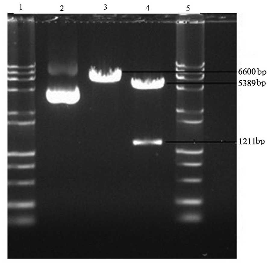
The results of identification by the NotI and
NotI+XbaI restriction enzyme reaction systems
demonstrated that the donor plasmid pcDNA3-BMP2 contained the
target BMP2 gene (1,211 bp). The sequencing results revealed that
the sequence of the BMP2 gene did not contain restriction sites of
XhoI, XbaI, PmeI, PacI or NotI
(Fig. 1). Following gene mutation,
the sequences after the translation termination codon were removed
and XhoI and XbaI BMP2 restriction sites were added.
The novel plasmid pcDNA3-BMP2+ was obtained.
Construction of the adenovirus shuttle
plasmid
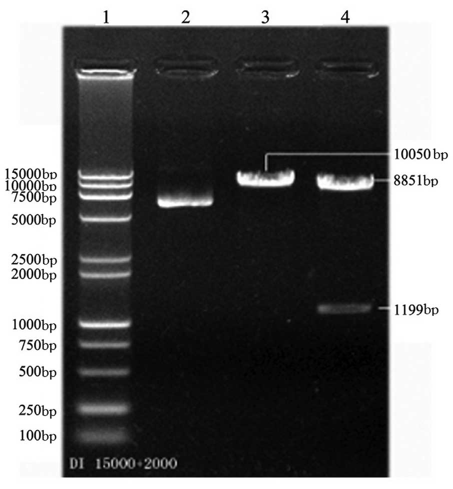
Following the NotI+XbaI restriction
enzyme reaction, two bands for pShuttle
CMV-BMP2+-IRES-hrGFP-1 were obtained by electrophoresis,
with molecular weights of 8,851 bp and 1,199 bp, respectively,
which were consistent with the theoretical molecular weights of the
shuttle vector and BMP2+. This indicated the successful
construction of pShuttle CMV-BMP2+-IRES-hrGFP-1
(Fig. 2).
Construction of the adenovirus
vector
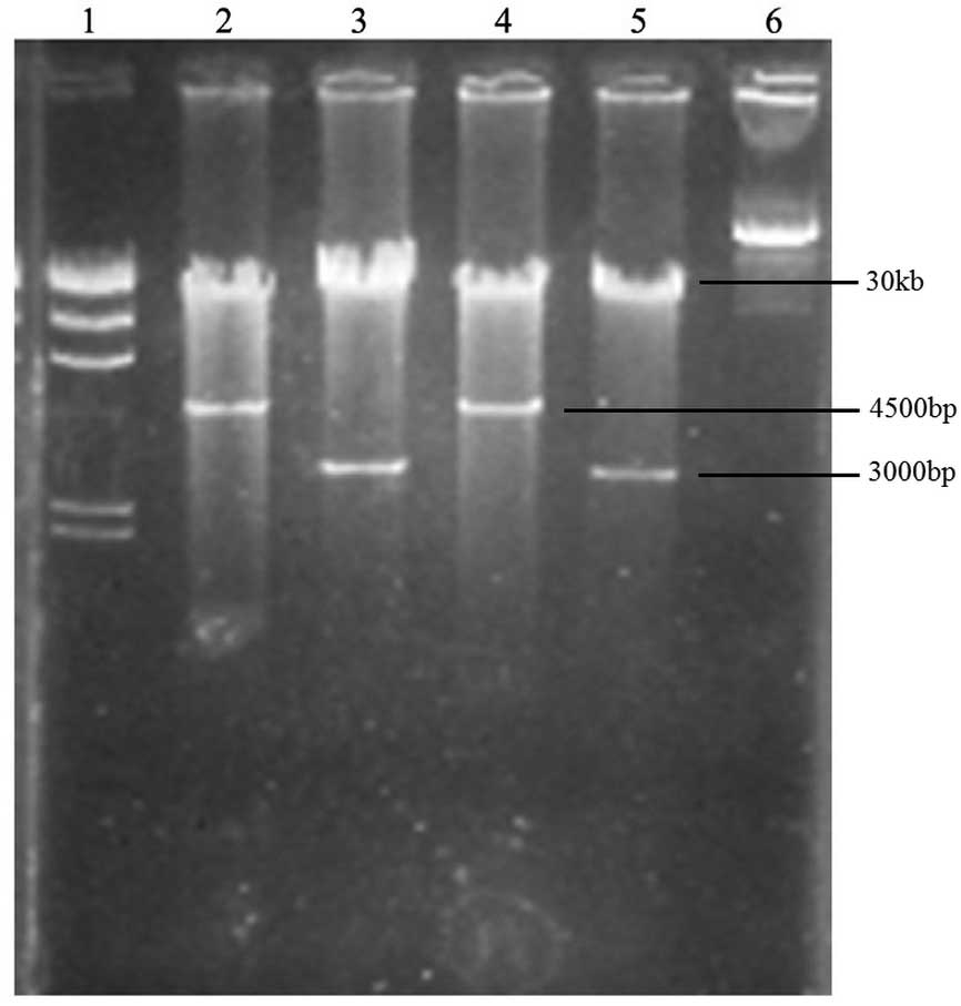
Following the PacI restriction enzyme
reaction and agarose gel electrophoresis, two results for the
recombinant adenovirus vector were obtained, as follows: i) two
bands, at 30 kb and 3,000 bp, respectively, and ii) two bands, at
30 kb and 4,500 bp, respectively. The two results originated from
different clones and were in accordance with the theoretical
results of the pAd Easy-1 adenovirus system, indicating the
successful construction of the adenovirus vector (Fig. 3).
Packaging of the adenovirus
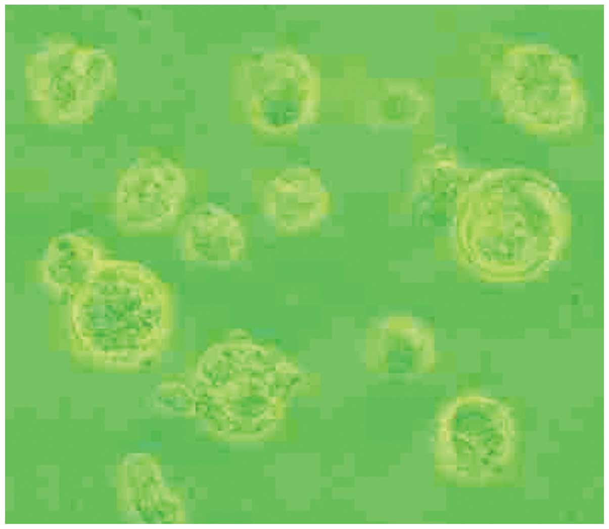
The successfully constructed recombinant adenovirus
vector was transfected into HEK293A cells for packaging. HEK293A
cells presented a cytopathic effect (CPE) with cell suspension,
tentacle contraction and swelling, as well as a round shape,
indicating successful adenovirus packaging (Fig. 4). Following four rounds of
amplification, the titers of the successfully packaged pAd
CMV-BMP2+-IRES-GFP-1 and pAd CMV-BMP2 were each
∼5×10⁸ pfu/ml.
Expression of BMP2 and GFP
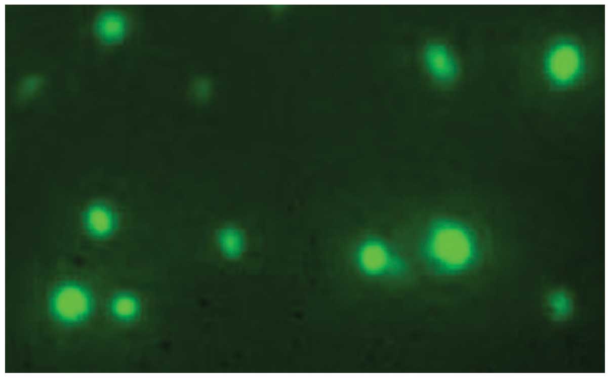
From 72 h after transfection of the adenovirus
solution into MSCs, fluorescence microscopy revealed high
expression levels of GFP, indicating successful transfection
(Fig. 5). RT-PCR results
demonstrated clear expression of BMP2 mRNA in MSCs in the
experimental and control groups, respectively, with no BMP2 mRNA
expression in the blank group. This further indicated successful
transfection (Fig. 6).
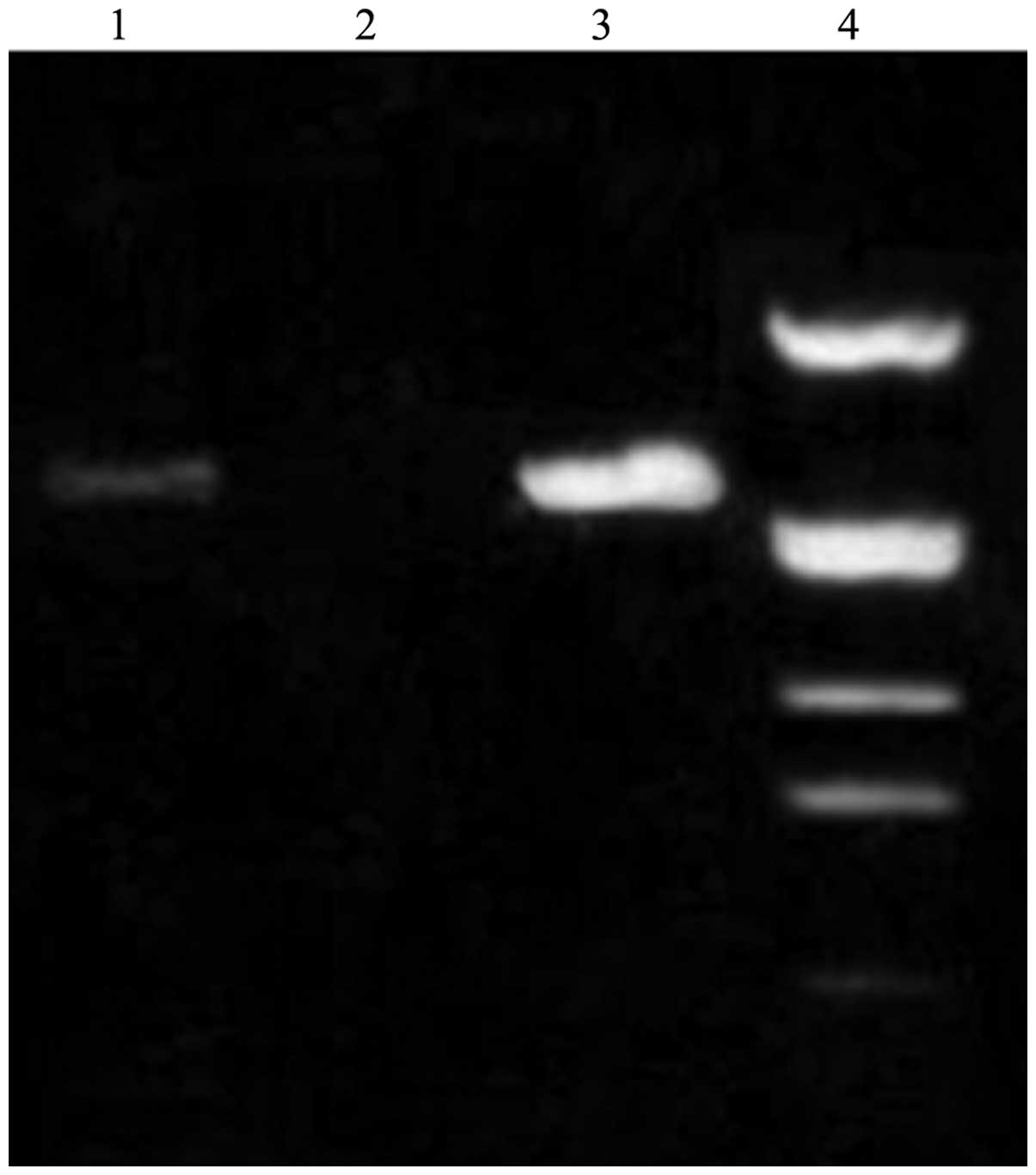 | Figure 6.Expression of BMP2 mRNA in adenovirus
transfected MSCs (RT-PCR). Lane 1, control group (pAd CMV-BMP2);
lane 2, blank group; lane 3, experimental group (pAd
CMV-BMP2+-IRES-hrGFP-1); lane 4, DL 2000). BMP2, bone
morphogenetic protein 2; RT-PCR, reverse transcription-polymerase
chain reaction; MSCs, marrow stromal cells; CMV, cytomegalovirus;
IRES, internal ribosome entry site; GFP, green fluorescent
protein. |
Detection of ALP activity
Significant differences in ALP activity were
observed between the experimental group and the blank group, and
between the control group and the blank group (P<0.05; Table I), with no significant difference
between the experimental and control groups (P>0.05). This
indicated that, compared with pAd-BMP2,
pAd-CMV-BMP2+-IRES-hrGFP-1 had the same function of
inducing osteoblast differentiation; however, this vector included
a GFP label, which previous vectors have lacked.
 | Table I.ALP activity at various time points
after transfection (U/l; mean ± SD). |
Table I.
ALP activity at various time points
after transfection (U/l; mean ± SD).
| Group | Day 6 | Day 8 | Day 10 | Day 12 |
|---|
| Experimental | 24.61±0.33 | 30.86±0.51 | 33.01±0.64 | 34.35±0.43 |
| Control | 24.50±0.42 | 29.61±0.53 | 33.11±0.31 | 34.01±0.23 |
| Blank | 15.68±0.01 | 16.01±0.54 | 16.55±0.23 | 17.67±0.47 |
Discussion
Homologous recombinant adenovirus vectors are an
efficient system for expressing the BMP2 gene (9). The constructed vector is essentially
a replication-defective recombinant adenovirus from which the E1
gene for DNA replication and viral packaging has been deleted. In
the current study, the shuttle vector pShuttle-CMV-IRES-hrGFP-1 was
used to construct the novel BMP2-expressing adenovirus vector.
There are several important structures, including multiple cloning
sites for connecting the target gene, FLAG epitope tag, IRES and
GFP behind the strong constitutive CMV promoter. The target gene
and the FLAG, IRES and GFP genes are transcribed in polycistronic
form at the same time; namely, the four transcripts are located in
the same mRNA. With the existence of IRES, the target genes are
translated into the target proteins and the ribosome is combined
with GFP mRNA, without being dropped from the polycistronic mRNA
chain. Therefore, when the target gene is introduced into MSCs, it
is simultaneously expressed with GFP (10,11).
GFP is easily detected by fluorescence microscopy,
immunohistochemistry and other intrusive or non-intrusive detection
methods, and it is a useful target gene reporting molecule
(12). FLAG is a small synthetic
polypeptide that is a type of highly specific antibody, with a
translation termination codon in its gene sequence. After removal
of the translation termination codon, FLAG may be fused with a
target protein and expressed as a type of antigen component of the
target protein (13). This has
enriched the detection methods of target genes.
In the present study, the restriction enzyme
reaction and sequencing were performed on the BMP2 gene of
pcDNA3-BMP2, and the restriction enzyme recognition sites were
analyzed. Results show that the length of the BMP2 gene is 1,211 bp
and the coding sequence is the same as the 318–1,528 base sequence
in human BMP2 mRNA (NM 001200) from Genbank; it is mainly composed
of the sequence from the translation initiation codon to the
translation termination codon (324–1,514 bp). As the BMP2 gene in
the donor plasmid has its own translation termination codon, with
no restriction site for cloning of the BMP2 gene into pShuttle
CMV-IRES-hrGFP-1, PCR is used for mutation of the BMP2 gene. The
sequence after the translation termination codon TAG (including
TAG) of the BMP2 gene was removed and XhoI and XbaI
were added. The mutated BMP2 gene was subcloned in the pcDNA3
vector and the new donor plasmid pcDNA3-BMP2+ was
prepared.
Following the successful preparation of the
recombinant adenovirus shuttle vector, according to the homologous
recombination mechanism in BJ5183 bacteria, the novel adenovirus
plasmid pAd CMV-BMP2+-IRES-hrGFP-1 was constructed. The
PacI restriction map shows that in different recombinant
positive clones in BJ5183, the shuttle plasmid and adenovirus
plasmid have the DNA prokaryotic replication origin sequence.
Following the PacI restriction enzyme reaction, the
constructed adenovirus plasmid should produce DNA fragments >23
kb and small fragments at 3.0–4.5 kb. In this study, these two DNA
fragments appear in the positive clone of the constructed pAd
CMV-BMP2+-IRES-hrGFP-1.
In the current study, homologous recombination in
bacteria was conducted to construct the adenovirus vector, which
saves time and cost. Compared with the widely used homologous
recombination method in mammalian cells, this method is simple,
fast, economical and efficient. In previous methods (14), homologous recombination has been
performed in HEK293A packaging cells and the cell state may greatly
affect the recombination efficiency, with a passage number of no
more than 45 generations. Occasionally, it has been necessary to
use HEK293A cells with a low number of generations. In addition,
HEK293A cells are not only the sites for homologous recombination,
but also for virus packaging. Following packaging, the virus
requires repeated screening, identification and purification. The
method in the present study uses bacteria, and the culture,
transformation and screening of the recombinant clone are more
convenient, economic and faster compared with a method using cells,
with more proficient processing. The time from plasmid
transformation to appearance of the recombinant clone was only
12–20 h and the total time, including screening time was only 36 h.
Most importantly, following identification, the recombinant DNA
(adenovirus plasmid) may be directly packaged in HEK293A cells,
without secondary screening and identification. In addition, the
virus titer and purity of this method are high.
In order to detect the expression of GFP and BMP2,
the constructed vector and control vector were transfected into
MSCs. After 3 days, the living cells were observed under a
fluorescence microscope. Results demonstrated that the MSCs
transfected by the constructed vector expressed GFP. Then, the
total mRNA of MSCs was extracted for RT-PCR detection. Results of
the electrophoresis of PCR products demonstrated that there were
DNA fragments with the same length amplified from the two
recombinant adenovirus vectors, with no DNA fragment in the blank
group. This indicated that the adenovirus vector expressed the BMP2
gene. In order to further confirm the osteogenic induction effect
of the expression product (BMP2+) and to investigate the
kinetics, the ALP activity in MSCs following vector transfection
was detected. Results showed that, on the sixth day after
transfection, the novel adenovirus vector and control vector
significantly increased the ALP activity level in MSCs (P<0.01),
with no significant difference between them (P>0.05). Thus, the
feasibility of BMP2 expression in a novel adenovirus vector is
demonstrated by the biological behavior of expression products.
The BMP2-expressing adenovirus vector constructed in
the current study is different from previously prepared
BMP2-expressing adenovirus vectors. It not only expresses the
target protein BMP2 with the epitope tag, but also expresses the
reporting molecule GFP, which provides greater advantages compared
with previous vectors. First, introduction of the reporting
molecule GFP enables the detection of BMP2 expression in living
cells. Therefore, BMP2 expression may be monitored and following
detection, the cells may still be used for subsequent experiments.
Secondly, the introduction of the FLAG epitope tag enriches the
detection means of the target gene, which is particularly important
in the absence of specific monoclonal antibodies of the target
gene.
Transgenic therapy using cytokines to induce
osteogenic differentiation has received widespread attention in the
past twenty years. It may cause a revolutionary transformation of
bone repair therapy and is likely to bring new hope for bone
orthopedic surgery (15–18). The successful construction of a
novel BMP2-expressing adenovirus vector is likely to lay a good
foundation for further investigation of transgenic therapy for
BMP2-induced osteogenic differentiation and bone tissue
engineering. This study is a preliminary attempt at the
construction of a BMP2-expressing adenovirus vector.
The main aim of this study was to demonstrate the
feasibility of the simultaneous expression of GFP and FLAG-labeled
BMP2, and to provide a foundation for the investigation of
osteogenic differentiation induced by adenovirus-mediated BMP2.
However, the newly constructed vector is an adenovirus vector
comprising recombinant BMP2 with deletion of the EI and E3 genes.
As the vast majority of the viral gene remains in the vector, the
immunogenicity and toxicity of the vector remains high. In
addition, during the vector packaging process or when the vector
enters the human cells with wild virus infection, a virus with
replication ability may be generated by homologous
recombination.
References
|
1.
|
Wozney JM: Bone morphogenetic proteins.
Prog Growth Factor Res. 1:267–280. 1989. View Article : Google Scholar
|
|
2.
|
Wozney JM: Overview of bone morphogenetic
proteins. Spine. (Phila Pa 1976). 27(Suppl 1): S2–S8. 2002.
View Article : Google Scholar : PubMed/NCBI
|
|
3.
|
Takahashi K: Bone morphogenetic protein
(BMP): from basic studies to clinical approaches. Nihon Yakurigaku
Zasshi. 116:232–240. 2000.(In Japanese).
|
|
4.
|
Harris SE, Guo D, Harris MA, Krishnaswamy
A and Lichtler A: Transcriptional regulation of BMP-2 activated
genes in osteoblasts using gene expression microarray analysis:
role of Dl×2 and Dl×5 transcription factors. Front Biosci.
8:s1249–s1265. 2003.
|
|
5.
|
Chen D, Zhao M and Mundy GR: Bone
morphogenetic proteins. Growth Factors. 22:233–241. 2004.
View Article : Google Scholar
|
|
6.
|
Lee SJ: Cytokine delivery and tissue
engineering. Yonsei Med J. 41:704–719. 2000. View Article : Google Scholar
|
|
7.
|
Alden TD, Varady P, Kallmes D, Jane J and
Helm G: Bone morphogenetic protein gene therapy. Spine. (Phila Pa
1976). 27(Suppl 1): S87–S93. 2002. View Article : Google Scholar : PubMed/NCBI
|
|
8.
|
Khan SN and Lane JM: The use of
recombinant human bone morphogenetic protein-2 (rhBMP-2) in
orthopaedic applications. Expert Opin Biol Ther. 4:741–748. 2004.
View Article : Google Scholar : PubMed/NCBI
|
|
9.
|
He TC, Zhou S, da Costa LT, Yu J, Kinzler
KW and Vogelstein B: A simplified system for generating recombinant
adenoviruses. Proc Natl Acad Sci USA. 95:2509–2514. 1998.
View Article : Google Scholar : PubMed/NCBI
|
|
10.
|
Lee JC, Wu TY, Huang CF, Yang FM, Shih SR
and Hsu JT: High-efficiency protein expression mediated by
enterovirus 71 internal ribosome entry site. Biotechnol Bioeng.
90:656–662. 2005. View Article : Google Scholar : PubMed/NCBI
|
|
11.
|
Naylor LH: Reporter gene technology: the
future looks bright. Biochem Pharmacol. 58:749–757. 1999.
View Article : Google Scholar : PubMed/NCBI
|
|
12.
|
Michel YM, Borman AM, Paulous S and Kean
KM: Eukaryotic initiation factor 4G-poly(A) binding protein
interaction is required for poly(A) tail-mediated stimulation of
picornavirus internal ribosome entry segment-driven translation but
not for X-mediated stimulation of hepatitis C virus translation.
Mol Cell Biol. 21:4097–4109. 2001. View Article : Google Scholar
|
|
13.
|
Prickett KS, Amberg DC and Hopp TP: A
calcium-dependent antibody for identification and purification of
recombinant proteins. Biotechniques. 7:580–589. 1989.PubMed/NCBI
|
|
14.
|
Wu C, Lei X, Wang J and Hung T: Generation
of a replication-deficient recombinant human adenovirus type 35
vector using bacteria-mediated homologous recombination. J Virol
Methods. 177:55–63. 2011. View Article : Google Scholar
|
|
15.
|
Pacifico MD, Floyd D and Wood SH: Tibial
stress fracture as a complication of free-fibula vascularised graft
for mandibular reconstruction. Br J Plast Surg. 56:832–834. 2003.
View Article : Google Scholar : PubMed/NCBI
|
|
16.
|
Scaduto AA and Lieberman JR: Gene therapy
for osteoinduction. Orthop Clin North Am. 30:625–633. 1999.
View Article : Google Scholar : PubMed/NCBI
|
|
17.
|
Ludwig SC and Boden SD: Osteoinductive
bone graft substitutes for spinal fusion: a basic science summary.
Orthop Clin North Am. 30:635–645. 1999. View Article : Google Scholar : PubMed/NCBI
|
|
18.
|
Zhang F, Fischer K and Lineaweaver WC: DNA
strand gene transfer and bone healing. J Long Term Eff Med
Implants. 12:113–123. 2002.PubMed/NCBI
|