Introduction
Epidermal cysts are the most common benign lesions.
They usually appear in hair-bearing skin areas, including the
scalp, face, neck, back and trunk (1). Conventional epidermal cysts are
generally small, slow-growing, non-tender, dome-shaped lesions. An
epidermal cyst is usually asymptomatic unless it becomes infected
or is enlarged to the extent that it causes damage to adjacent
anatomical structures. A number of authors have previously reported
giant epidermal cysts with diameters >5 cm (2,3).
However, few cases of giant epidermal cysts in the neck have been
reported. The present study concerns a giant epidermal cyst present
in the posterior neck, which grew to an extremely large size for
>40 years without inflammation or rupture, and was misdiagnosed
as a large soft tissue neoplasm. The patient exhibited depression
and developed social anxiety due to the negative cosmetic
consequences of the huge mass. To the best of our knowledge, a
giant epidermal cyst growing for >40 years has not previously
been reported. The study was approved by the Ethics Committee of
Chonbuk National University Hospital. Informed consent was obtained
from the patient and the family of the patient.
Case Report
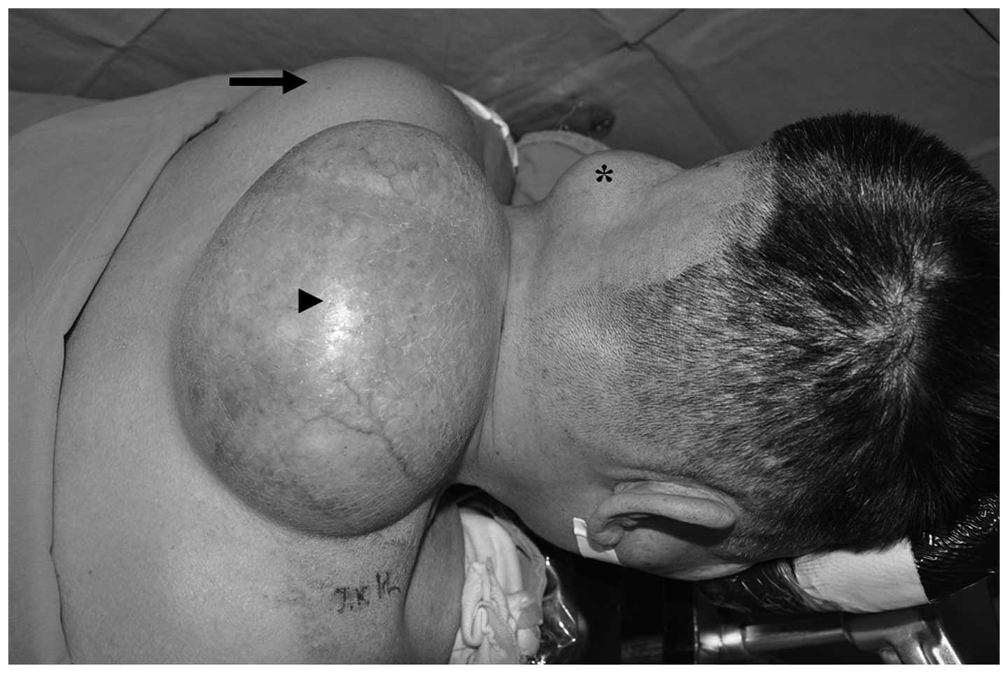
A 66-year-old male presented to The Department of
Orthopaedic Surgery, Chonbuk National University Medical School
(Jeonju, Korea) with three soft painless masses in the posterior
neck, left upper back and left scalp. The patient initially became
aware of the large mass in the midline of the posterior neck ~40
years previously and the other masses ~5 years previously. The
masses had gradually become enlarged. Clinical examination revealed
that all masses were soft, movable and well defined (Fig. 1). There was no history of trauma or
any previous surgery. The patient presented with discomfort due to
compression from the extremely large tumor. Moreover, the patient
exhibited depression and had developed social anxiety as a result
of the cosmetic problems caused by the large mass in the posterior
neck. Consequently, the patient was prescribed several
antidepressants and anti-anxiety drugs by an outpatient psychiatric
clinic.
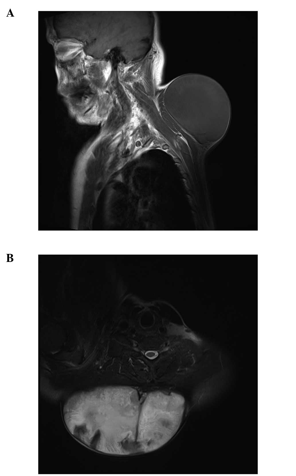
Magnetic resonance imaging (MRI) revealed a large
encapsulated homogeneous mass measuring 18×21×15 cm in the midline
of the posterior neck. The mass had low signal intensity on
T1-weighted images, while in T2-weighted images, its signals were
of high signal intensity with multiple occurrences of focal low
signal intensity debris (Fig. 2).
The masses in the left upper back (13×12×6 cm) and the left scalp
(5×5×4 cm) were diagnosed as subcutaneous lipomas based on MRI
findings.
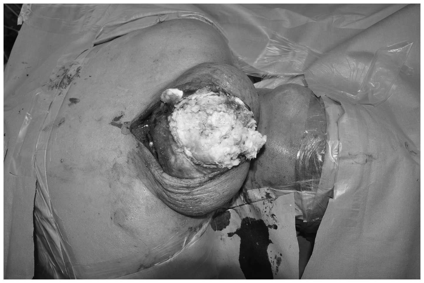
The patient underwent excision of all masses under
general anesthesia. The mass in the posterior neck was well defined
with an elastic texture and contained a cream-colored fluid with
the consistency of butter (Fig.
3). The other masses contained well-capsulated mature fat
tissue. A skin graft of superficial thickness was performed to
correct skin defects following the excision of friable skin from
the epidermal cyst of the posterior neck.
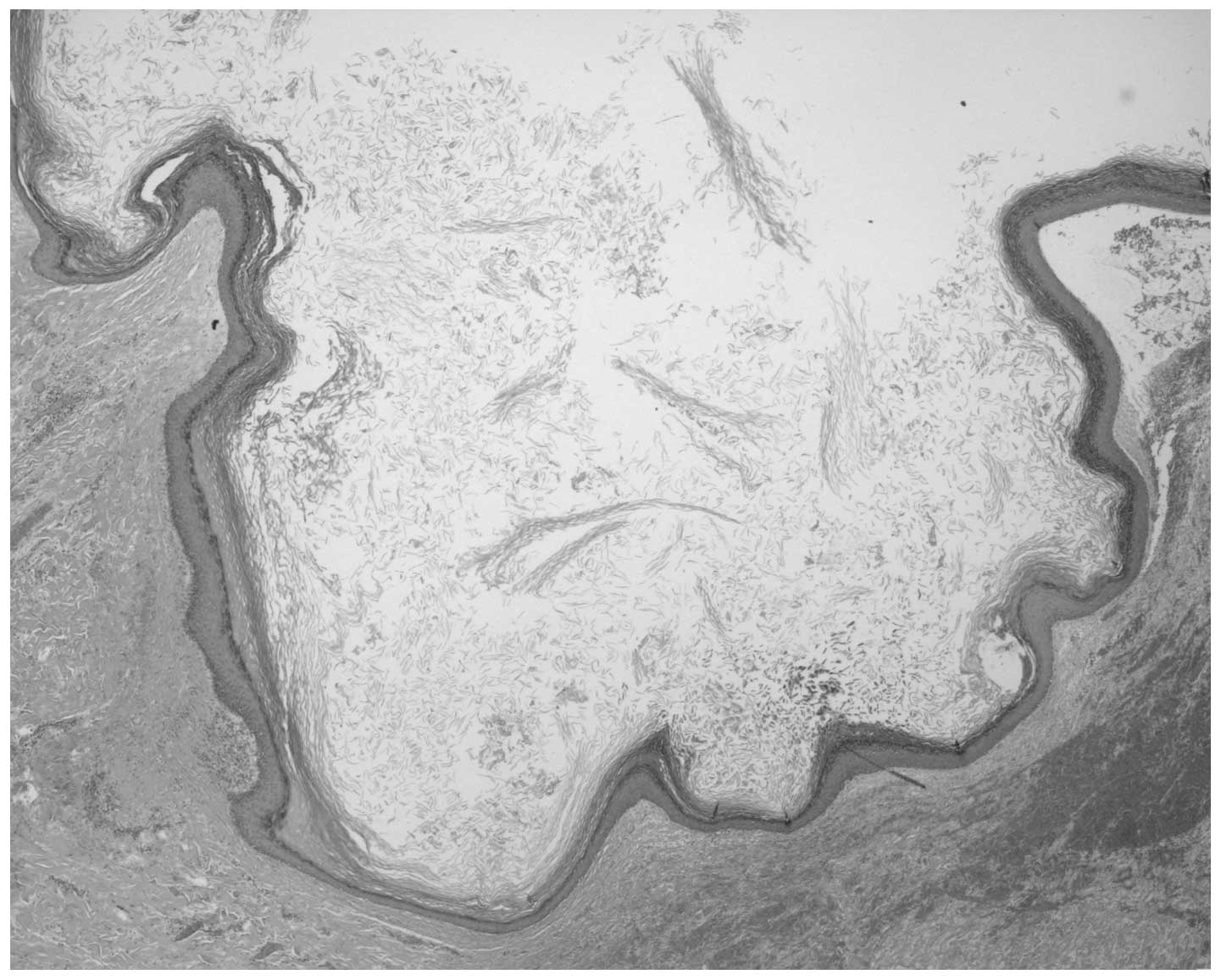
Histopathological examination revealed an epidermal
cyst wall with a thin layer of benign stratified squamous
epithelium and lamellated keratin debris present within the cyst
(Fig. 4).
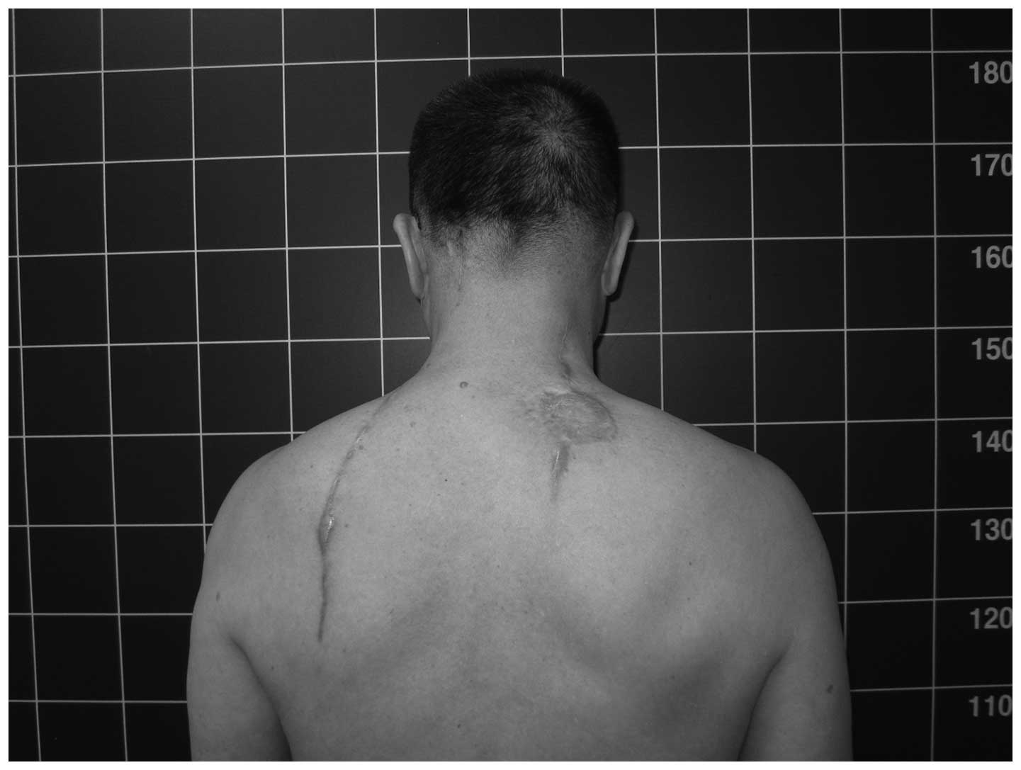
At the follow-up examination two years
postoperatively, there were no local recurrences of the lesions and
the psychiatric symptoms of the patient were completely resolved
(Fig. 5).
Discussion
Epidermoid cysts are characteristically observed in
the trunk, neck and face, and are likely to be derived from the
inflammation of pilosebaceous structures (2). Cysts on the acral surfaces of the
skin are considered to arise from the implantation of epidermis
into the dermis through trauma. These cysts usually grow through
the accumulation of epithelial and keratinous debris (3,4).
However, an epidermal cyst with a diameter ≥5 cm is rare. Giant
epidermal cysts in the head and neck present several problems.
Firstly, they are easy to rupture and this may induce infection.
Secondly, giant epidermal cysts may compress adjacent organs,
including major arteries, veins and nerves (2,4).
Furthermore, giant masses may cause cosmetic problems due to the
high visibility of the head and neck. The patient in the present
case exhibited pressure symptoms as well as psychiatric symptoms,
including depression and social anxiety, caused by the cosmetic
appearance of the large mass.
A proliferative epidermal cyst >5 cm in size is
locally aggressive and may potentially be a malignant tumor. The
development of squamous cell carcinoma in epidermal cysts is rare
and few cases have been reported (5). In a proliferative epidermal cyst,
epithelial proliferation from the cyst wall projects into the lumen
(5). Generally, squamous cell
carcinomas in epidermal cysts have a low malignant potential
(5). No malignant transformations
were identified in this case.
The present case of a giant epidermal cyst was
associated with two large adjacent lipomas. There was no clear
association between the epidermal cyst and the lipomas. To the best
of our knowledge, this case is the first report of a giant
epidermal cyst associated with two separate large lipomas in the
head and neck.
In conclusion, the present case demonstrates that
giant epidermal cysts may grow for long durations of time and
produce adverse effects due to the pressure exerted on surrounding
structures, as well as serious cosmetic problems that may require
psychiatric medication. Therefore, early surgical excision is
recommended for patients exhibiting giant epidermal cysts.
Acknowledgements
This study was supported by the Basic Science
Research Program through the National Research Foundation of Korea
(NRF) funded by the Ministry of Education, Science and Technology
(2010-0021514).
References
|
1
|
Polychronidis A, Perente S, Botaitis S,
Sivridis E and Simopoulos C: Giant multilocular epidermoid cyst on
the left buttock. Dermatol Surg. 31:1323–1324. 2005. View Article : Google Scholar : PubMed/NCBI
|
|
2
|
Kang SG, Kim CH, Cho HK, Park MY, Lee YJ
and Cho MK: Two cases of giant epidermal cyst occurring in the
neck. Ann Dermatol. 23(Suppl 1): S135–S138. 2011. View Article : Google Scholar : PubMed/NCBI
|
|
3
|
Golshan Momeni M, Anavim A, Lin F and
Tehranzadeh J: Giant epidermal inclusion cyst of buttock. Skeletal
Radiol. 35:864–866. 2006.PubMed/NCBI
|
|
4
|
Kim C, Park MC, Seo SJ, Yoo YM, Jang YJ
and Lee IJ: Giant epidermoid cyst of the posterior neck. J
Craniofac Surg. 22:1142–1144. 2011. View Article : Google Scholar : PubMed/NCBI
|
|
5
|
Sau P, Graham JH and Helwig EB:
Proliferating epithelial cysts. Clinicopathological analysis of 96
cases. J Cutan Pathol. 22:394–406. 1995. View Article : Google Scholar : PubMed/NCBI
|