Introduction
Hepatocellular carcinoma (HCC) is one of the most
common malignancies in the world, ranking as the third most common
cause of mortality from cancer (1,2) and
the fifth most prevalent global malignancy (3). Since the majority of cases of HCC
arise from livers diseased with chronic hepatitis and liver
cirrhosis, caused by infection with hepatitis C virus (HCV) and
hepatitis B virus (HBV), regular screening of these patients using
ultrasonography (US) and tumor markers, including α-fetoprotein
(AFP) and protein induced by vitamin K absence or antagonist-II
(PIVKA-II), is required for the detection of HCC at an early stage
(4). Despite advancements in the
treatment of HCC and the early detection of HCC, the prognosis of
patients with HCC remains unsatisfactory due to a high recurrence
rate following treatment (5). In
order to improve the outcomes of patients with chronic liver
diseases, novel chemopreventive and therapeutic compounds that may
be utilized for patients at a high risk of HCC and that exhibit no
systemic toxicity are required.
Silymarin is a polyphenolic mixture of
flavonoligands derived from the seeds of the milk thistle plant
(Silybum marianum). Silymarin has been clinically applied
for the treatment of liver diseases as an encapsulated,
standardized extract, since it is not water-soluble (6). Silymarin has been indicated to
possess hepatoprotective (7,8) and
antihepatocarcinogenic (9–11) properties. The mechanisms of the
antihepatocarcinogenic effects induced by silymarin include the
inhibition of cell proliferation and the stimulation of apoptosis
(12).
Diethylnitrosamine (DEN), which is present in
tobacco smoke, cured and fried meals, cosmetics and pharmaceutical
agents, has been established to be a powerful hepatocarcinogen in
rats (13). The proposed
mechanisms of DEN-induced hepatocarcinogenesis include the
alteration of DNA structure, the formation of alkyl DNA adducts and
the induction of chromosomal aberrations and micronuclei in the
liver (14,15). In addition to a single injection of
DEN followed by partial hepatectomy and coupled with
2-acetyl-aminofluorene (2-AAF) (16), the sequential administration of DEN
for several weeks has been demonstrated to induce HCC in rodents
(17,18). In the present study, severe and
mild models of HCC were generated by the intraperitoneal
administration of 40 mg/kg DEN once a week for 18 weeks and 100
mg/kg DEN every 2 weeks for 6 weeks in male Wistar rats,
respectively. By establishing these DEN-induced rat models, the aim
of the study was to evaluate the antihepatocarcinogenic effects of
silymarin, with the aim of enabling the future administration of
silymarin to patients with chronic liver diseases who are at high
risk of HCC.
Materials and methods
Chemicals
Silymarin, DEN and an anti-β-actin antibody were
purchased from Sigma Aldrich (St. Louis, MO, USA) and pentobarbital
was obtained from Dainippon Sumitomo Pharma Co., Ltd. (Osaka,
Japan). Antibodies against proliferating cell nuclear antigen
(PCNA) and glutathione S-transferase (GST) P were purchased
from Santa Cruz Biotechnology Inc. (Santa Cruz, CA, USA) and Assay
Designs, Inc. (Ann Arbor, MI, USA), respectively. Secondary
anti-mouse and anti-rabbit horseradish peroxidase (HRP) antibodies
for western blot analysis were obtained from GE Healthcare Ltd.
(Buckinghamshire, UK). All other chemicals and solvents used in
this study were of analytical grade.
Animals, treatments and tissue
collection
Male Wistar rats (weight, ~200 g) were obtained from
Japan SLC, Inc. (Hamamatsu, Japan). The rats were housed two per
cage with rice husks for bedding in an air-ventilated room under a
12-h light/dark cycle. The temperature (22ºC) and humidity (55%)
were kept constant. The animals were allowed free access to food
and tap water ad libitum during the experiment. All animals
received humane care and protocols were approved by the Animal
Ethics Committee of Tottori University (Yonago, Japan). Two models
were employed to evaluate the antihepatocarcinogenic effects of
silymarin.
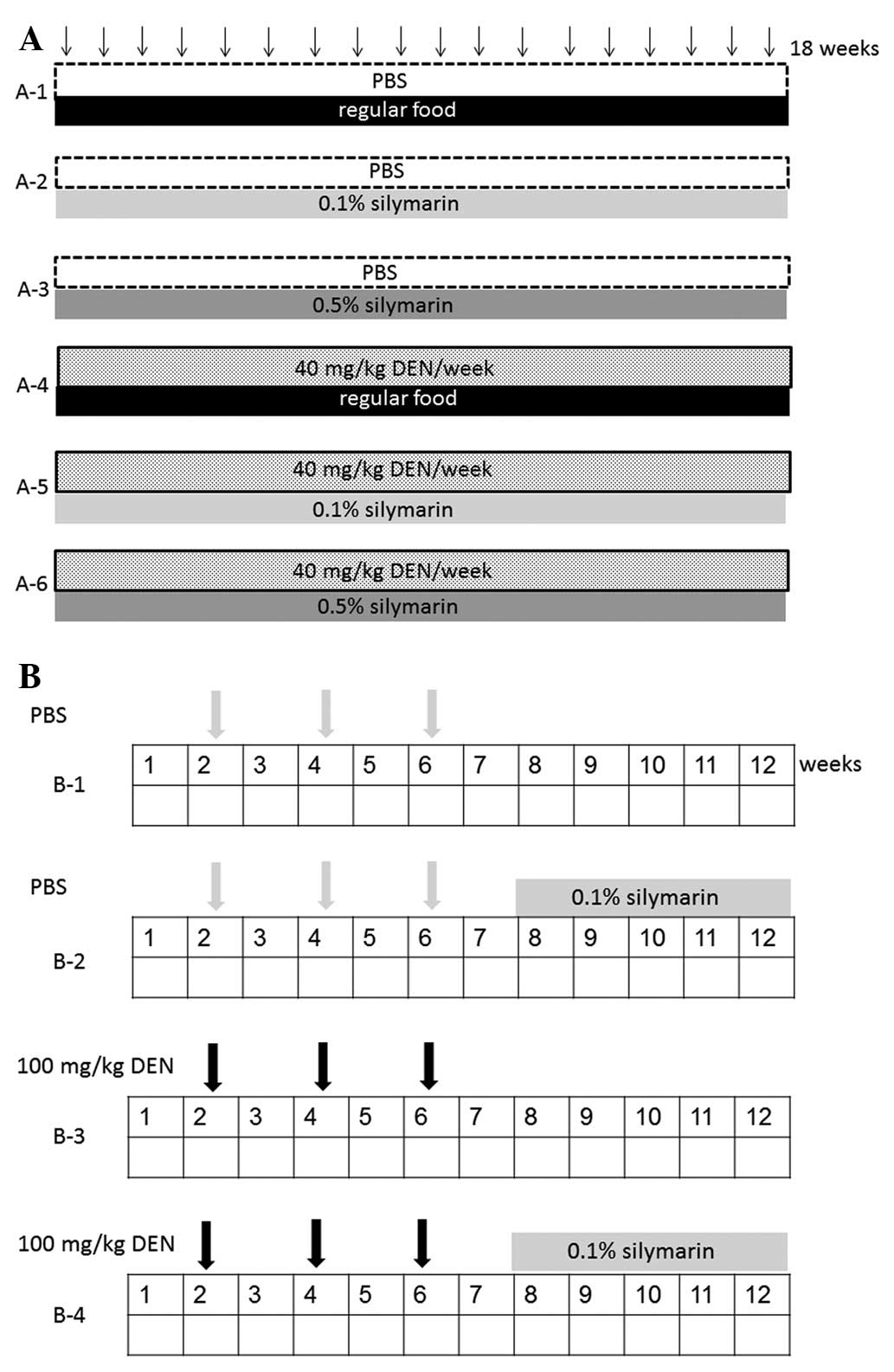
The study utilized severe (model A) and mild (model
B) models of HCC, in which the animals were randomized and divided
into six (Fig. 1A) and four
(Fig. 1B) groups, respectively. In
model A, the animals were intraperitoneally injected with 300 μl
phosphate-buffered saline (PBS) (groups A-1, A-2 and A-3; n=4) or
DEN (40 mg/kg body weight) dissolved in PBS weekly for 18 weeks
(groups A-4, A-5 and A-6; n=4). In order to examine the preventive
effects of silymarin on hepatocarcinogenesis, the rats were fed
with 0.1% silymarin (groups A-2 and A-5) or 0.5% silymarin (groups
A-3 and A-6) in powder form for 18 weeks. One week subsequent to
the 18-week treatments, animals were sacrificed by cardiac puncture
under anesthesia using pentobarbital.
In model B, the animals were intraperitoneally
injected with 300 μl PBS (groups B-1 and B-2; n=8) or DEN (100
mg/kg body weight) dissolved in PBS (groups B-3 and B-4; n=8) once
every 2 weeks on experimental weeks 2, 4 and 6. In groups B-2 and
B-4, the rats were fed with 0.1% silymarin in powder form during
experimental weeks 8 to 12 to examine the therapeutic effects of
silymarin on the DEN-induced hepatocarcinogenesis. One week
subsequent to the final treatments, the animals were sacrificed
under anesthesia using pentobarbital. Blood samples were obtained
via cardiac puncture and serum samples were stored at −30ºC until
analysis. Immediately following the excision of the livers, the
livers were divided into two sections for histological examination
in 10% neutral buffered formalin and for protein studies at
−80ºC.
Measurement of serum transaminase
levels
Serum aspartate aminotransferase (AST), alanine
aminotransferase (ALT) and alkaline phosphatase (ALP) levels were
measured at SRL, Inc. (Tokyo, Japan).
Total protein preparation and western
blotting
The liver samples were homogenized using a
BioMasher® (Nippi Inc., Tokyo, Japan) and lysed in
radioimmunoprecipitation (RIPA) buffer (Millipore Corp., Bedford,
MA, USA) supplemented with 1 mM sodium orthovanadate, 1 mM
phenylmethylsulfonyl fluoride (PMSF) and a protease inhibitor
mixture tablet (Roche Diagnostics, Basel, Switzerland) for 10 min
on ice. Total protein samples (5 μg) were separated using sodium
lauryl sulfate (SDS)-polyacrylamide gel electrophoresis (PAGE;
SuperSep; Wako Pure Chemical Industries, Ltd., Osaka, Japan) and
transferred to a polyvinylidene difluoride (PVDF) membrane
(Immobilon-P; Millipore Corp.). Subsequent to the membranes being
blocked in 5% non-fat milk (Santa Cruz Biotechnology Inc.) in 10 mM
Tris, 150 mM NaCl (pH 8.0) and 0.1% Tween 20 (TBST) for 1 h at room
temperature, they were probed with primary antibodies overnight at
4ºC, washed three times in TBST and incubated with anti-mouse or
anti-rabbit HRP antibody in TBST for 1 h at room temperature.
Following this, the signals were developed with a chemiluminescence
solution (ECL; GE Healthcare Ltd.), visualized and quantified using
an image analyzer (LAS-3000 mini; Fujifilm Co., Tokyo, Japan).
Histology and immunohistochemistry
The rat liver tissues were fixed in 10% neutral
buffered formalin and paraffin-embedded. For the histological
analysis, serial sections (5-μm) were stained with hematoxylin and
eosin (H&E). Neoplastic nodules and HCC were classified on the
basis of the published criteria (19). For immunohistochemistry with the
PCNA and GST-P antibodies, Histofine® Simple Stain Rat
MAX PO (Nichirei Biosciences Inc., Tokyo, Japan) was employed.
Briefly, following routine dewaxing with xylene and hydration
through a graded ethanol series, the sections were incubated with
1.5% hydrogen peroxide solution for 15 min at room temperature to
terminate the endogenous peroxidase activity. The sections were
subsequently washed in PBS, blocked with 1.5% serum solution and
incubated with primary antibodies overnight at 4ºC. Following this,
the sections were rinsed with PBS and incubated with biotinylated
secondary antibody for 30 min at room temperature. In addition,
HRP-conjugated avidin biotin complex (ABC) solution (Vector
Laboratories, Inc., Burlingame, CA, USA) was applied for 30 min at
room temperature. The peroxidase activity was developed using
3,3′-diaminobenzidine (DAB) solution (Vector Laboratories, Inc.).
Counterstaining was performed using hematoxylin. The PCNA labeling
indices were represented as the percentage of positively stained
nuclei by counting 1,000 cells in a field at ×200 magnification.
The GST-P-positive area was measured on images captured by a Charge
Coupled Device (CCD) camera on a Windows® computer.
Statistical analysis
Values are expressed as the mean ± standard
deviation. Values between two groups were compared using the
Mann-Whitney U-test. P<0.05 was considered to indicate a
statistically significant difference.
Results
Relative liver weight and serum
transaminase levels
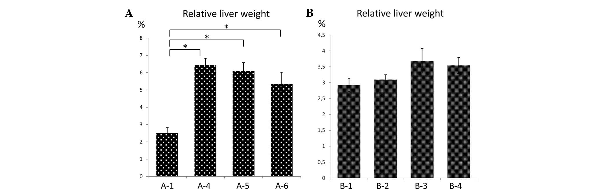
In model A, two rats in group A-4 died during the
experimental period. The relative liver weight (liver weight/body
weight) was significantly higher in the DEN groups (A-4, A-5 and
A-6) than in A-1, which was presumably due to the development of
liver tumors. No significant differences were identified in
relative liver weight among groups A-4, A-5 and A-6, irrespective
of the silymarin treatment (Fig.
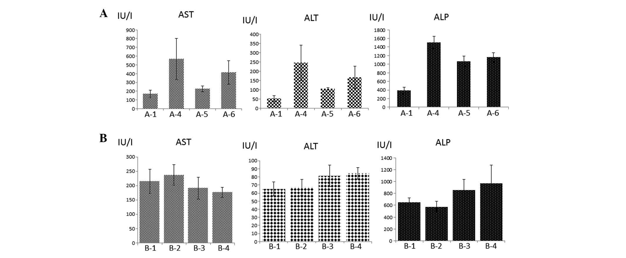
2A). Serum transaminase (AST and ALT) and ALP levels were
higher in the DEN groups (A-4, A-5 and A-6) than in A-1, which most
likely reflected the hepatic injury induced by DEN (Fig. 3A). No significant differences were
identified in serum transaminase and ALP levels among groups A-4,
A-5 and A-6. Relative liver weight and serum transaminase and ALP
levels in groups A-2 and A-3 were not significantly different
compared with the values in group A-1 (data not shown). In model B,
one rat in group B-4 died during the experimental period. No
significant differences were identified in the relative liver
weight or serum transaminase and ALP levels among the four groups,
B-1, B-2, B-3 and B-4 (Fig. 2B and
3B).
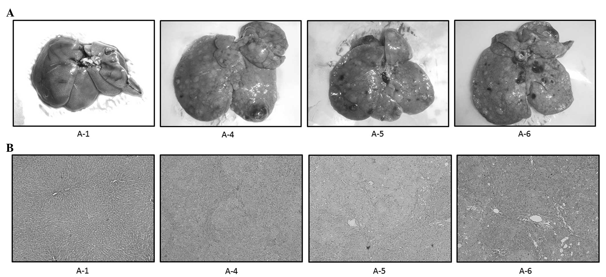
Macroscopic and histological
examinations
Macroscopic and microscopic features of the liver
were evaluated in the two models. As expected, in control rats
without DEN treatment (A-1, A-2, A-3, B-1 and B-2), no tumors were
observed (Figs. 4A and 5A for A1 and B1, respectively; data not
shown for A-2, A-3 and B-2) and the liver histology showed a normal
appearance (Figs. 4B and 5B for A1 and B1, respectively; data not
shown for A-2, A-3 and B-2). In model A, multiple white nodules
were macroscopically observed in the groups treated with DEN
(Fig. 4A, group A-4). The gross
appearance of the livers treated with DEN and 0.1 and 0.5%
silymarin was predominantly identical to that of the liver treated
with DEN alone, with no significant differences in the number of
nodules among groups A-4, A-5 and A-6 (Fig. 4A). In the histological analysis,
the white nodules were demonstrated to be HCC. Consistent with the
macroscopic findings, the HCC area was not significantly modified
by silymarin treatment (Fig.
4B).
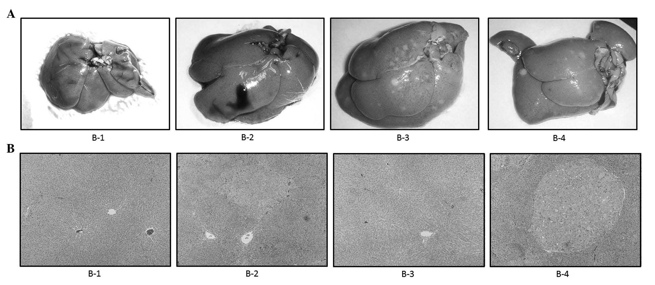
In model B, a number of white nodules, although
fewer than in model A, were macroscopically observed in the groups
treated with DEN (Fig. 5A, group
B-3). In the histological analysis, hyperplastic nodules were shown
to have developed following DEN treatment (Fig. 5B, group B-3). The gross appearance
of the liver treated with DEN and 0.1% silymarin was predominantly
identical to that of the liver treated with DEN alone, with no
significant difference in the number of nodules between B-3 and B-4
(Fig. 5B). Consistent with the
macroscopic findings, the nodular area was not significantly
modified by the silymarin treatment (Fig. 5B). These results indicated that
silymarin did not have a significant impact on hepatitis or
hepatocarcinogenesis induced by DEN in the severe or mild model of
hepatocarcinogenesis.
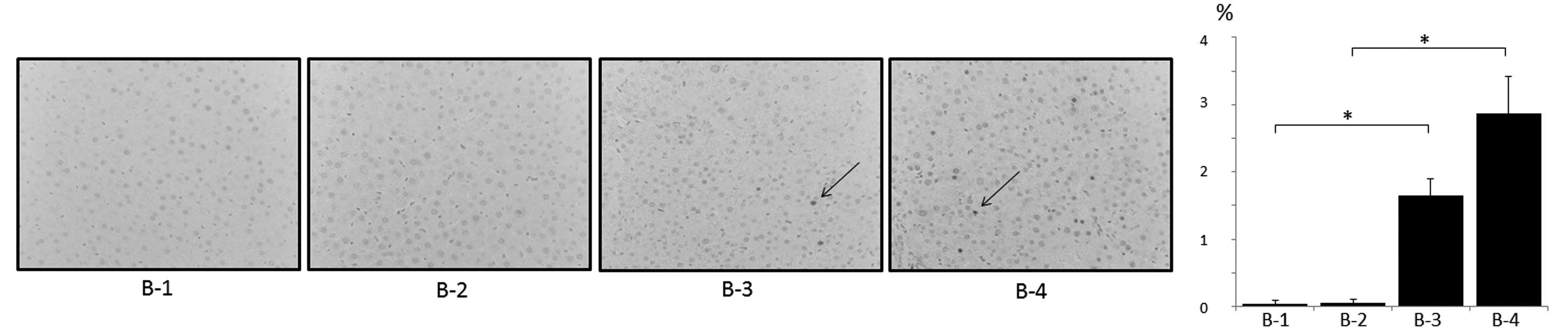
Expression levels of PCNA and GST P
PCNA is an essential regulator of the cell cycle and
its expression is a useful tool for the study of cell
proliferation, including in the liver (20). The expression levels of PCNA in the
liver among the treatment groups of models A and B were examined.
Immunohistochemical analysis revealed that PCNA-positive cells were
scarcely observed in the control livers without DEN treatment (A-1,
A-2, A-3, B-1 and B-2; Figs. 6A
and 7 and data not shown).
Following treatment with DEN, the number of PCNA-positive cells was
significantly increased in the two models (Figs. 6A and 7). The number of PCNA-positive cells was
not significantly altered following treatment with silymarin in
model A (Fig. 6A), which was
demonstrated using western blot analysis (Fig. 6B). However, treatment with
silymarin in model B appeared to increase the number of
PCNA-positive cells; the mechanisms for this are unknown (Fig. 7).
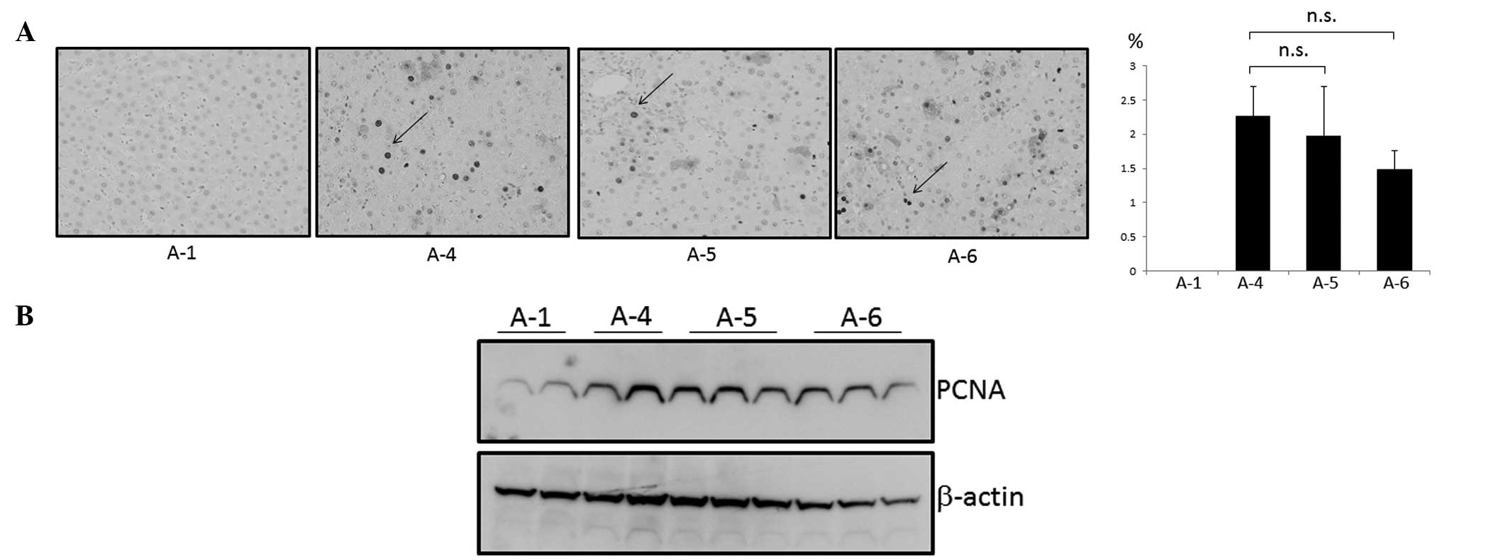 | Figure 6Expression of proliferating cell
nuclear antigen (PCNA) in the livers of model A rats. Expression
levels were evaluated using (A) immunohistochemical and (B) western
blot analyses. (A) Representative liver tissues immunostained with
anti-PCNA antibody in the control (A-1), diethylnitrosamine (DEN;
A-4), DEN with 0.1% silymarin (A-5) and DEN with 0.5% silymarin
(A-6) groups (top left panel). Arrows indicate the representative
PCNA-positive cells. Percentages of PCNA-positive cells in A-1,
A-4, A-5 and A-6 were 0, 2.3, 2.0 and 1.5%, respectively (top right
panel); original magnification, ×400. (B) Representative liver
samples from groups A-1, A-4, A-5 and A-6 were probed with
anti-PCNA antibody (top lane). The membrane was reprobed with
anti-β-actin antibody (bottom lane). n.s., not significant. |
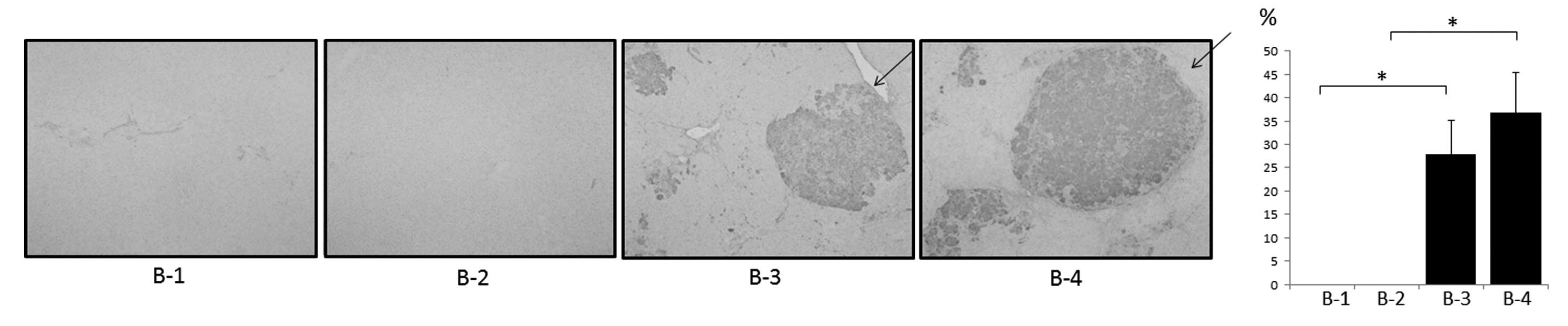
Among the glutathione S-transferases (GSTs),
a family of detoxification enzymes catalyzing the conjugation of
glutathione with a large number of carcinogens, placental GST (GST
P) is specifically expressed during rat hepatocarcinogenesis and
has been used as a reliable tumor marker for experimental
hepatocarcinogenesis in rats (21). As a result of this, the expression
levels of GST P in the livers of the rats in the model A and B
treatment groups were examined. Immunohistochemical and western
blot analyses revealed that a GST P-positive area appeared in the
DEN-treated livers (Figs. 8 and
9). However, expression levels of
GST P were not significantly modified by the treatment with
silymarin at any condition (Figs.
8 and 9). The combined results
indicate that silymarin is not a potent compound useful for either
the prevention or treatment of HCC.
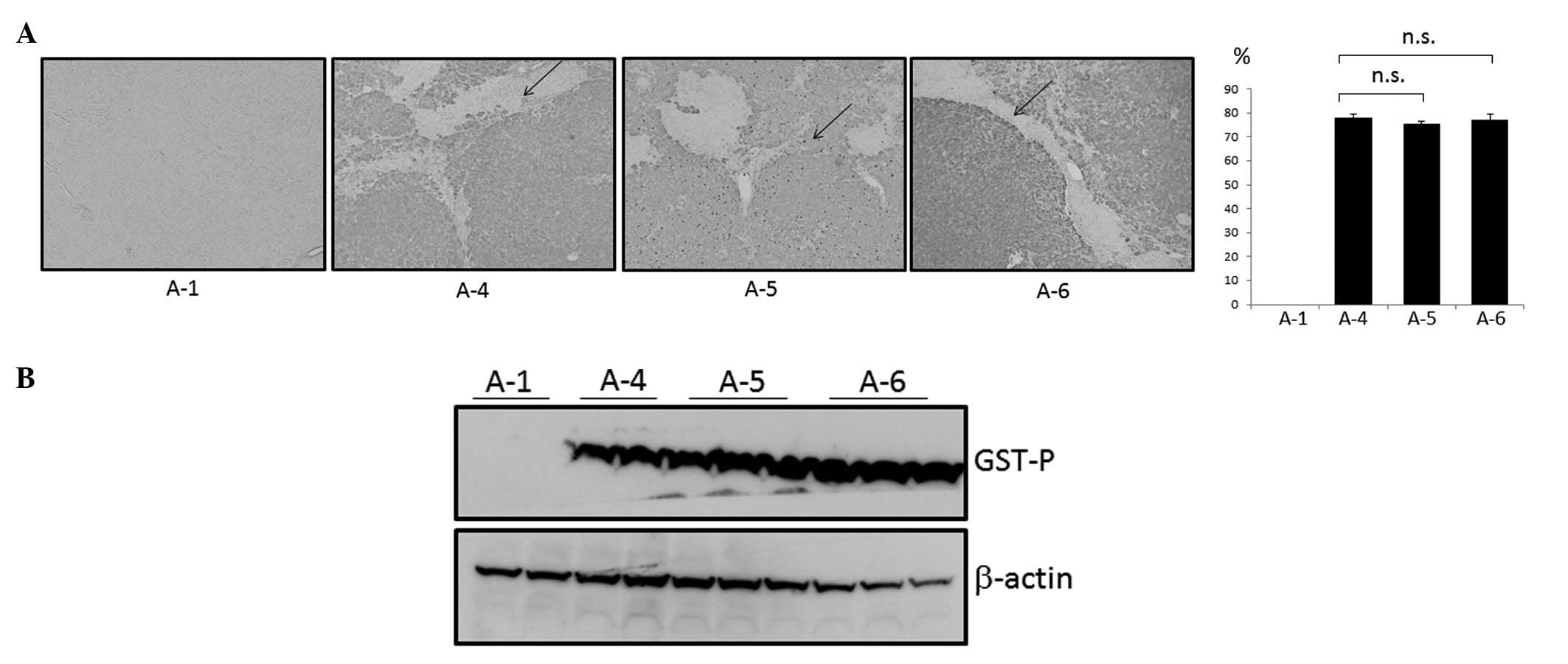 | Figure 8Expression of glutathione
S-transferase P (GST P) in the livers of model A rats.
Expression levels were evaluated using (A) immunohistochemical and
(B) western blot analyses. (A) Representative liver tissues
immunostained with anti-GST P antibody in the control (A-1),
diethylnitrosamine (DEN; A-4), DEN with 0.1% silymarin (A-5) and
DEN with 0.5% silymarin (A-6) groups (top left panel). Arrows
indicate the representative GST P-positive area. Percentages of GST
P-positive areas in A-1, A-4, A-5 and A-6 were 0, 78.2, 75.5 and
77.4%, respectively (top right panel); original magnification,
×100. (B) Representative liver samples from groups A-1, A-4, A-5
and A-6 were probed with anti-GST P antibody (top lane). The
membrane was reprobed with anti-β-actin antibody (bottom lane).
n.s., not significant. |
Discussion
Based on the fact that the majority of cases of HCC
are complicated by chronic liver diseases, including chronic
hepatitis and liver cirrhosis associated with HBV and HCV
infection, the regular screening of patients who are at high risk
of HCC, using US and computed tomography (CT), has been proposed in
Japan to enable the early detection of HCC (4). However, HCC is often diagnosed at an
advanced stage due to the inefficiency of US instrument operators,
dropouts among the patients targeted by the screening program and
an increasing incidence of non-B non-C HCC, which is difficult to
include in the screening program (22,23).
In addition to these complications hindering the early diagnosis of
HCC, sorafenib, a multi-kinase inhibitor, has been demonstrated to
exert marginally beneficial effects on the survival of patients
with advanced HCC (24). Since the
prognosis remains poor for patients with HCC, particularly at
advanced stages, novel preventive and therapeutic approaches for
these patients are urgently required.
Cancer chemoprevention is defined as ‘the use of
specific natural or synthetic chemical agents to reverse or
suppress carcinogenesis and prevent the development of invasive
cancer using physiological pathways’ (25). Phytochemicals, which are
plant-derived chemicals contained in fruits, vegetables and grains,
have been subject to investigation due to their apparent antitumor
activity against a variety of cancers, including HCC (26). Possible compounds with a potential
to exert chemopreventive effects on the liver include caffeine,
capsaicin, cinnamaldehyde, curcumin, epigallocatechin-3-gallate
(EGCG), resveratrol, sulforaphane (SFN) and silymarin (27).
Among these phytochemicals, silymarin is a
polyphenolic mixture of flavonoligands derived from the seeds of
the milk thistle plant (Silybum marianum). Silymarin is a
complex of five major compounds, silibinin, silychristin,
isosilychristin, silydianin and taxifolin (8). Silymarin has been widely used as a
natural remedy for the treatment of liver diseases since the time
of the Ancient Greeks (12). The
Hepatitis C Antiviral Long-Term Treatment against Cirrhosis
(HALT-C) trial was designed to evaluate the efficacy of long-term
treatment with low-dose peginterferon α-2a in patients with
fibrosis and cirrhosis associated with hepatitis C, who had failed
previous peginterferon and ribavirin therapy. Nearly one-third of
the patients in the trial were former or current users of silymarin
(28). Silymarin has been
demonstrated to exhibit anticancer effects against cancers at a
variety of sites, including the prostate (29), urinary bladder (30) and colon (31). Furthermore, silymarin has been
indicated to display hepatoprotective (7,8) and
antihepatocarcinogenic (9–11) properties in the liver.
In the present study, the antihepatocarcinogenic
effects of silymarin in severe (model A) and mild (model B) models
of HCC were explored. In model A, a low concentration of DEN (40
mg/kg body weight) was intraperitoneally administered weekly in a
long-term course (18 weeks), which generated multiple HCCs in the
rats. Treatment with silymarin was initiated from the beginning of
DEN administration, in order to investigate the preventive effect
of silymarin on HCC. In model B, a high concentration of DEN (100
mg/kg body weight) was intraperitoneally administered once every 2
weeks and silymarin was administered during experimental weeks 8 to
12, in order to explore the therapeutic effects of silymarin on the
DEN-induced hepatocarcinogenesis. The doses of silymarin were
selected to be 0.1 and 0.5% based on previous studies (12,32,33)
and a preliminary experiment in which we tested the administration
of 1% silymarin. Food intake and body weight gain with 1% silymarin
were poor (data not shown). In the present study, under the
selected experimental conditions, the results did not demonstrate
the antihepatocarcinogenic effects of silymarin. Furthermore,
silymarin appeared to accelerate the DEN-induced
hepatocarcinogenesis (Fig. 7).
There are several plausible explanations for these results,
including: (i) the serum concentrations of silymarin attained were
not high enough to exert biological effects, due to a low
bioavailability of silymarin, although this was not measured; (ii)
the hepatocarcinogenic models were too harsh to observe the
antihepatocarcinogenic effects of silymarin; and (iii) silymarin
does not possess antihepatocarcinogenic potential. Certain previous
studies have revealed results consistent with the null
anticarcinogenic effect of silymarin on DEN-induced
hepatocarcinogenesis in rats that was observed in the present study
(28,34). Silymarin potentiated
ethanol-dependent HCC progression in mice (34), while users of silymarin had similar
serum transaminase and HCV levels to those of nonusers in the
HALT-C trial (28). In order to
provide firm conclusions concerning the role of silymarin on
hepatocarcinogenesis, further intensive investigations are
required.
Abbreviations:
|
ALP
|
alkaline phosphatase
|
|
ALT
|
alanine aminotransferase
|
|
AST
|
aspartate aminotransferase
|
|
DEN
|
diethylnitrosamine
|
|
GST
|
glutathione S-transferase
|
|
H&E
|
hematoxylin and eosin
|
|
HBV
|
hepatitis B virus
|
|
HCC
|
hepatocellular carcinoma
|
|
HCV
|
hepatitis C virus
|
|
PCNA
|
proliferating cell nuclear antigen
|
References
|
1
|
Bosch FX, Ribes J and Borràs J:
Epidemiology of primary liver cancer. Semin Liver Dis. 19:271–285.
1999. View Article : Google Scholar
|
|
2
|
El-Serag HB and Rudolph KL: Hepatocellular
carcinoma: epidemiology and molecular carcinogenesis.
Gastroenterology. 132:2557–2576. 2007. View Article : Google Scholar : PubMed/NCBI
|
|
3
|
Farazi PA and DePinho RA: Hepatocellular
carcinoma pathogenesis: from genes to environment. Nat Rev Cancer.
6:674–687. 2006. View
Article : Google Scholar : PubMed/NCBI
|
|
4
|
Makuuchi M, Kokudo N, Arii S, et al:
Development of evidence-based clinical guidelines for the diagnosis
and treatment of hepatocellular carcinoma in Japan. Hepatol Res.
38:37–51. 2008. View Article : Google Scholar : PubMed/NCBI
|
|
5
|
Avila MA, Berasain C, Sangro B and Prieto
J: New therapies for hepatocellular carcinoma. Oncogene.
25:3866–3884. 2006. View Article : Google Scholar : PubMed/NCBI
|
|
6
|
Luper S: A review of plants used in the
treatment of liver disease: part 1. Altern Med Rev. 3:410–421.
1998.PubMed/NCBI
|
|
7
|
Freedman ND, Curto TM, Morishima C, Seeff
LB, Goodman ZD, Wright EC, Sinha R and Everhart JE; HALT-C Trial
Group. Silymarin use and liver disease progression in the Hepatitis
C Antiviral Long-Term Treatment against Cirrhosis trial. Aliment
Pharmacol Ther. 33:127–137. 2011. View Article : Google Scholar : PubMed/NCBI
|
|
8
|
Polyak SJ, Morishima C, Lohmann V, Pal S,
Lee DY, Liu Y, Graf TN and Oberlies NH: Identification of
hepatoprotective flavonolignans from silymarin. Proc Natl Acad Sci
USA. 107:5995–5999. 2010. View Article : Google Scholar : PubMed/NCBI
|
|
9
|
Bousserouel S, Bour G, Kauntz H, Gossé F,
Marescaux J and Raul F: Silibinin inhibits tumor growth in a murine
orthotopic hepatocarcinoma model and activates the TRAIL apoptotic
signaling pathway. Anticancer Res. 32:2455–2462. 2012.
|
|
10
|
Féher J and Lengyel G: Silymarin in the
prevention and treatment of liver diseases and primary liver
cancer. Curr Pharm Biotechnol. 13:210–217. 2012.PubMed/NCBI
|
|
11
|
Varghese L, Agarwal C, Tyagi A, Singh RP
and Agarwal R: Silibinin efficacy against human hepatocellular
carcinoma. Clin Cancer Res. 11:8441–8448. 2005. View Article : Google Scholar : PubMed/NCBI
|
|
12
|
Gopalakrishnan R, Sundaram J, Sattu K,
Pandi A and Thiruvengadam D: Dietary supplementation of silymarin
is associated with decreased cell proliferation, increased
apoptosis, and activation of detoxification system in
hepatocellular carcinoma. Mol Cell Biochem. 377:163–176. 2013.
View Article : Google Scholar : PubMed/NCBI
|
|
13
|
El-Shahat M, El-Abd S, Alkafafy M and
El-Khatib G: Potential chemoprevention of
diethylnitrosamine-induced hepatocarcinogenesis in rats: myrrh
(Commiphora molmol) vs. turmeric (Curcuma longa).
Acta Histochem. 114:421–428. 2012. View Article : Google Scholar : PubMed/NCBI
|
|
14
|
Al-Rejaie SS, Aleisa AM, Al-Yahya AA,
Bakheet SA, Alsheikh A, Fatani AG, Al-Shabanah OA and Sayed-Ahmed
MM: Progression of diethylnitrosamine-induced hepatic
carcinogenesis in carnitine-depleted rats. World J Gastroenterol.
15:1373–1380. 2009. View Article : Google Scholar : PubMed/NCBI
|
|
15
|
Verna L, Whysner J and Williams GM:
N-nitrosodiethylamine mechanistic data and risk assessment:
bioactivation, DNA-adduct formation, mutagenicity, and tumor
initiation. Pharmacol Ther. 71:57–81. 1996. View Article : Google Scholar : PubMed/NCBI
|
|
16
|
Solt DB, Cayama E, Tsuda H, Enomoto K, Lee
G and Farber E: Promotion of liver cancer development by brief
exposure to dietary 2-acetylaminofluorene plus partial hepatectomy
or carbon tetrachloride. Cancer Res. 43:188–191. 1983.PubMed/NCBI
|
|
17
|
Chuang SE, Kuo ML, Hsu CH, Chen CR, Lin
JK, Lai GM, Hsieh CY and Cheng AL: Curcumin-containing diet
inhibits diethylnitrosamine-induced murine hepatocarcinogenesis.
Carcinogenesis. 21:331–335. 2000. View Article : Google Scholar : PubMed/NCBI
|
|
18
|
Shiota G, Harada K, Ishida M, Tomie Y,
Okubo M, Katayama S, Ito H and Kawasaki H: Inhibition of
hepatocellular carcinoma by glycyrrhizin in
diethylnitrosamine-treated mice. Carcinogenesis. 20:59–63. 1999.
View Article : Google Scholar : PubMed/NCBI
|
|
19
|
Squire RA and Levitt MH: Report of a
workshop on classification of specific hepatocellular lesions in
rats. Cancer Res. 35:3214–3223. 1975.PubMed/NCBI
|
|
20
|
Alenzi FQ, El-Nashar EM, Al-Ghamdi SS,
Abbas MY, Hamad AM, El-Saeed OM, Wyse RK and Lotfy M: Investigation
of Bcl-2 and PCNA in hepatocellular carcinoma: relation to chronic
HCV. J Egypt Natl Canc Inst. 22:87–94. 2010.PubMed/NCBI
|
|
21
|
Sakai M and Muramatsu M: Regulation of
glutathione transferase P: a tumor marker of hepatocarcinogenesis.
Biochem Biophys Res Commun. 357:575–578. 2007. View Article : Google Scholar : PubMed/NCBI
|
|
22
|
Abe R, Okano J, Imamoto R, Fujise Y, Koda
M and Murawaki Y: Evaluation of the surveillance program for
hepatocellular carcinoma. Nihon Shokakibyo Gakkai Zasshi.
109:741–750. 2012.(In Japanese).
|
|
23
|
Hatanaka K, Kudo M, Fukunaga T, Ueshima K,
Chung H, Minami Y, Sakaguchi Y, Hagiwara S, Orino A and Osaki Y:
Clinical characteristics of Non-BNonC- HCC: Comparison with HBV and
HCV related HCC. Intervirology. 50:24–31. 2007. View Article : Google Scholar : PubMed/NCBI
|
|
24
|
Lee JK and Abou-Alfa GK: An update on
clinical trials in the treatment of advanced hepatocellular
carcinoma. J Clin Gastroenterol. 47(Suppl): S16–S19. 2013.
View Article : Google Scholar : PubMed/NCBI
|
|
25
|
Hong WK and Lippman SM: Cancer
chemoprevention. J Natl Cancer Inst Monogr. 17:49–53. 1995.
|
|
26
|
Zhang Y and Tang L: Discovery and
development of sulforaphane as a cancer chemopreventive
phytochemical. Acta Pharmacol Sin. 28:1343–1354. 2007. View Article : Google Scholar : PubMed/NCBI
|
|
27
|
Okano J, Fujise Y, Abe R, Imamoto R and
Murawaki Y: Chemoprevention against hepatocellular carcinoma. Clin
J Gastroenterol. 4:185–197. 2011. View Article : Google Scholar
|
|
28
|
Seeff LB, Curto TM, Szabo G, et al; HALT-C
Trial Group. Herbal product use by persons enrolled in the
hepatitis C Antiviral Long-Term Treatment Against Cirrhosis
(HALT-C) Trial. Hepatology. 47:605–612. 2008. View Article : Google Scholar : PubMed/NCBI
|
|
29
|
Deep G, Oberlies NH, Kroll DJ and Agarwal
R: Identifying the differential effects of silymarin constituents
on cell growth and cell cycle regulatory molecules in human
prostate cancer cells. Int J Cancer. 123:41–50. 2008. View Article : Google Scholar : PubMed/NCBI
|
|
30
|
Tyagi A, Raina K, Singh RP, Gu M, Agarwal
C, Harrison G, Glode LM and Agarwal R: Chemopreventive effects of
silymarin and silibinin on N-butyl-N-(4-hydroxybutyl) nitrosamine
induced urinary bladder carcinogenesis in male ICR mice. Mol Cancer
Ther. 6:3248–3255. 2007. View Article : Google Scholar : PubMed/NCBI
|
|
31
|
Rajamanickam S, Velmurugan B, Kaur M,
Singh RP and Agarwal R: Chemoprevention of intestinal tumorigenesis
in APCmin/+mice by silibinin. Cancer Res. 70:2368–2378.
2010. View Article : Google Scholar : PubMed/NCBI
|
|
32
|
Ramakrishnan G, Raghavendran HR,
Vinodhkumar R and Devaki T: Suppression of N-nitrosodiethylamine
induced hepatocarcinogenesis by silymarin in rats. Chem Biol
Interact. 161:104–114. 2006. View Article : Google Scholar : PubMed/NCBI
|
|
33
|
Ramakrishnan G, Augustine TA, Jagan S,
Vinodhkumar R and Devaki T: Effect of silymarin on
N-nitrosodiethylamine induced hepatocarcinogenesis in rats. Exp
Oncol. 29:39–44. 2007.PubMed/NCBI
|
|
34
|
Brandon-Warner E, Eheim AL, Foureau DM,
Walling TL, Schrum LW and McKillop IH: Silibinin (Milk Thistle)
potentiates ethanol-dependent hepatocellular carcinoma progression
in male mice. Cancer Lett. 326:88–95. 2012. View Article : Google Scholar : PubMed/NCBI
|