Introduction
Hemangiomas mainly occur as benign tumors or
arteriovenous malformation of the head and neck area (1). Removing these tumors is difficult and
the surgical risks during resection are high. Intratumoral
pingyangmycin (PY) injection has been demonstrated to be effective
in treating tumors in clinical studies. The local reactions
following PY injection were mild and the tumors spontaneously
regressed (2–5). Previous studies have confirmed that
PY induces apoptosis in malignant cells (6,7).
Therefore, we speculated that PY may affect hemangioma endothelial
cells through a similar mechanism, although no studies have
confirmed this presumption. In the present study, hemangiomas were
simulated using rat splenic tissues with extensive cavernous
sinusoids. The possible mechanism of action and therapeutic targets
of PY in hemangiomas were investigated. This study may provide a
basis for further studies investigating potential treatments for
hemangiomas. According to the research of Mulliken et
al(1). Cavernous hemangiomas
belongs to vascular malformations. The incidence in the head and
the neck region is approximately 60% of the whole body (8). Pingyangmycin is a type of antitumor
antibiotic which was first separated from the soil in Pingyang
county of Zhejiang province, China in the last century, and its
effects on the treatment of hemangioma are confirmed, but its
mechanism of action remains unclear. It is suggested that
pingyangmycin may effect the endothelial cells of the vessels. The
major active component of pingyangmycin is bleomycin A5 (9). Pingyangmycin intralesion injection is
a most widely used treatment for cavernous hemangioma (5,10).
Material and methods
Experimental animal model
A total of 54 Sprague-Dawley rats (male, n=27;
female, n−27; weight 250–300 g) were provided by the Experimental
Animal Center of the Second Military Medical University (Shanghai,
China). The rats were randomly divided into nine groups (n=6 per
group). The blank control group was not injected. The saline
treatment group was injected with normal saline on days 2, 5, 8 and
14. Rats in the treatment group (PY group) were injected with 8 mg
PY hydrochloride (Tianjin Taihe Pharmaceutical Co., Ltd., Hebei,
China) in 5 ml normal saline on days 2, 5, 8 and 14. The animals
were anesthetized with ketamine and diazepam (1:1) prior to
injection. The skin on the left upper abdomen was disinfected, the
abdominal wall was cut and the stomach was flipped over to show its
dorsal side. The spleen was gently exposed and fixed in the
abdominal cavity. Normal saline or PY solution (0.5 ml) was
injected into the spleen along the longitudinal axis using a 1 ml
syringe. The syringe was quickly retrieved following injection and
slight pressure was applied to stop the bleeding. The wound was
closed and the animals were placed in cages with 6 rats in each
cage. The rats were maintained at 22–26°C in light and ventilated
cages with water ad libitum. At 2, 5, 8 and 14 days after PY
injection, rats were anesthetized, incisions were made along the
original incisions and the spleen was removed by cutting the
splenic vessels after ligation. Pathological changes of the spleen
were observed through direct visualization. The head and tail of
the spleen were removed, and the middle spleen was sectioned and
fixed in 10% neutral formalin and glutaraldehyde. This study was
carried out in strict accordance with the recommendations in the
Guide for the Care and Use of Laboratory Animals of the National
Institutes of Health (8th edition, 2012). The animal use protocol
was reviewed and approved by the Institutional Animal Care and Use
Committee (IACUC) of the 118th Hospital of Chinese PLA (Wenzhou,
China).
Tissue section preparation
The tissue specimens were fixed in 10% neutral
formalin for 24 h, and subsequently dehydrated, cleared and
embedded in paraffin. The tissue sections were stained with
hematoxylin and eosin, and observed under a microscope (Leica ATC
2000; Beijing Guanpujia Technology Co., Ltd., Beijing, China).
Terminal deoxynucleotidyl transferase dUTP nick end
labeling (TUNEL) was used to label the apoptotic cells. The TUNEL
kit was provided by Roche Diagnostics (Shanghai) Co., Ltd.
(Shanghai, China):
The paraffinized tissue sections (4 ml) were dewaxed
with xylene, treated with 0.3% H2O2, digested
with 20 μg/ml Proteinase K (Beyotime company, Shanghai, China) and
labeled with TUNEL mixture for 30 min. Sections were washed with
phosphate-buffered saline and the paraffinized sections were sealed
with neutral resin (Shanghai Hualing Rehabilitation Equipment
Factory, Shanghai, China).
Immunohistochemical analysis of caspase-3
expression
Caspase-3 kit was provided by BD Biosciences
Pharmingen (San Diego, CA, USA). The paraffinized sections (4 ml)
were dewaxed using conventional xylene and subjected to antigen
retrieval. The primary (Bcl-2 1:100, PcNA 1:200, F8 1:100, VEGF
1:60) and secondary antibodies (EnVision System) were added
(Zhengzhou Biosail Technology and Trade Co., Ltd., Zhengzhou,
China). The sections were stained with DAB (3,3′-dimethylbenzidine)
and counterstained with hematoxylin. Tissue sections were
differentiated using hydrochloric acid in ethanol, blued by washing
with water and sealed with conventional resin.
Microscopic image analysis
The paraffin sections subjected to TUNEL labeling
and caspase-3 immunohistochemical staining were analyzed and
photographed under a microscope (Axioplan 2 Imaging microscope and
image analyzer; Carl Zeiss Microscopy GmbH, Göttingen, Germany).
The measurement parameters were selected. The positive and strongly
positive rates were calculated from three replicates in each group.
Three visual fields were examined for each section.
Transmission electron microscopy
(TEM)
Specimens were fixed with glutaraldehyde solution
for 2 h, refixed with 1% osmium tetroxide, dehydrated with ethanol
and acetone, and embedded in Epon 812 epoxy resin (Hede
Biotechnology Go., Ltd., Beijing, China). A 50–70-nm thin section
was obtained and subsequently dyed with uranium lead staining.
Apoptotic cells were observed and photographed under an XP-201
transmission electron microscope (Chongqing Mic Photoelectric
Instrument Co., Ltd., Chongqing, China).
Statistical analysis
The mean of multiple samples were analyzed using
SPSS software, version 11.0 (SPSS Inc., Chicago, IL, USA).
P<0.05 was considered to indicate a statistically significant
result.
Results
Morphological observations
The spleens of the rats in the control group were
dark red, with a smooth surface and evident swelling. The spleens
of the rats in the saline group did not significantly differ from
those of the control group. The splenic tissues from the PY day 2
group were dark red and showed slight swelling. The splenic tissues
from the PY day 5 group were dark red and the surface was slightly
concave with mild swelling. The splenic tissues from the PY day 8
group exhibited atrophy, with jagged edges, a white scar on the
surface and adhesion in the surrounding tissues. The splenic tissue
from the PY day 14 group showed atrophy, with a white scarring and
depressions on the surface, and abundant adhesion in the
surrounding tissues.
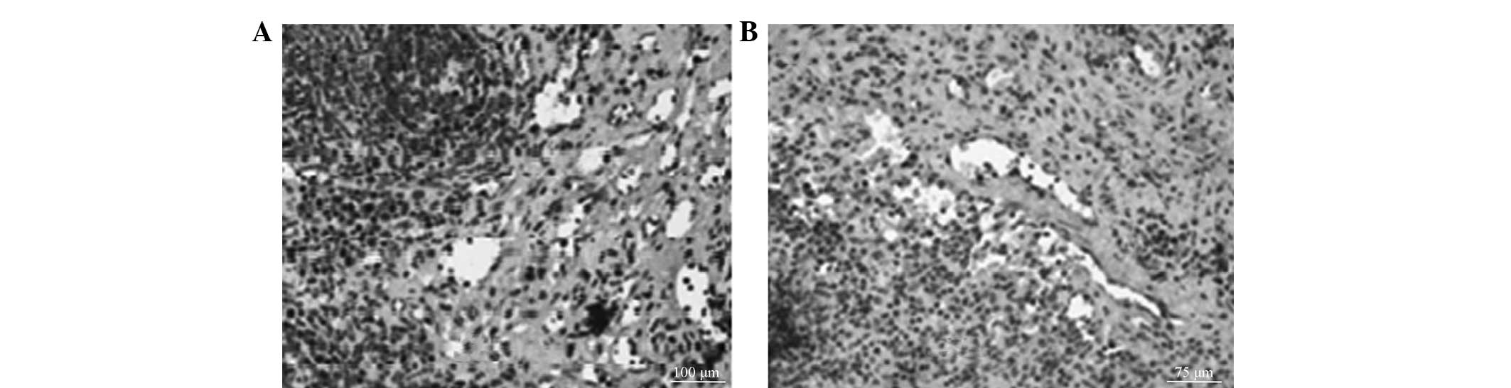
Light microscope observations
The appearance of the splenic tissues in the control
and saline groups were the essentially the same with no significant
histological differences. The splenic sinuses in the PY day 2 group
were dilated and congested, with several degenerated sinusoidal
endothelial cells and splenic cord fiber cells. The sinusoidal
endothelial cells were swollen with eosinophilic changes of nuclear
condensation and fragmentation. The structure of the splenic cord
was slightly blurred with infiltrating inflammatory cells and
histiocytic hyperplasia. The splenic tissue from the PY day 5 group
was congested with significant expansion. A number of sinusoidal
endothelial cells and splenic cord fiber cells were degenerated and
the sinusoidal endothelial cells were swollen with eosinophilic
changes. Numerous cells showed nuclear condensation, fragmentation
and disintegration, and the structure of the splenic cord was
indistinct. Many inflammatory cells showed infiltration and
histiocytosis. The spleen sinus endothelial cell structure was not
observed in the PY day 8 group, but nuclear dissolution, fibrin
exudation and partial shrinkage of splenic bodies were observed.
The edges were congested with bleeding. The splenic corpuscles were
atrophied in the PY day 14 group, with dead endothelial cells and
fibrous tissue proliferation. The splenic capsule was thickened and
interstitial hemosiderin deposition was observed.
TUNEL assay and caspase-3
immunohistochemical analysis
TUNEL labeling identified positive staining in the
cytoplasm (Fig. 1A). The control
and saline groups showed a small degree of positive staining. As
the PY exposure was prolonged, the splenic sinus endothelial cell
apoptosis increased. Caspase-3 immunohistochemistry indicated
positive staining in the cytoplasm (Fig. 1B). Caspase-3 expression was also
observed in the sinusoidal endothelial cells of the control and
saline groups. Following PY treatment, increased staining was
observed in the splenic stromal cells, lymphocytes and vascular
endothelial cells. The control group was selected for comparison
with the day 2 and day 5 saline groups, as well as the day 2 and
day 5 PY groups as the sinusoids were destroyed on days 8 and 14.
The results of the microscopic image analysis showed no significant
differences between the day 2 and day 5 saline groups and the
control group, whereas the PY day 2 and day 5 groups showed
significantly higher staining than was present in the control group
(P<0.01; Table I).
 | Table IChanges in the rate of apoptosis and
caspase-3 expression levels following PY injection (%). |
Table I
Changes in the rate of apoptosis and
caspase-3 expression levels following PY injection (%).
| Apoptosis | Caspase-3 |
|---|
|
|
|
|---|
| Group | Positive rate | Strongly positive
rate | Positive rate | Strongly positive
rate |
|---|
| Control | 13.71±2.97 | 5.89±1.26 | 24.52±1.43 | 4.34±1.92 |
| Saline (day 2) | 15.35±1.14 | 6.57±0.79 | 23.70±1.88 | 4.78±0.78 |
| Saline (day 5) | 14.08±2.96 | 5.75±1.04 | 23.61±2.97 | 4.92±1.45 |
| PY (day 2) | 27.91±3.18a–c | 16.16±2.41a–c | 59.76±3.37a–c | 32.03±0.31a–c |
| PY (day 5) | 32.50±4.07a–c | 19.00±3.49a–c | 63.23±3.27a–c | 30.91±3.23a–c |
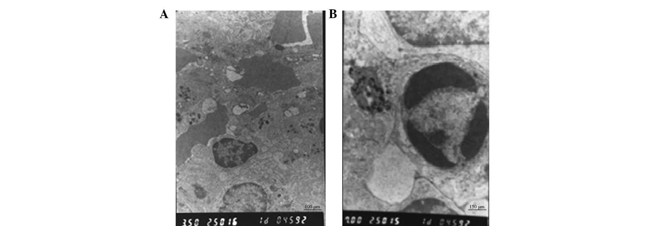
TEM observations
The structure of the splenic sinusoids was normal in
the control and saline groups at the various time-points. In the PY
day 2 group, the splenic sinus was dilated and congested, with
several degenerated sinusoidal endothelial cells and splenic cord
fiber cells. The sinusoidal endothelial cells were swollen with
eosinophilic changes, and the nucleus showed condensation and
fragmentation. The structure of the splenic cord was indistinct and
a few infiltrating inflammatory cells with histiocytic hyperplasia
were observed. The splenic tissues from the PY day 5 group were
significantly congested, and the splenic sinuses were expanded and
congested, with numerous degenerated sinusoidal endothelial cells
and splenic cord fiber cells. The sinusoidal endothelial cells were
swollen with eosinophilic changes, and nuclear condensation,
disintegration and fragmentation were observed (Fig. 2). The structure of the splenic cord
was indistinct and a few infiltrating inflammatory cells with
histiocytic hyperplasia were observed. The splenic sinusoids and
cell structure of the PY day 8 group were not present,
demonstrating nuclear dissolution. Several nuclei were observed
along the edge of the cells, with fibrin exudates and shrinkage of
certain splenic bodies. Additionally, fibrous tissues indicated
proliferation with congestion and hemorrhage along the edge. The
splenic bodies in the PY day 14 group showed atrophy, the splenic
sinuses were collapsed, the endothelial cells and interstitial
cells were necrotic, with fibrous proliferation, the spleen capsule
was thick and interstitial hemosiderin deposition were
observed.
Discussion
No animal model of cavernous hemangioma is available
for study. In previous studies, rats have been injected with tumor
endothelial cells to induce the formation of vascular tumors, but
the resulting vascular tumors significantly differed from
hemangiomas (11,12). The spleen exhibits extensive
sinusoids, which are large and irregular with mutual connections
and filled with blood. The sinus walls consist of rod-shaped
endothelial cells arranged along the longitudinal axis of the
sinusoids and lined with venous sinusoids of the cavernous
hemangioma. These features are similar to those of normal venous
endothelial structures. Thus, the spleen was chosen as a model for
studying the mechanism of action of PY for treating cavernous
hemangioma. In the present study, morphological and TEM
ultrastructural observations indicated that the splenic sinus
endothelial cells and other cells of the spleen were impaired under
PY treatment and the splenic sinuses were gradually destroyed. The
complete splenic sinus structure was unclear under light microscopy
and electron microscopy following PY injection on days 8 and 14.
Among the methods for analyzing apoptotic cells, observation of
morphological changes under an electron microscope was the most
credible (13). Apoptotic cells
were observed at varied PY treatment times under an electron
microscope; however, apoptotic cells were not identified in the
control group. A certain degree of necrosis was observed by light
and electron microscopy. Marginal fibroblast proliferation was
observed 8 and 14 days after PY injection. The morphological
changes may be associated with the splenic capsule thickening and
fibrosis proliferation observed by light microscopy.
TUNEL assay is the most sensitive, rapid and
specific method for detecting apoptosis in situ(14,15).
TUNEL assays are widely used for detecting apoptosis due to the
specificity of the results and the accurate expression of
morphological characteristics, localization and distribution of
apoptotic cells. Furthermore, small concentrations of apoptotic
cells may be detected (16). In
the present study, the analysis of splenic sinus vascular
endothelial cell apoptosis using a TUNEL assay showed that
apoptosis increased in the spleen sinusoidal endothelial cells,
stromal cells and lymphocytes on days 2 and 5 following PY
treatment. Image analysis indicated that the apoptotic rate was
significantly higher than that in the control and saline groups.
These results are consistent with the gradual destruction of the
spleen structure observed by light and electron microscopy and
demonstrated that apoptosis is the main cause of structural damage
in splenic tissues treated with PY.
Caspase-3 is a member of the caspase family and an
important initiator and executor of apoptosis (17). Caspase-3 triggers the
characteristic nuclear changes of apoptosis, such as chromatin
condensation and DNA cleavage (18). The apoptosis of hemangioma
endothelial cell has been identified to be caspase-dependent in a
clinical study (19). In the
present study, quantitative analysis showed that caspase-3
expression was positive in the control group, which may be
associated with the presence of a few apoptotic cells in the spleen
(20). However, caspase-3
expression gradually increased with the extension of PY action
time, which corresponded with the increased rate of PY-induced
apoptosis.
In conclusion, PY was observed to induce the
apoptosis of endothelial cells in the splenic sinuses, which was
accompanied by a certain degree of necrosis and fibroblast
proliferation, ultimately resulting in the destruction of splenic
sinuses, splenic atrophy and scarring. Apoptosis induced by PY
treatment is associated with increased caspase-3 activity.
Considering cell necrosis usually does not exhibit caspase-3
activation characteristics (21),
the results indicated that PY induced apoptosis through the
caspase-3 activation pathway.
The clinical treatment of cavernous hemangioma with
PY requires repeated injections. Therefore, the aforementioned
effects may be more intense in the clinic.
The induction of apoptosis may become the primary
method for treating tumors (22,23).
As caspase activation induces apoptosis (24,25),
the caspase-3-dependent induction of apoptosis in spleen tissues by
PY may provide a novel method for treating hemangiomas. Caspase-3
was specifically activated to trigger apoptosis and the apoptotic
efficiency of tumor vascular endothelial cells was further improved
(26). Hemangioma regression was
performed to avoid the repeated use of chemotherapy agents that may
result in adverse reactions.
References
|
1
|
Mulliken JB and Glowacki J: Hemangiomas
and vascular malformations in infants and children: a
classification based on endothelial characteristics. Plast Reconstr
Surg. 69:412–422. 1982. View Article : Google Scholar
|
|
2
|
Yang Y, Sun M, Hou R, et al: Preliminary
study of fibrin glue combined with pingyangmycin for the treatment
of venous malformations in the oral and maxillofacial region. J
Oral Maxillofac Surg. 66:2219–2225. 2008. View Article : Google Scholar : PubMed/NCBI
|
|
3
|
Liu XJ, Qin ZP and Tai MZ: Angiographic
classification and sclerotic therapy of maxillofacial cavernous
haemangiomas: a report of 204 cases. J Int Med Res. 37:1285–1292.
2009. View Article : Google Scholar : PubMed/NCBI
|
|
4
|
Luo Q and Zhao F: How to use bleomycin A5
for infantile maxillofacial haemangiomas: clinical evaluation of 82
consecutive cases. J Craniomaxillofac Surg. 39:482–486. 2011.
View Article : Google Scholar
|
|
5
|
Hou J, Wang M, Tang H, Wang Y and Huang H:
Pingyangmycin sclerotherapy for infantile hemangiomas in oral and
maxillofacial regions: an evaluation of 66 consecutive patients.
Int J Oral Maxillofac Surg. 40:1246–1251. 2011. View Article : Google Scholar : PubMed/NCBI
|
|
6
|
Gong JH, Liu XJ, Li Y and Zhen YS:
Pingyangmycin downregulates the expression of EGFR and enhances the
effects of cetuximab on esophageal cancer cells and the xenograft
in athymic mice. Cancer Chemother Pharmacol. 69:1323–1332. 2012.
View Article : Google Scholar : PubMed/NCBI
|
|
7
|
Chen P, Liu B and Hu M: The effect of
hydroxycamptothecin and pingyangmycin on human squamous cell
carcinoma of the tongue. Oncol Lett. 5:947–952. 2013.PubMed/NCBI
|
|
8
|
Martines F, Bentivegna D, Maira E, et al:
Cavernous haemangioma of the external auditory canal: clinical case
and review of the literature. Acta Otorhinolaryngol Ital. 32:54–57.
2012.PubMed/NCBI
|
|
9
|
Gu L, Huang DY, Fu CJ, et al:
Intralesional injection of Pingyangmycin plus corticosteroids may
be an effective treatment for cheilitis granulomatosa. Med
Hypotheses. 81:729–730. 2013. View Article : Google Scholar : PubMed/NCBI
|
|
10
|
Yue H, Qian J, Elner VM, et al: Treatment
of orbital vascular malformations with intralesional injection of
pingyangmycin. Br J Ophthalmol. 97:739–745. 2013. View Article : Google Scholar : PubMed/NCBI
|
|
11
|
Mabeta P and Pepper MS: Inhibition of
hemangioma development in a syngeneic mouse model correlates with
bcl-2 suppression and the inhibition of Akt kinase activity.
Angiogenesis. 15:131–139. 2012. View Article : Google Scholar : PubMed/NCBI
|
|
12
|
Gordillo G, Fang H, Khanna S, Harper J,
Phillips G and Sen CK: Oral administration of blueberry inhibits
angiogenic tumor growth and enhances survival of mice with
endothelial cell neoplasm. Antioxid Redox Signal. 11:47–58. 2009.
View Article : Google Scholar : PubMed/NCBI
|
|
13
|
Otsuki Y: Various methods of apoptosis
detection. Acta Histochem Cytochem. 33:235–241. 2000. View Article : Google Scholar
|
|
14
|
Tang J, Li J, Zeng G, et al: Antisense
oligonucleotide suppression of human IGF-1R inhibits the growth and
survival of in vitro cultured epithelial ovarian cancer cells. J
Ovarian Res. 6:712013. View Article : Google Scholar
|
|
15
|
Farivar TN, Najafipour R and Johari P:
Nano - drug delivery of apoptosis activator 2 to AGS cells by
liposomes conjugated with anti-TROP2 antibody. N Am J Med Sci.
4:582–585. 2012. View Article : Google Scholar : PubMed/NCBI
|
|
16
|
Hewitson TD, Bisucci T and Darby IA:
Histochemical localization of apoptosis with in situ labeling of
fragmented DNA. Methods Mol Biol. 326:227–234. 2006.PubMed/NCBI
|
|
17
|
Chang HY and Yang X: Proteases for cell
suicide: functions and regulation of caspases. Microbiol Mol Biol
Rev. 64:821–846. 2000. View Article : Google Scholar
|
|
18
|
Woo M, Hakem R, Soengas MS, et al:
Essential contribution of caspase 3/CPP32 to apoptosis and its
associated nuclear changes. Genes Dev. 12:806–819. 1998. View Article : Google Scholar : PubMed/NCBI
|
|
19
|
Takagi Y, Hattori I, Nozaki K, Ishikawa M
and Hashimoto N: DNA fragmentation in central nervous system
vascular malformations. Acta Neurochir (Wien). 142:987–994. 2000.
View Article : Google Scholar : PubMed/NCBI
|
|
20
|
Zhang Y, Chong E and Herman B:
Age-associated increases in the activity of multiple caspases in
Fisher 344 rat organs. Exp Gerontol. 37:777–789. 2002. View Article : Google Scholar : PubMed/NCBI
|
|
21
|
Vanden Berghe T, Denecker G, Brouckaert G,
Vadimovisch Krysko D, D’Herde K and Vandenabeele P: More than one
way to die: methods to determine TNF-induced apoptosis and
necrosis. Methods Mol Med. 98:101–126. 2004.PubMed/NCBI
|
|
22
|
Piro LD: Apoptosis, Bcl-2 antisense, and
cancer therapy. Oncology (Williston Park). 18:5–10. 2004.PubMed/NCBI
|
|
23
|
Kalas W, Swiderek E, Switalska M, Wietrzyk
J, Rak J and Strzadala L: Thrombospondin-1 receptor mediates
autophagy of RAS-expressing cancer cells and triggers tumour growth
inhibition. Anticancer Res. 33:1429–1438. 2013.PubMed/NCBI
|
|
24
|
Adrain C, Murphy BM and Martin SJ:
Molecular ordering of the caspase activation cascade initiated by
the cytotoxic T lymphocyte/natural killer (CTL/NK) protease
granzyme B. J Biol Chem. 280:4663–4673. 2005. View Article : Google Scholar
|
|
25
|
Sarkars R, Mukherjee S and Roy M:
Targeting heat shock proteins by phenethyl isothiocyanate results
in cell-cycle arrest and apoptosis of human breast cancer cells.
Nutr Cancer. 65:480–493. 2013. View Article : Google Scholar : PubMed/NCBI
|
|
26
|
Frejlich E, Rudno-Rudzińska J, Janiszewski
K, et al: Caspases and their role in gastric cancer. Adv Clin Exp
Med. 22:593–602. 2013.
|