Introduction
Complex regional pain syndrome (CRPS) is a
neuropathic pain syndrome characterized by pain beyond the area of
injury and impairment of the autonomic nervous system and motor
function. The pathophysiological mechanisms leading to neuropathic
pain in CRPS are considered to be complex and multifactorial, and
certain aspects of the mechanism(s) remain to be elucidated
(1).
A considerable amount of evidence implicates
oxidative stress in the pathophysiology of CRPS type I (2,3).
Eisenberg et al(2)
demonstrated significant increases in the quantities of lipid
peroxidation products and the antioxidant parameters in the serum
and saliva of patients with CRPS I. It has also been observed that
vitamin C, an antioxidant, reduces the prevalence of CRPS in humans
following wrist fractures (3).
Consistent with the clinical data, Coderre et al(4) demonstrated that painful
hypersensitivity was reduced by free-radical scavengers and
antioxidant therapy in animals with chronic post-ischemic pain
(CPIP), which is a model of CRPS I. As an animal model for CRPS I,
rats with CPIP display several symptoms that mimic human CRPS,
including edema and chronic mechanical and cold allodynia with no
direct nerve injury (4).
Furthermore, the direct contributions of superoxide
(O2•−) and nitric oxide (NO) were
demonstrated as allopurinol, which inhibits xanthine oxidase
(XO)-mediated superoxide production, or superoxide dismutase (SOD)
and N-nitro-L-arginine methyl ester (L-NAME), which remove
O2•− and inhibit the synthesis of NO,
significantly reduced mechanical allodynia in CPIP model rats
(5).
Peroxynitrite (ONOO−), formed from the
diffusion-controlled reaction of O2•− with
NO, is a highly toxic reactive oxygen species (ROS). It has been
proposed that a number of the toxic effects of NO are due to the
subsequent generation of ONOO−(6,7).
ONOO− is cytotoxic via several mechanisms, including the
initiation of lipid peroxidation, the direct inhibition of
mitochondrial respiratory chain enzymes, the inactivation of
membrane sodium channels, the modifications of oxidative proteins,
and the inhibition of antioxidant enzymes (7–9). Due
to its toxic nature, ONOO− may be involved in a number
of inflammatory conditions (10,11),
cardiovascular diseases (12) and
neurodegenerative diseases (13).
Peroxynitrite has been implicated in several
pathophysiological pain processes, such as thermal hyperalgesia
associated with inflammation and nerve injury (14,15),
opioid-induced hyperalgesia and antinociceptive tolerance (16,17),
and spinal activation of the N-methyl-D-aspartate receptor (NMDAR)
(18). Increased spinal NMDAR
activity, as reflected by increased phosphorylation of NMDAR
subunit 1 (NR1), is critically involved in the development of
central sensitization as a basis of chronic pain (19–21).
However, although it is conceivable that
nitroxidative stress may contribute to CRPS pathophysiology, the
mechanism and significance of ONOO− in CRPS have not yet
been specifically explored. Thus, in the present study, the aim was
to establish whether ONOO- was involved in the development of
allodynia and NMDAR-mediated processes in a CPIP animal model that
mimics the symptoms of human CRPS I. The effect of ONOO−
was examined by removing ONOO− through the
administration of a peroxynitrite decomposition catalyst, FeTMPyP
[5,10,15,20-tetrakis(N-methyl-4′-pyridyl)porphyrinato iron (III)]
pre-reperfusion and subsequently focusing on the preventative
action of FeTMPyP in the ischemia/reperfusion (I/R) injury-induced
CRPS I model.
Materials and methods
Animals
Male Sprague-Dawley rats (280–320 g) were used in
the present study (Central Lab. Animal Inc., Seoul, Korea).
Following their arrival, the rats were acclimated in their cages
for three days prior to the experiment. All housing conditions and
experimental procedures were conducted according to the National
Institutes of Health (Bethesda, MD, USA) guidelines on laboratory
animal welfare and under protocols approved by the Institutional
Animal Care and Use Committee at the Kyungpook National University,
Daegu, Korea.
Induction of the CPIP model by hind paw
I/R
Male Sprague-Dawley rats (n=20) were randomly
allocated to one of five groups (n=4): i) Sham (sham surgery
control group); ii) vehicle (CPIP control group); and CPIP rats
treated with iii) 1, iv) 3 or v) 10 mg/kg FeTMPyP. All rats were
treated at 30 min prior to reperfusion intraperitoneally. The CPIP
model was induced as described by Coderre et al(4). Following induction of anesthesia with
sodium pentobarbital, a Nitrile 70 Durometer O-ring (O-Rings West,
Seattle, WA, USA) was placed around each rat’s left ankle joint for
3 h and then the O-ring was cut to allow reperfusion. The rats in
the sham surgery control group underwent only anesthesia, similar
to the CPIP animals, without an O-ring. For the CPIP control group,
normal saline (the vehicle used to deliver FeTMPyP) was
administered. The dosages of FeTMPyP (1, 3 and 10 mg/kg) were
chosen based on previous publications (14,16,17).
All the chemicals were from Sigma Chemical Co. (St. Louis, MO, USA)
and freshly dissolved in normal saline immediately prior to the
experiment.
Hind paw mechanical allodynia
To assess the mechanical thresholds of the
ipsilateral and contralateral hind paws, rats were acclimatized to
a transparent acrylic box installed on a wire net for 15 min. A
Dynamic Plantar Aesthesiometer (Ugo Basile, Comerio, Italy),
operated in an automated von Frey device, was used for the
measurement of mechanical allodynia. A von Frey filament (steel rod
of 0.5 mm diameter) was pushed against the plantar surface of the
hind paw with an ascending force of 0–50 g. The force at which the
animal withdrew its paw was recorded. Animals were subjected to
four consecutive trials with a minimum 10-sec interval and the
average threshold was calculated. Rats were tested for mechanical
allodynia prior to the I/R injury (baseline value) and on the third
day following reperfusion when mechanical allodynia was at the
maximum (5) by an observer blinded
to the treatments.
Western blot analysis
Animals were rapidly sacrificed. At the time of
sacrifice, rats were deeply anesthetized by sodium pentobarbital
(50 mg/kg, i.p.) and then perfused quickly with cold saline. The
L4–6 section of the spinal cord was immediately harvested,
separated into the left (ipsilateral) and right (contralateral)
sides of the cord and frozen with liquid nitrogen. Subsequently,
the spinal cord was dissolved in lysis buffer solution containing
20 mM Tris-HCl pH 8.0, 150 mM NaCl, 1 mM EDTA, 2 mM
Na3VO4, 0.5 mM DTT, 10% glycerol, 1% Nonidet
P-40 and protease inhibitor cocktail tablet (Roche Diagnostics,
Mannheim, Germany). The samples were centrifuged at 13,800 × g for
20 min at 4°C, the supernatants were separated and the proteins
were quantified by the Bradford method (Bio-Rad Protein Assay kit
I; Bio-Rad, Hercules, CA, USA). Protein samples (50 μg) from each
group were resolved in a buffer solution (0.1 M Tris-HCl, 10%
glycerol, 2% SDS and 0.1% bromophenol blue). The samples were
heated at 100°C for 5 min, loaded onto a 10% SDS-polyacrylamide gel
electrophoresis gel, and then transferred onto nitrocellulose
membrane (Whatman GmbH, Dassel. Germany). The membranes were
blocked with Tris-buffered saline (50 mM Tris pH 7.4 and 10 mM
NaCl) in 3% non-fat milk at room temperature for 1 h and incubated
with a phosphorylated NRI (pNR1) antibody (Upstate Biotechnology,
Inc., Temecula, CA, USA) at 4°C overnight. After washing with
Tris-buffered saline (50 mM Tris pH 7.4, 10 mM NaCl), the membranes
were incubated with anti-rabbit or anti-mouse horseradish
peroxidase-conjugated secondary antibodies (Cell signaling
Technology, Inc., Danvers, MA, USA) (1:2,000) for 1 h at room
temperature prior to identifying the proteins with an ECL system
(Amersham Biosciences, Buckinghamshire, UK). The densities of
protein blots were quantified using LabWorks 4.5 software
(Ultra-Violet Products, Cambridge, UK).
Statistical analysis
The measured data are presented as the mean ±
standard deviation. The data were analyzed with one-way analysis of
variance, followed by post-hoc comparisons (Tukey’s HSD method)
with SPSS, software, version 12.0 (SPSS, Inc., Chicago, IL, USA).
P<0.05 was considered to indicate a statistically significant
difference.
Results
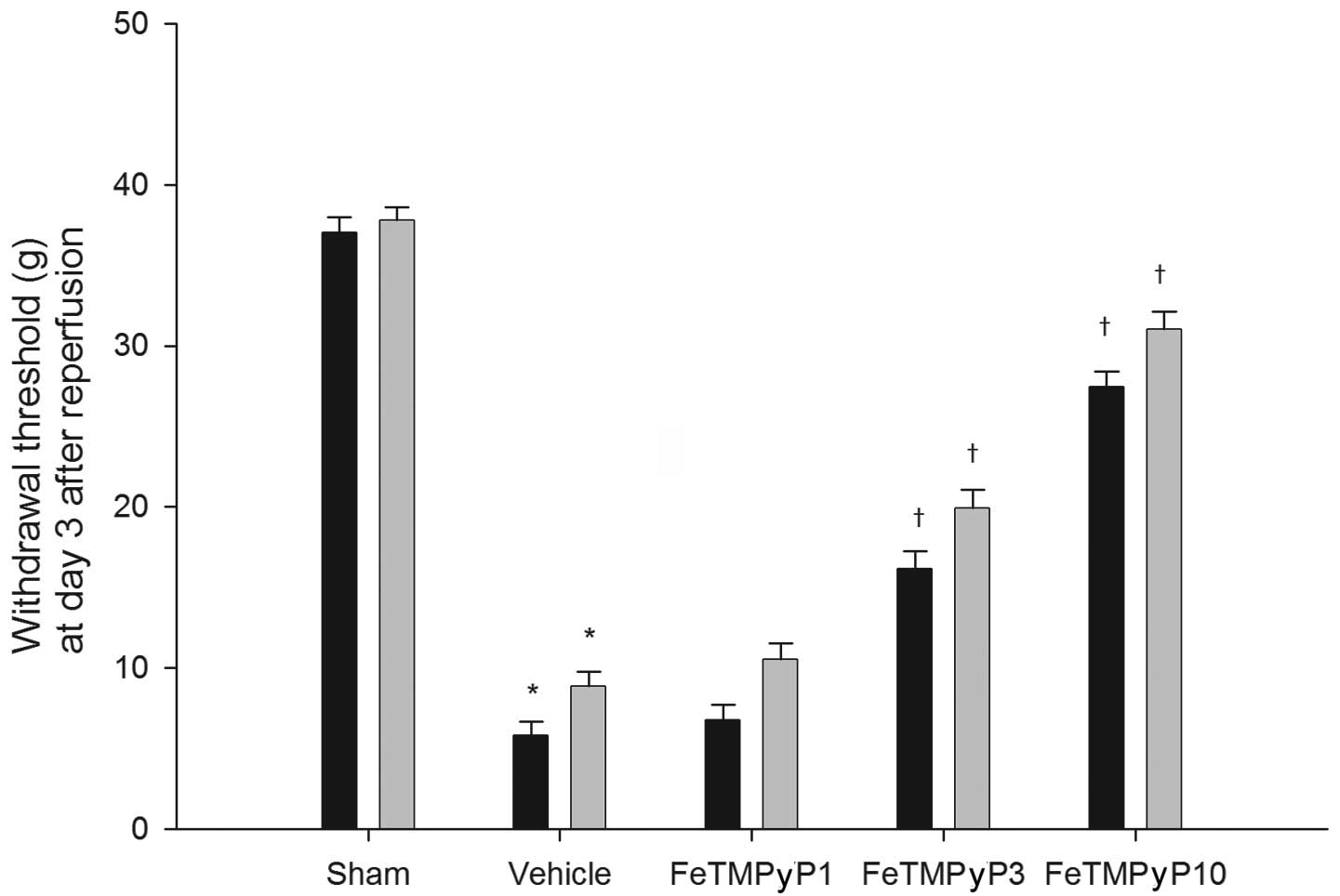
Hindpaw mechanical allodynia
CPIP control rats exhibited significant reductions
in their ipsilateral and contralateral paw withdrawal thresholds
compared with those of the sham control rats. Treatment with 1
mg/kg FeTMPyP was not associated with a significant attenuation of
the ipsilateral and contralateral paw withdrawal thresholds,
whereas treatment with 3 or 10 mg/kg of FeTMPyP was associated with
significant protective effects (Fig.
1).
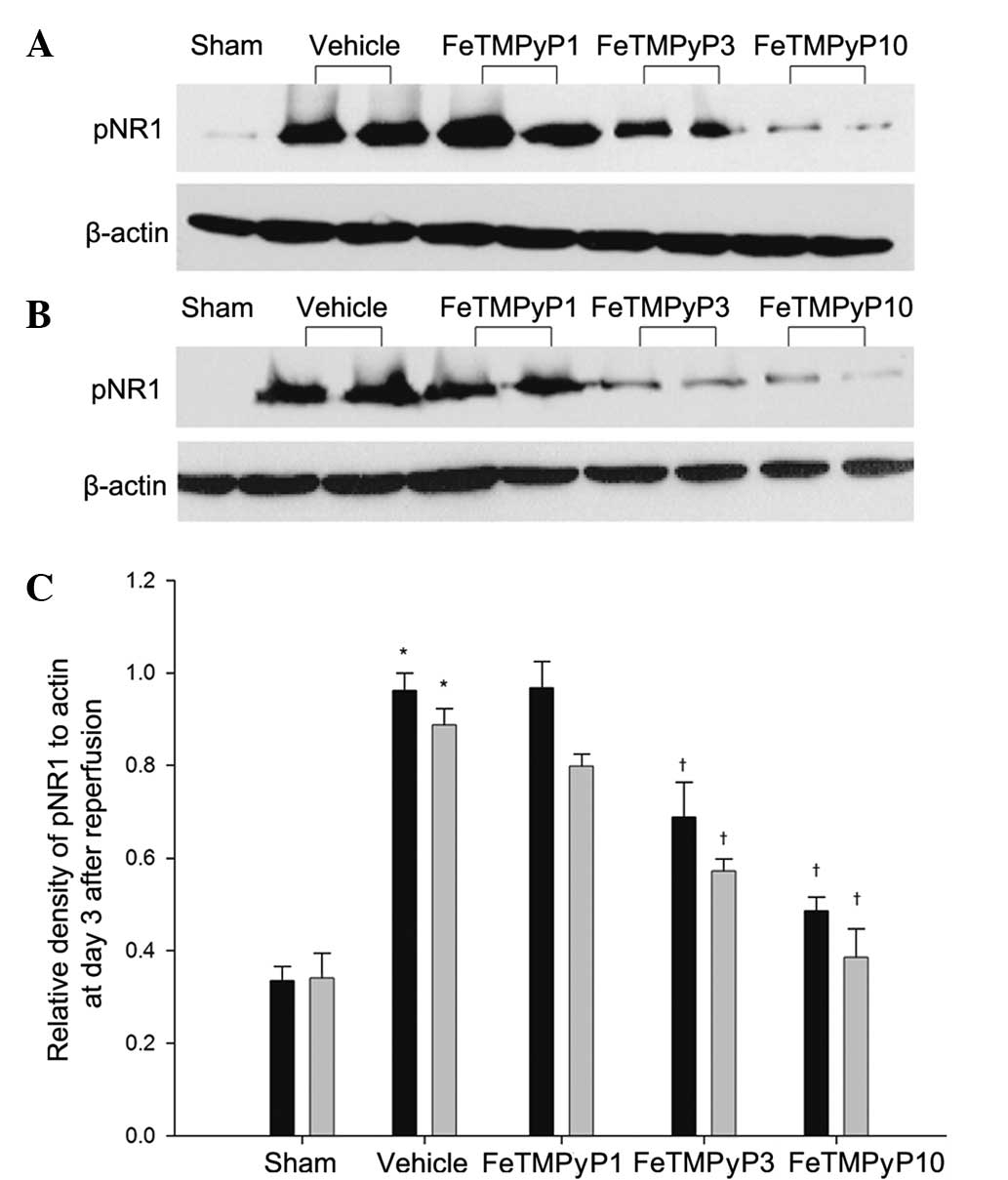
Measurement of pNR1
The degrees of ipsilateral and contralateral
phosphorylation of NR1 were demonstrated to be significantly
increased in the spinal cords of the CPIP control rats compared
with those of the sham control rats. Treatment with 3 or 10 mg/kg
FeTMPyP significantly decreased the ipsilateral and contralateral
pNR1 levels. A degree of reduction was also observed in the 1 mg/kg
FeTMPyP-treated group, but the change was not statistically
significant (Fig. 2).
Discussion
ONOO− is well established as a very
reactive species, inducing cytotoxicity in various diseases, but
its involvement in the pain associated with CRPS is not well
understood. Using a CPIP rat model exhibiting symptoms that
resembled those of humans with CRPS, the results of the present
study demonstrated that ONOO− is a determinant of pain
in this setting.
CRPS I is a chronic pain syndrome that occurs
following relatively benign injuries, such as sprains, fractures,
tissue trauma and ischemia, with no definable nerve lesion
(22). The CPIP model was
developed by Coderre et al(4) and was used to study the mechanisms
that potentially underlie CRPS I. Coderre et al(4) demonstrated that 3-h I/R of the hind
paw induced several symptoms that mimicked human CRPS I, including
edema, as well as chronic mechanical and cold allodynia, with no
direct nerve injury.
I/R injury is the tissue damage caused when the
blood supply returns to a tissue following a period of ischemia.
Prolonged ischemia leads to the accumulation of oxidases, which are
enzymes that produce free radicals. The reintroduction of molecular
O2 into ischemic tissue upon reperfusion leads to the
overproduction of ROS. A cascade of ROS formation is initiated by
the generation of O2•−, which is generated by
XO. NO and O2•− may react together to produce
significant amounts of a much more oxidatively active molecule,
ONOO−, which is a potent oxidizing agent that causes
post-hypoxic cellular injury (23). A study has observed XO-mediated
O2•− production in a CPIP model and
demonstrated that O2•− and NO mediate I/R
injury-induced chronic pain (5).
O2•− and NO are highly reactive and unstable
(24); their reaction is ~3 times
faster than the dismutation of O2•− by SOD.
Additionally, ONOO− is a strong oxidant and is more
stable than NO or O2•−(25). The stability of ONOO− is
sufficient to allow it to cross several cell diameters to reach
target cells prior to becoming protonated and decomposing (26). Evidence supports the major
involvement of ONOO− in the development of tissue damage
during inflammation (27,28), as well as in the pain of several
etiologies (29).
Previous studies have provided concrete evidence
implicating peroxynitrite in the development of certain aspects of
chronic pain (30,31). Peroxynitrite has been reported to
contribute to the development of chronic pain by means of
peripheral and central actions. These studies identified that the
peripheral formation of ONOO− contributes to
hyperalgesia by favoring the production of several proinflammatory
cytokines and by increasing the production of prostaglandin E2.
Peripheral administration of ONOO− or ONOO−
precursors induces inflammatory hyperalgesia (14). In a neuropathic pain model, the
daily administration of uric acid decreased
ONOO−-mediated nitration in peripheral nerves and
alleviated thermal hyperalgesia and Wallerian degeneration
(15). These effects may also
occur in CNS regions responsible for pain processing through the
mediation of central sensitization. ONOO− is considered
to contribute to central sensitization through the alteration of
NMDAR activation by nitrating proteins that are important in the
maintenance of normal nociceptive processing, such as MnSOD
(32,33), glutamate transporters (GTs) and
glutamine synthase (GS) (34,35).
The ONOO−-mediated nitration of MnSOD inactivates the
enzyme, which results in increased O2 and
ONOO− levels, leading to enhanced postsynaptic neuronal
responsiveness that contributes to central sensitization (18,29,32,33).
Nitration of GLT-1 and GS disrupts glutamate homeostasis and
increases glutamate neurotransmission, and the resulting signaling
events underlie central sensitization. Glutamate is the primary
endogenous ligand for the NMDAR. Extracellular glutamate
concentrations have to be maintained low enough to terminate
glutamate receptor activation. When GTs are nitrated by
ONOO−, their inactivation results in increased glutamate
concentrations and altered synaptic transmission (34). GS, which catalyzes the conversion
of glutamate and ammonia to glutamine, is also inactivated via
nitration by ONOO−(35).
In the present study, it was demonstrated that the
ONOO− decomposition catalyst FeTMPyP prevented NMDAR
activation, suggesting another mechanism for ONOO− in
the spinal cord in addition to its ability to block mechanical
allodynia. It is possible that I/R injury induces pathological
responses in the CNS similar to those induced by mechanical or
inflammatory nerve damage through ONOO−-induced
nitrosative processes. Although the results suggest
ONOO− is potentially involved in mechanical allodynia
and central sensitization, the site of action has not been
identified as the drugs used were administered systemically. To
this end, and as ONOO− is produced in peripheral
nociceptors and also in central nociceptors, it is likely that
mechanical allodynia is associated with increased ONOO−
formation in peripheral and central pathways.
In effect, counteracting the damage associated with
ONOO− may be accomplished by decomposing
ONOO− and also by preventing the formation of
ONOO−. In a previous study, in which the formation of SO
and NO, as a precursor of ONOO−, was prevented in
NMDA-mediated central sensitization using L-NAME and SOD in CPIP
rats, it was indirectly observed that ONOO− from these
reactive species may be a causative factor in allodynia in CPIP
rats, since the prevention of ONOO− formation centers on
inhibition of the formation of SO and NO. However, NO is often
considered to be pro-nociceptive by generating nitrogen free
radicals and causing vasodilatation, thereby facilitating
inflammatory processes. NO-induced vasodilatation may relieve
pain-producing vasospasm in the CPIP model. Thus, decomposing
ONOO− appears to be an improved strategy for reducing
free radical-mediated toxicity as it preserves the positive effects
of NO.
In conclusion, the results of the present study
indicate that ONOO− is critically involved in the
pathogenesis of I/R injury-induced neuropathy and the formation of
ONOO− in the spinal cord in response to NMDAR
activation, and that ONOO− contributes to the
development of central sensitization. A ONOO−
decomposition catalyst demonstrated a protective effect against
CPIP, as evidenced by the improvement in mechanical allodynia and
central sensitization. The present study supports the potential of
ONOO− as a novel target for pain management in CRPS
patients.
Acknowledgements
This study was supported by a BioMedical Research
Institute grant from Kyungpook National University Hospital
(2010).
References
|
1
|
Bruehl S: An update on the pathophysiology
of complex regional pain syndrome. Anesthesiology. 113:713–725.
2010.PubMed/NCBI
|
|
2
|
Eisenberg E, Shtahl S, Geller R, et al:
Serum and salivary oxidative analysis in complex regional pain
syndrome. Pain. 138:226–232. 2008. View Article : Google Scholar : PubMed/NCBI
|
|
3
|
Zollinger PE, Tuinebreijer WE, Breederveld
RS and Kreis RW: Can vitamin C prevent complex regional pain
syndrome in patients with wrist fractures? A randomized,
controlled, multicenter dose-response study. J Bone Joint Surg Am.
89:1424–1431. 2007. View Article : Google Scholar : PubMed/NCBI
|
|
4
|
Coderre TJ, Xanthos DN, Francis L and
Bennett GJ: Chronic post-ischemia pain (CPIP): a novel animal model
of complex regional pain syndrome-type I (CRPS-I; reflex
sympathetic dystrophy) produced by prolonged hindpaw ischemia and
reperfusion in the rat. Pain. 112:94–105. 2004. View Article : Google Scholar
|
|
5
|
Kwak KH, Han CG, Lee SH, et al: Reactive
oxygen species in rats with chronic post-ischemia pain. Acta
Anaesthesiol Scand. 53:648–656. 2009. View Article : Google Scholar : PubMed/NCBI
|
|
6
|
Rodrigo J, Fernandez AP, Serrano J,
Peinado MA and Martinez A: The role of free radicals in cerebral
hypoxia and ischemia. Free Radic Biol Med. 39:26–50. 2005.
View Article : Google Scholar : PubMed/NCBI
|
|
7
|
Szabó C: Multiple pathways of
peroxynitrite cytotoxicity. Toxicol Lett. 140–141:105–112.
2003.
|
|
8
|
Beckman JS, Chen J, Crow JP and Ye YZ:
Reactions of nitric oxide, superoxide and peroxynitrite with
superoxide dismutase in neurodegeneration. Prog Brain Res.
103:371–380. 1994. View Article : Google Scholar : PubMed/NCBI
|
|
9
|
Radi R, Beckman JS, Bush KM and Freeman
BA: Peroxynitrite-induced membrane lipid peroxidation: the
cytotoxic potential of superoxide and nitric oxide. Arch Biochem
Biophys. 288:481–487. 1991. View Article : Google Scholar : PubMed/NCBI
|
|
10
|
Mabley JG, Liaudet L, Pacher P, Southan
GJ, Groves JT, Salzman AL and Szabó C: Part II: beneficial effects
of the peroxynitrite decomposition catalyst FP15 in murine models
of arthritis and colitis. Mol Med. 8:581–590. 2002.PubMed/NCBI
|
|
11
|
Cuzzocrea S, Mazzon E, Di Paola R,
Esposito E, Macarthur H, Matuschak GM and Salvemini D: A role for
nitric oxide-mediated peroxynitrite formation in a model of
endotoxin-induced shock. J Pharmacol Exp Ther. 319:73–81. 2006.
View Article : Google Scholar : PubMed/NCBI
|
|
12
|
Pacher P, Beckman JS and Liaudet L: Nitric
oxide and peroxynitrite in health and disease. Physiol Rev.
87:315–424. 2007. View Article : Google Scholar : PubMed/NCBI
|
|
13
|
Torreilles F, Salman-Tabcheh S, Guérin M
and Torreilles J: Neurodegenerative disorders: the role of
peroxynitrite. Brain Res Brain Res Rev. 30:153–163. 1999.
View Article : Google Scholar : PubMed/NCBI
|
|
14
|
Ndengele MM, Cuzzocrea S, Esposito E,
Mazzon E, Di Paola R, Matuschak GM and Salvemini D: Cyclooxygenases
1 and 2 contribute to peroxynitrite-mediated inflammatory pain
hypersensitivity. FASEB J. 22:3154–3164. 2008. View Article : Google Scholar : PubMed/NCBI
|
|
15
|
Liu T, Knight KR and Tracey DJ:
Hyperalgesia due to nerve injury-role of peroxynitrite.
Neuroscience. 97:125–131. 2000. View Article : Google Scholar
|
|
16
|
Muscoli C, Cuzzocrea S, Ndengele MM, et
al: Therapeutic manipulation of peroxynitrite attenuates the
development of opiate-induced antinociceptive tolerance in mice. J
Clin Invest. 117:3530–3539. 2007. View
Article : Google Scholar : PubMed/NCBI
|
|
17
|
Ndengele MM, Cuzzocrea S, Masini E, et al:
Spinal ceramide modulates the development of morphine
antinociceptive tolerance via peroxynitrite-mediated nitroxidative
stress and neuroimmune activation. J Pharmacol Exp Ther. 329:64–75.
2009. View Article : Google Scholar
|
|
18
|
Muscoli C, Mollace V, Wheatley J, Masini
E, Ndengele M, Wang ZQ and Salvemini D: Superoxide-mediated
nitration of spinal manganese superoxide dismutase: a novel pathway
in N-methyl-D-aspartate-mediated hyperalgesia. Pain. 111:96–103.
2004. View Article : Google Scholar
|
|
19
|
Guo W, Zou S, Guan Y, Ikeda T, Tal M,
Dubner R and Ren K: Tyrosine phosphorylation of the NR2B subunit of
the NMDA receptor in the spinal cord during the development and
maintenance of inflammatory hyperalgesia. J Neurosci. 22:6208–6217.
2002.PubMed/NCBI
|
|
20
|
Zou X, Lin Q and Willis WD: Enhanced
phosphorylation of NMDA receptor 1 subunits in spinal cord dorsal
horn and spinothalamic tract neurons after intradermal injection of
capsaicin in rats. J Neurosci. 20:6989–6997. 2000.
|
|
21
|
Gao X, Kim HK, Chung JM and Chung K:
Reactive oxygen species (ROS) are involved in enhancement of
NMDA-receptor phosphorylation in animal models of pain. Pain.
131:262–271. 2007. View Article : Google Scholar : PubMed/NCBI
|
|
22
|
Stanton-Hicks M, Jänig W, Hassenbusch S,
Haddox JD, Boas R and Wilson P: Reflex sympathetic dystrophy:
changing concepts and taxonomy. Pain. 63:127–133. 1995. View Article : Google Scholar : PubMed/NCBI
|
|
23
|
Beckman JS, Beckman TW, Chen J, Marshall
PA and Freeman BA: Apparent hydroxyl radical production by
peroxynitrite: implications for endothelial injury from nitric
oxide and superoxide. Proc Natl Acad Sci USA. 87:1620–1624. 1990.
View Article : Google Scholar : PubMed/NCBI
|
|
24
|
Goldstein S and Czapski G: The reaction of
NO• with O2•− and
HO2•: a pulse radiolysis study. Free Radic
Biol Med. 19:505–510. 1995.
|
|
25
|
Beckman JS and Crow JP: Pathological
implications of nitric oxide, superoxide and peroxynitrite
formation. Biochem Soc Trans. 21:330–334. 1993.PubMed/NCBI
|
|
26
|
Crow JP and Beckman JS: The role of
peroxynitrite in nitric oxide-mediated toxicity. Curr Top Microbiol
Immunol. 196:57–73. 1995.PubMed/NCBI
|
|
27
|
Cuzzocrea S, Zingarelli B, Hake P, Salzman
AL and Szabó C: Antiinflammatory effects of mercaptoethylguanidine,
a combined inhibitor of nitric oxide synthase and peroxynitrite
scavenger, in carrageenan-induced models of inflammation. Free
Radic Biol Med. 24:450–459. 1998. View Article : Google Scholar
|
|
28
|
Salvemini D, Wang ZQ, Wyatt PS, Bourdon
DM, Marino MH, Manning PT and Currie MG: Nitric oxide: a key
mediator in the early and late phase of carrageenan-induced rat paw
inflammation. Br J Pharmacol. 118:829–838. 1996. View Article : Google Scholar : PubMed/NCBI
|
|
29
|
Salvemini D, Little JW, Doyle T and
Neumann WL: Roles of reactive oxygen and nitrogen species in pain.
Free Radic Biol Med. 51:951–966. 2011. View Article : Google Scholar : PubMed/NCBI
|
|
30
|
Drel VR, Pacher P, Vareniuk I, et al: A
peroxynitrite decomposition catalyst counteracts sensory neuropathy
in streptozotocin-diabetic mice. Eur J Pharmacol. 569:48–58. 2007.
View Article : Google Scholar : PubMed/NCBI
|
|
31
|
Obrosova IG, Drel VR, Oltman CL, Mashtalir
N, Tibrewala J, Groves JT and Yorek MA: Role of nitrosative stress
in early neuropathy and vascular dysfunction in
streptozotocin-diabetic rats. Am J Physiol Endocrinol Metab.
293:E1645–E1655. 2007. View Article : Google Scholar : PubMed/NCBI
|
|
32
|
Radi R: Nitric oxide, oxidants, and
protein tyrosine nitration. Proc Natl Acad Sci USA. 101:4003–4008.
2004. View Article : Google Scholar : PubMed/NCBI
|
|
33
|
Wang Z, Porreca F, Cuzzocrea S, et al: A
newly identified role for superoxide in inflammatory pain. J
Pharmacol Exp Ther. 309:869–878. 2004. View Article : Google Scholar : PubMed/NCBI
|
|
34
|
Trotti D, Rossi D, Gjesdal O, Levy LM,
Racagni G, Danbolt NC and Volterra A: Peroxynitrite inhibits
glutamate transporter subtypes. J Biol Chem. 271:5976–5979. 1996.
View Article : Google Scholar : PubMed/NCBI
|
|
35
|
Görg B, Wettstein M, Metzger S, Schliess F
and Häussinger D: Lipopolysaccharide-induced tyrosine nitration and
inactivation of hepatic glutamine synthetase in the rat.
Hepatology. 41:1065–1073. 2005.PubMed/NCBI
|