Contradictory imaging and EEG results in resection surgery of bitemporal lobe epilepsy: A case report

  • Authors:
    • Kang Yang
    • Jing Su
    • Yue Lang
    • Shu‑Ping Liu
    • Jian Yin
  • View Affiliations

  • Published online on: December 27, 2013     https://doi.org/10.3892/etm.2013.1462
  • Pages: 731-733
Metrics: Total Views: 0 (Spandidos Publications: | PMC Statistics: )
Total PDF Downloads: 0 (Spandidos Publications: | PMC Statistics: )


Abstract

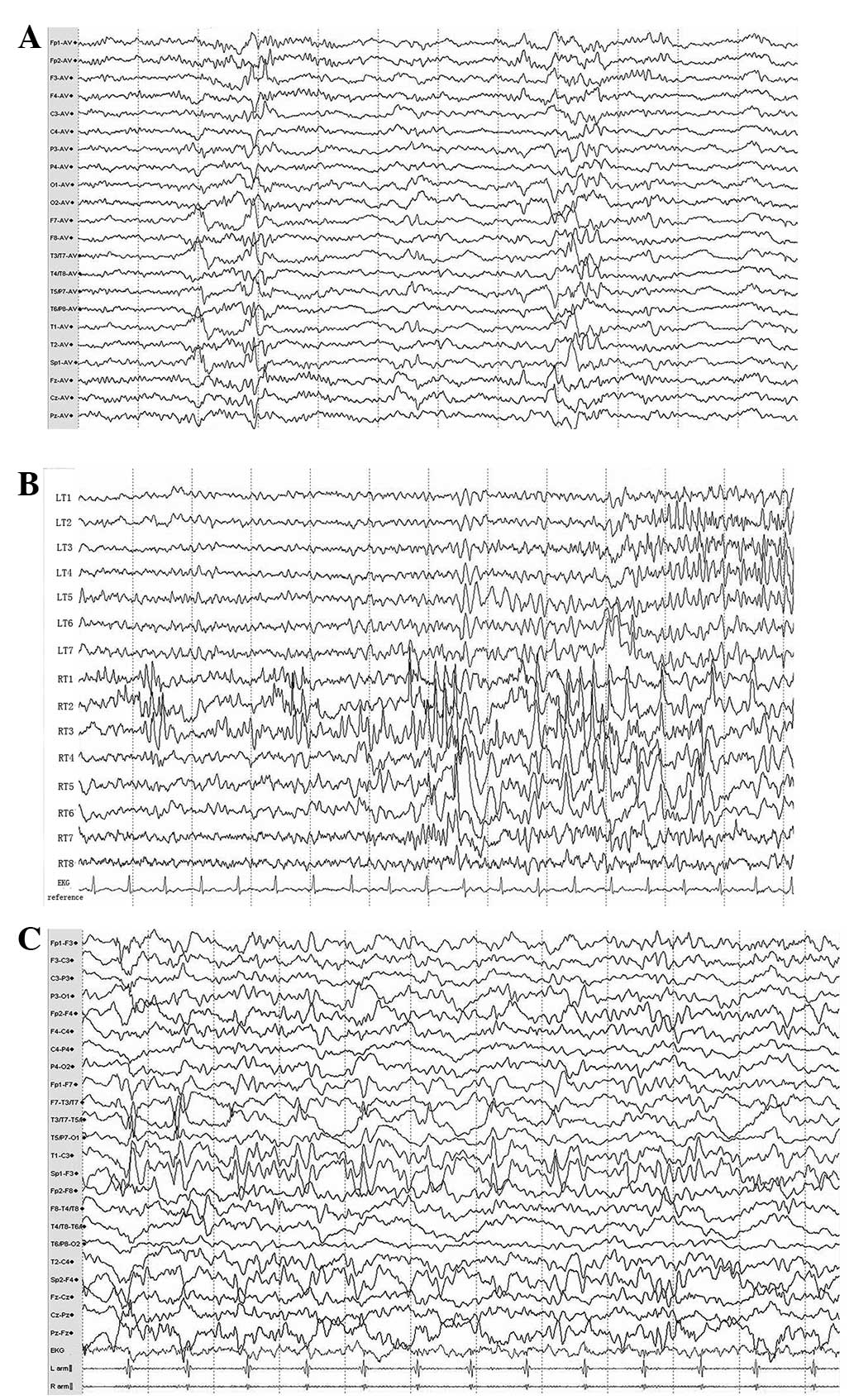
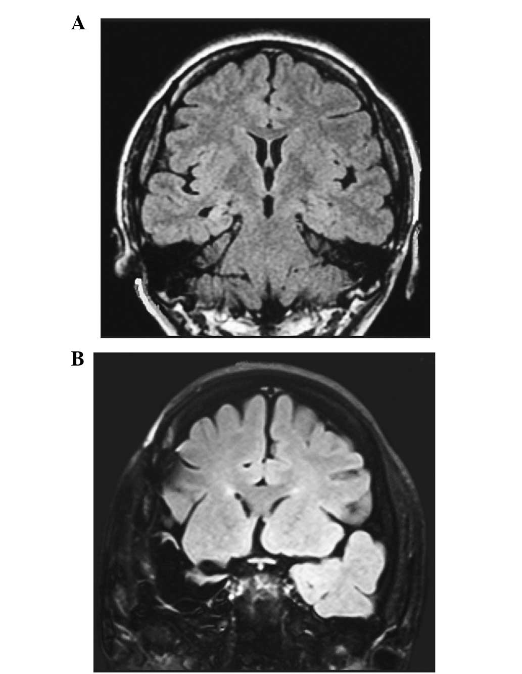
The present study describes the case of a 27‑year‑old, right‑handed female with bilateral mesial temporal lobe epilepsy. Electroencephalogram (EEG) monitoring from implanted electrodes displayed two different and independent onsets on the two sides of the mesial temporal structures, which specifically included clinical generalized tonic clonic seizure (GTCS) discharges originating from the left mesial temporal lobe, as well as complex partial seizure (CPS) discharges arising from the right mesial region. However, fluid‑attenuated inversion recovery magnetic resonance imaging (FLAIR MRI) showed a unilateral abnormality, as in right mesial temporal lobe sclerosis. A decision was made to resect one side of the mesial temporal lobe, in order to avoid memory function impairment, and, relying on the MRI results, the right side was selected. However, surgery did not leave the patient seizure‑free. The CPSs gradually eased, while the GTCSs originating from the left side became severely aggravated. In describing this case, the drawbacks of current epileptic diagnostic methods and surgical strategies for bitemporal lobe epilepsy are discussed, and the requirement for more treatment options is emphasized.
View Figures
View References

Related Articles

Journal Cover

2014-March
Volume 7 Issue 3

Print ISSN: 1792-0981
Online ISSN:1792-1015

Sign up for eToc alerts

Recommend to Library

Copy and paste a formatted citation
x
Spandidos Publications style
Yang K, Su J, Lang Y, Liu SP and Yin J: Contradictory imaging and EEG results in resection surgery of bitemporal lobe epilepsy: A case report. Exp Ther Med 7: 731-733, 2014
APA
Yang, K., Su, J., Lang, Y., Liu, S., & Yin, J. (2014). Contradictory imaging and EEG results in resection surgery of bitemporal lobe epilepsy: A case report. Experimental and Therapeutic Medicine, 7, 731-733. https://doi.org/10.3892/etm.2013.1462
MLA
Yang, K., Su, J., Lang, Y., Liu, S., Yin, J."Contradictory imaging and EEG results in resection surgery of bitemporal lobe epilepsy: A case report". Experimental and Therapeutic Medicine 7.3 (2014): 731-733.
Chicago
Yang, K., Su, J., Lang, Y., Liu, S., Yin, J."Contradictory imaging and EEG results in resection surgery of bitemporal lobe epilepsy: A case report". Experimental and Therapeutic Medicine 7, no. 3 (2014): 731-733. https://doi.org/10.3892/etm.2013.1462